Advertisement

Main menu

  • Home
  • Content
    • Current Issue
    • Accepted Manuscripts
    • Article Preview
    • Past Issue Archive
    • Video Articles
    • AJNR Case Collection
    • Case of the Week Archive
    • Case of the Month Archive
    • Classic Case Archive
  • Special Collections
    • AJNR Awards
    • Low-Field MRI
    • Alzheimer Disease
    • ASNR Foundation Special Collection
    • Photon-Counting CT
    • View All
  • Multimedia
    • AJNR Podcasts
    • AJNR SCANtastic
    • Trainee Corner
    • MRI Safety Corner
    • Imaging Protocols
  • For Authors
    • Submit a Manuscript
    • Submit a Video Article
    • Submit an eLetter to the Editor/Response
    • Manuscript Submission Guidelines
    • Statistical Tips
    • Fast Publishing of Accepted Manuscripts
    • Graphical Abstract Preparation
    • Imaging Protocol Submission
    • Author Policies
  • About Us
    • About AJNR
    • Editorial Board
    • Editorial Board Alumni
  • More
    • Become a Reviewer/Academy of Reviewers
    • Subscribers
    • Permissions
    • Alerts
    • Feedback
    • Advertisers
    • ASNR Home

User menu

  • Alerts
  • Log in

Search

  • Advanced search
American Journal of Neuroradiology

American Journal of Neuroradiology

ASHNR American Society of Functional Neuroradiology ASHNR American Society of Pediatric Neuroradiology ASSR
  • Alerts
  • Log in

Advanced Search

  • Home
  • Content
    • Current Issue
    • Accepted Manuscripts
    • Article Preview
    • Past Issue Archive
    • Video Articles
    • AJNR Case Collection
    • Case of the Week Archive
    • Case of the Month Archive
    • Classic Case Archive
  • Special Collections
    • AJNR Awards
    • Low-Field MRI
    • Alzheimer Disease
    • ASNR Foundation Special Collection
    • Photon-Counting CT
    • View All
  • Multimedia
    • AJNR Podcasts
    • AJNR SCANtastic
    • Trainee Corner
    • MRI Safety Corner
    • Imaging Protocols
  • For Authors
    • Submit a Manuscript
    • Submit a Video Article
    • Submit an eLetter to the Editor/Response
    • Manuscript Submission Guidelines
    • Statistical Tips
    • Fast Publishing of Accepted Manuscripts
    • Graphical Abstract Preparation
    • Imaging Protocol Submission
    • Author Policies
  • About Us
    • About AJNR
    • Editorial Board
    • Editorial Board Alumni
  • More
    • Become a Reviewer/Academy of Reviewers
    • Subscribers
    • Permissions
    • Alerts
    • Feedback
    • Advertisers
    • ASNR Home
  • Follow AJNR on Twitter
  • Visit AJNR on Facebook
  • Follow AJNR on Instagram
  • Join AJNR on LinkedIn
  • RSS Feeds

AJNR Awards, New Junior Editors, and more. Read the latest AJNR updates

Research ArticleNeurointervention

How to Size Intracranial Aneurysms: A Phantom Study of Invasive and Noninvasive Methods

D. Behme, N. Amelung, T. Khakzad and M.-N. Psychogios
American Journal of Neuroradiology December 2018, 39 (12) 2291-2296; DOI: https://doi.org/10.3174/ajnr.A5866
D. Behme
aFrom the Department of Diagnostic and Interventional Neuroradiology, University Medical Center Göttingen, Göttingen, Germany.
  • Find this author on Google Scholar
  • Find this author on PubMed
  • Search for this author on this site
  • ORCID record for D. Behme
N. Amelung
aFrom the Department of Diagnostic and Interventional Neuroradiology, University Medical Center Göttingen, Göttingen, Germany.
  • Find this author on Google Scholar
  • Find this author on PubMed
  • Search for this author on this site
  • ORCID record for N. Amelung
T. Khakzad
aFrom the Department of Diagnostic and Interventional Neuroradiology, University Medical Center Göttingen, Göttingen, Germany.
  • Find this author on Google Scholar
  • Find this author on PubMed
  • Search for this author on this site
  • ORCID record for T. Khakzad
M.-N. Psychogios
aFrom the Department of Diagnostic and Interventional Neuroradiology, University Medical Center Göttingen, Göttingen, Germany.
  • Find this author on Google Scholar
  • Find this author on PubMed
  • Search for this author on this site
  • ORCID record for M.-N. Psychogios
  • Article
  • Figures & Data
  • Info & Metrics
  • Responses
  • References
  • PDF
Loading

Article Figures & Data

Figures

  • Tables
  • Fig 1.
    • Download figure
    • Open in new tab
    • Download powerpoint
    Fig 1.

    Experimental setup of an aneurysm model inside a human skull on the biplane Artis Q angiosuite.

  • Fig 2.
    • Download figure
    • Open in new tab
    • Download powerpoint
    Fig 2.

    Examples of the different image techniques applied: MPR and VRT of MDCTA images (A and B), MPR and VRT images of FDCTA images (C and D), MPR and VRT images obtained from a 3D-DSA run (E and F), short object-to-detector distance DSA image no optimal projection (G), and optimized projection DSA image with short object-to-detector distance (H, DSA rotation near).
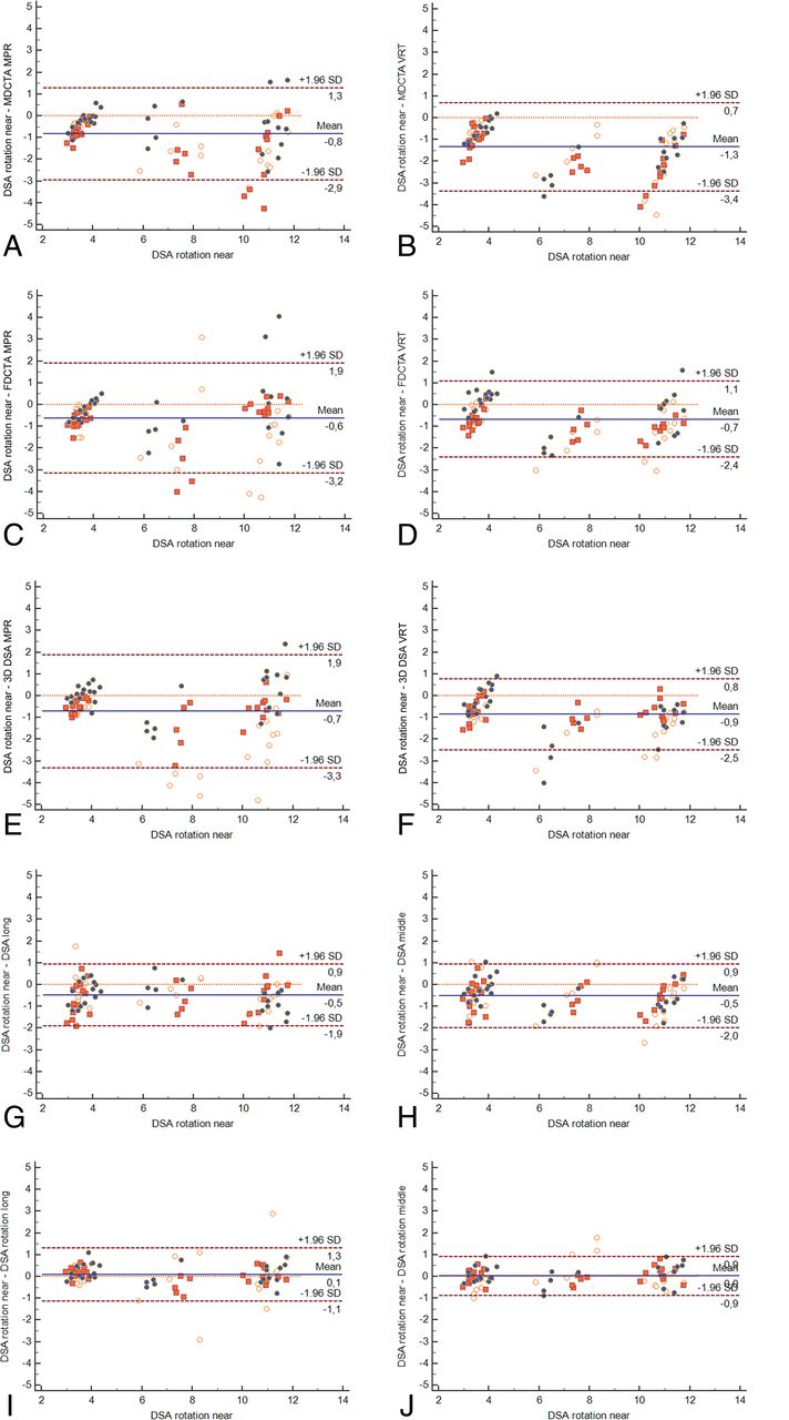
  • Fig 3.
    • Download figure
    • Open in new tab
    • Download powerpoint
    Fig 3.

    Bland-Altman plots comparing all techniques with DSA rotation near MDCTA MPR (A) versus DSA rotation near MDCTA VRT (B) versus DSA rotation near FDCTA MPR (C) versus DSA rotation near FDCTA VRT (D) versus DSA rotation near 3D DSA MPR (E) versus DSA rotation near 3D DSA VRT (F) versus DSA rotation near DSA long (G) versus DSA rotation near DSA middle (H) versus DSA rotation near DSA rotation long (I) versus DSA rotation near DSA rotation middle versus DSA rotation near (J).

Tables

  • Figures
    • View popup
    Table 1:

    Examination parameters

    MDCTAFDCTA3D DSADSA
    FOV (cm)10 × 1010 × 1010 × 1015 × 15
    Matrix size512 × 512512 × 512512 × 5121024 × 1024
    In-plane resolutions (mm)0.20.20.290.15
    Cumulative dose (mGy)3040953
    • View popup
    Table 2:

    Aneurysm model 1—mean of differences in known aneurysm sizes (all 3 dimensions combined)

    Statistics/TechniqueMean (mm)SDSE of MeanLower 95% CIUpper 95% CI
    FDCTA MPR0.751.640.240.261.24
    FDCTA VRT0.990.690.100.781.20
    MDCTA MPR1.151.260.190.771.52
    MDCTA VRT1.910.690.101.702.11
    DSA long0.500.840.130.250.75
    DSA middle0.650.520.080.490.80
    DSA near0.660.710.110.450.87
    3D DSA MPR1.101.550.230.631.56
    3D DSA VRT1.190.690.100.981.40
    DSA rotation long−0.051.710.25−0.560.47
    DSA rotation middle−0.150.640.09−0.340.04
    DSA rotation near−0.070.610.09−0.250.18
    • Note:—SE indicates standard error.

    • View popup
    Table 3:

    Aneurysm model 2—mean of differences in known aneurysm sizes (all 3 dimensions combined)

    Statistics/TechniqueMean (mm)SDSE of MeanLower 95% CIUpper 95% CI
    FDCTA MPR0.620.600.090.440.79
    FDCTA VRT0.400.480.070.250.54
    MDCTA MPR0.540.310.050.450.63
    MDCTA VRT0.830.370.060.720.94
    DSA long0.570.690.100.370.78
    DSA middle0.510.660.100.310.70
    DSA near0.630.730.110.410.85
    3D DSA MPR0.410.400.060.290.53
    3D DSA VRT0.680.490.070.530.82
    DSA rotation long−0.050.340.05−0.150.05
    DSA rotation middle0.170.390.060.0520.28
    DSA rotation near0.120.250.040.050.20
    • Note:—SE indicates standard error.

    • View popup
    Table 4:

    Statistical analyses of the Bland-Altman plots comparing all techniques against DSA rotation “near” images

    Statistics/TechniqueArithmetic Mean Differences (95% CI)Lower Limit (95% CI)Upper Limit (95% CI)
    FDCTA MPR−0.64 (−0.91 to −0.37)−3.14 (−3.60 to −2.68)1.86 (1.40–2.32)
    FDCTA VRT−0.67 (−0.86 to −0.49)−2.37 (−2.68 to −2.06)1.02 (0.71–1.33)
    MDCTA MPR−0.84 (−1.05 to −0.62)−2.89 (−3.27 to −2.52)1.22 (0.85–1.60)
    MDCTA VRT−1.34 (−1.56 to −1.13)−3.37 (−3.74 to −2.99)0.68 (0.31–1.05)
    DSA long−0.48 (−0.63 to −0.33)−1.90 (−2.16 to −1.64)0.94 (0.68–1.20)
    DSA middle−0.51 (−0.67 to −0.36)−1.98 (−2.25 to −1.71)0.95 (0.68–1.22)
    DSA near−0.58 (−0.75 to −0.41)−2.15 (−2.44 to −1.86)0.99 (0.70–1.28)
    3D DSA MPR−0.72 (−0.98 to −0.46)−3.19 (−3.64 to −2.74)1.75 (1.30–2.20)
    3D DSA VRT−0.86 (−1.03 to −0.69)−2.49 (−2.9 to −2.19)0.77 (0.47–1.07)
    DSA rotation long0.89 (−0.04 to −0.21)−1.13 (−1.36 to −0.91)1.31 (1.09–1.54)
    DSA rotation middle0.01 (−0.08 to −11)−0.89 (−1.05 to −0.71)0.91 (0.75–1.08)
    • View popup
    Table 5:

    Interclass correlation coefficient for all techniques

    TechniqueICC95% CI
    FDCTA MPR0.88010.7932–0.9366
    FDCTA VRT0.95180.9078–0.9759
    MDCTA MPR0.95320.9065–0.9772
    MDCTA VRT0.98490.9712–0.9925
    DSA long0.96520.9377–0.9820
    DSA middle0.98380.9705–0.9917
    DSA near0.98060.9648–0.9901
    3D DSA MPR0.90630.7781–0.9577
    3D DSA VRT0.86380.7676–0.9275
    DSA rotation long0.86380.7676–0.9275
    DSA rotation middle0.98340.9700–0.9915
    DSA rotation near0.98550.9737–0.9926
    • Note:—ICC indicates interclass correlation coefficient.

PreviousNext
Back to top

In this issue

American Journal of Neuroradiology: 39 (12)
American Journal of Neuroradiology
Vol. 39, Issue 12
1 Dec 2018
  • Table of Contents
  • Index by author
  • Complete Issue (PDF)
Advertisement
Print
Download PDF
Email Article

Thank you for your interest in spreading the word on American Journal of Neuroradiology.

NOTE: We only request your email address so that the person you are recommending the page to knows that you wanted them to see it, and that it is not junk mail. We do not capture any email address.

Enter multiple addresses on separate lines or separate them with commas.
How to Size Intracranial Aneurysms: A Phantom Study of Invasive and Noninvasive Methods
(Your Name) has sent you a message from American Journal of Neuroradiology
(Your Name) thought you would like to see the American Journal of Neuroradiology web site.
CAPTCHA
This question is for testing whether or not you are a human visitor and to prevent automated spam submissions.
Cite this article
D. Behme, N. Amelung, T. Khakzad, M.-N. Psychogios
How to Size Intracranial Aneurysms: A Phantom Study of Invasive and Noninvasive Methods
American Journal of Neuroradiology Dec 2018, 39 (12) 2291-2296; DOI: 10.3174/ajnr.A5866

Citation Manager Formats

  • BibTeX
  • Bookends
  • EasyBib
  • EndNote (tagged)
  • EndNote 8 (xml)
  • Medlars
  • Mendeley
  • Papers
  • RefWorks Tagged
  • Ref Manager
  • RIS
  • Zotero
0 Responses
Respond to this article
Share
Bookmark this article
How to Size Intracranial Aneurysms: A Phantom Study of Invasive and Noninvasive Methods
D. Behme, N. Amelung, T. Khakzad, M.-N. Psychogios
American Journal of Neuroradiology Dec 2018, 39 (12) 2291-2296; DOI: 10.3174/ajnr.A5866
del.icio.us logo Twitter logo Facebook logo Mendeley logo
  • Tweet Widget
  • Facebook Like
  • Google Plus One
Purchase

Jump to section

  • Article
    • Abstract
    • ABBREVIATIONS:
    • Materials and Methods
    • Results
    • Discussion
    • Conclusions
    • Footnotes
    • References
  • Figures & Data
  • Info & Metrics
  • Responses
  • References
  • PDF

Related Articles

  • No related articles found.
  • PubMed
  • Google Scholar

Cited By...

  • Comparing Morphology and Hemodynamics of Stable-versus-Growing and Grown Intracranial Aneurysms
  • Crossref (6)
  • Google Scholar

This article has been cited by the following articles in journals that are participating in Crossref Cited-by Linking.

  • Deep Learning-Based Magnetic Resonance Imaging in Diagnosis and Treatment of Intracranial Aneurysm
    Xiubing Lei, Yang Yang, Ahmed Faeq Hussein
    Computational and Mathematical Methods in Medicine 2022 2022
  • Intra- and inter-observer variability in intracranial aneurysm segmentation: comparison between CT angiography (semi-automated segmentation software stroke VCAR) and digital subtraction angiography (3D rotational angiography)
    F. D’Argento, A. Pedicelli, C. Ciardi, E. Leone, M. Scarabello, A. Infante, A. Alexandre, E. Lozupone, I. Valente, C. Colosimo
    La radiologia medica 2021 126 3
  • Treatment of a middle cerebral artery bifurcation aneurysm with the novel Contour Neurovascular System compatible with 0.021″ catheters
    Maximilian Thormann, Anastasios Mpotsaris, Daniel Behme
    The Neuroradiology Journal 2022 35 3
  • Accuracy of uncalibrated 2D digital subtraction angiography measurements on a novel biplane system compared to computed tomography angiography
    Lovisa Landström, Johan A Wasselius
    Interventional Neuroradiology 2023
  • AngioSuite‐Assisted Volume Calculation and Coil Use Prediction in the Endovascular Treatment of Tiny Volume Intracranial Aneurysms
    Zhihua Du, Bin Lv, Xiangyu Cao, Xinfeng Liu, Rongju Zhang, Hui Su, Jun Wang, Silvia Todros
    BioMed Research International 2021 2021 1
  • Robust optimization of geometrical properties of flow diverter stents for treating cerebral aneurysm: A proof-of-concept study
    Zahra Darbandi, Mahkame Sharbatdar, Mehrdad Raisee
    Computer Methods and Programs in Biomedicine Update 2024 6

More in this TOC Section

  • Rescue Reentry in Carotid Near-Occlusion
  • Contour Neurovascular System: Five Year Follow Up
  • Effect of SARS-CoV2 on Endovascular Thrombectomy
Show more NEUROINTERVENTION

Similar Articles

Advertisement

Indexed Content

  • Current Issue
  • Accepted Manuscripts
  • Article Preview
  • Past Issues
  • Editorials
  • Editor's Choice
  • Fellows' Journal Club
  • Letters to the Editor
  • Video Articles

Cases

  • Case Collection
  • Archive - Case of the Week
  • Archive - Case of the Month
  • Archive - Classic Case

More from AJNR

  • Trainee Corner
  • Imaging Protocols
  • MRI Safety Corner
  • Book Reviews

Multimedia

  • AJNR Podcasts
  • AJNR Scantastics

Resources

  • Turnaround Time
  • Submit a Manuscript
  • Submit a Video Article
  • Submit an eLetter to the Editor/Response
  • Manuscript Submission Guidelines
  • Statistical Tips
  • Fast Publishing of Accepted Manuscripts
  • Graphical Abstract Preparation
  • Imaging Protocol Submission
  • Evidence-Based Medicine Level Guide
  • Publishing Checklists
  • Author Policies
  • Become a Reviewer/Academy of Reviewers
  • News and Updates

About Us

  • About AJNR
  • Editorial Board
  • Editorial Board Alumni
  • Alerts
  • Permissions
  • Not an AJNR Subscriber? Join Now
  • Advertise with Us
  • Librarian Resources
  • Feedback
  • Terms and Conditions
  • AJNR Editorial Board Alumni

American Society of Neuroradiology

  • Not an ASNR Member? Join Now

© 2025 by the American Society of Neuroradiology All rights, including for text and data mining, AI training, and similar technologies, are reserved.
Print ISSN: 0195-6108 Online ISSN: 1936-959X

Powered by HighWire