Advertisement

Main menu

  • Home
  • Content
    • Current Issue
    • Accepted Manuscripts
    • Article Preview
    • Past Issue Archive
    • Video Articles
    • AJNR Case Collection
    • Case of the Week Archive
    • Case of the Month Archive
    • Classic Case Archive
  • Special Collections
    • AJNR Awards
    • Low-Field MRI
    • Alzheimer Disease
    • ASNR Foundation Special Collection
    • Photon-Counting CT
    • View All
  • Multimedia
    • AJNR Podcasts
    • AJNR SCANtastic
    • Trainee Corner
    • MRI Safety Corner
    • Imaging Protocols
  • For Authors
    • Submit a Manuscript
    • Submit a Video Article
    • Submit an eLetter to the Editor/Response
    • Manuscript Submission Guidelines
    • Statistical Tips
    • Fast Publishing of Accepted Manuscripts
    • Graphical Abstract Preparation
    • Imaging Protocol Submission
    • Author Policies
  • About Us
    • About AJNR
    • Editorial Board
    • Editorial Board Alumni
  • More
    • Become a Reviewer/Academy of Reviewers
    • Subscribers
    • Permissions
    • Alerts
    • Feedback
    • Advertisers
    • ASNR Home

User menu

  • Alerts
  • Log in

Search

  • Advanced search
American Journal of Neuroradiology

American Journal of Neuroradiology

ASHNR American Society of Functional Neuroradiology ASHNR American Society of Pediatric Neuroradiology ASSR
  • Alerts
  • Log in

Advanced Search

  • Home
  • Content
    • Current Issue
    • Accepted Manuscripts
    • Article Preview
    • Past Issue Archive
    • Video Articles
    • AJNR Case Collection
    • Case of the Week Archive
    • Case of the Month Archive
    • Classic Case Archive
  • Special Collections
    • AJNR Awards
    • Low-Field MRI
    • Alzheimer Disease
    • ASNR Foundation Special Collection
    • Photon-Counting CT
    • View All
  • Multimedia
    • AJNR Podcasts
    • AJNR SCANtastic
    • Trainee Corner
    • MRI Safety Corner
    • Imaging Protocols
  • For Authors
    • Submit a Manuscript
    • Submit a Video Article
    • Submit an eLetter to the Editor/Response
    • Manuscript Submission Guidelines
    • Statistical Tips
    • Fast Publishing of Accepted Manuscripts
    • Graphical Abstract Preparation
    • Imaging Protocol Submission
    • Author Policies
  • About Us
    • About AJNR
    • Editorial Board
    • Editorial Board Alumni
  • More
    • Become a Reviewer/Academy of Reviewers
    • Subscribers
    • Permissions
    • Alerts
    • Feedback
    • Advertisers
    • ASNR Home
  • Follow AJNR on Twitter
  • Visit AJNR on Facebook
  • Follow AJNR on Instagram
  • Join AJNR on LinkedIn
  • RSS Feeds

AJNR Awards, New Junior Editors, and more. Read the latest AJNR updates

Research ArticlePediatric Neuroimaging
Open Access

The Impact of Persistent Leukoencephalopathy on Brain White Matter Microstructure in Long-Term Survivors of Acute Lymphoblastic Leukemia Treated with Chemotherapy Only

N.D. Sabin, Y.T. Cheung, W.E. Reddick, D. Bhojwani, W. Liu, J.O. Glass, T.M. Brinkman, S.N. Hwang, D. Srivastava, C.-H. Pui, L.L. Robison, M.M. Hudson and K.R. Krull
American Journal of Neuroradiology October 2018, 39 (10) 1919-1925; DOI: https://doi.org/10.3174/ajnr.A5791
N.D. Sabin
aFrom the Departments of Diagnostic Imaging (N.D.S., W.E.R., J.O.G., S.N.H.)
  • Find this author on Google Scholar
  • Find this author on PubMed
  • Search for this author on this site
  • ORCID record for N.D. Sabin
Y.T. Cheung
bEpidemiology and Cancer Control (Y.T.C., T.M.B., L.L.R., M.M.H., K.R.K.)
  • Find this author on Google Scholar
  • Find this author on PubMed
  • Search for this author on this site
  • ORCID record for Y.T. Cheung
W.E. Reddick
aFrom the Departments of Diagnostic Imaging (N.D.S., W.E.R., J.O.G., S.N.H.)
  • Find this author on Google Scholar
  • Find this author on PubMed
  • Search for this author on this site
  • ORCID record for W.E. Reddick
D. Bhojwani
fChildren's Center for Cancer and Blood Diseases (D.B.), Children's Hospital Los Angeles, Los Angeles, California.
  • Find this author on Google Scholar
  • Find this author on PubMed
  • Search for this author on this site
  • ORCID record for D. Bhojwani
W. Liu
cBiostatistics (W.L., D.S.)
  • Find this author on Google Scholar
  • Find this author on PubMed
  • Search for this author on this site
  • ORCID record for W. Liu
J.O. Glass
aFrom the Departments of Diagnostic Imaging (N.D.S., W.E.R., J.O.G., S.N.H.)
  • Find this author on Google Scholar
  • Find this author on PubMed
  • Search for this author on this site
  • ORCID record for J.O. Glass
T.M. Brinkman
bEpidemiology and Cancer Control (Y.T.C., T.M.B., L.L.R., M.M.H., K.R.K.)
dPsychology (T.M.B., K.R.K.)
  • Find this author on Google Scholar
  • Find this author on PubMed
  • Search for this author on this site
  • ORCID record for T.M. Brinkman
S.N. Hwang
aFrom the Departments of Diagnostic Imaging (N.D.S., W.E.R., J.O.G., S.N.H.)
  • Find this author on Google Scholar
  • Find this author on PubMed
  • Search for this author on this site
  • ORCID record for S.N. Hwang
D. Srivastava
cBiostatistics (W.L., D.S.)
  • Find this author on Google Scholar
  • Find this author on PubMed
  • Search for this author on this site
  • ORCID record for D. Srivastava
C.-H. Pui
eOncology (C.-H.P., M.M.H.), St. Jude Children's Research Hospital, Memphis, Tennessee
  • Find this author on Google Scholar
  • Find this author on PubMed
  • Search for this author on this site
  • ORCID record for C.-H. Pui
L.L. Robison
bEpidemiology and Cancer Control (Y.T.C., T.M.B., L.L.R., M.M.H., K.R.K.)
  • Find this author on Google Scholar
  • Find this author on PubMed
  • Search for this author on this site
  • ORCID record for L.L. Robison
M.M. Hudson
bEpidemiology and Cancer Control (Y.T.C., T.M.B., L.L.R., M.M.H., K.R.K.)
eOncology (C.-H.P., M.M.H.), St. Jude Children's Research Hospital, Memphis, Tennessee
  • Find this author on Google Scholar
  • Find this author on PubMed
  • Search for this author on this site
  • ORCID record for M.M. Hudson
K.R. Krull
bEpidemiology and Cancer Control (Y.T.C., T.M.B., L.L.R., M.M.H., K.R.K.)
dPsychology (T.M.B., K.R.K.)
  • Find this author on Google Scholar
  • Find this author on PubMed
  • Search for this author on this site
  • ORCID record for K.R. Krull
  • Article
  • Figures & Data
  • Supplemental
  • Info & Metrics
  • Responses
  • References
  • PDF
Loading

Article Figures & Data

Figures

  • Tables
  • Fig 1.
    • Download figure
    • Open in new tab
    • Download powerpoint
    Fig 1.

    Examples of leukoencephalopathy grading. Left, mild hyperintensity in the bilateral periatrial white matter in this survivor of childhood ALL is compatible with grade 1 leukoencephalopathy according to the Common Terminology Criteria for Adverse Events (Version 4.0). Right, more extensive and confluent hyperintensity in the periventricular white matter that extends into the bilateral supraventricular frontoparietal white matter is considered grade 2 leukoencephalopathy.
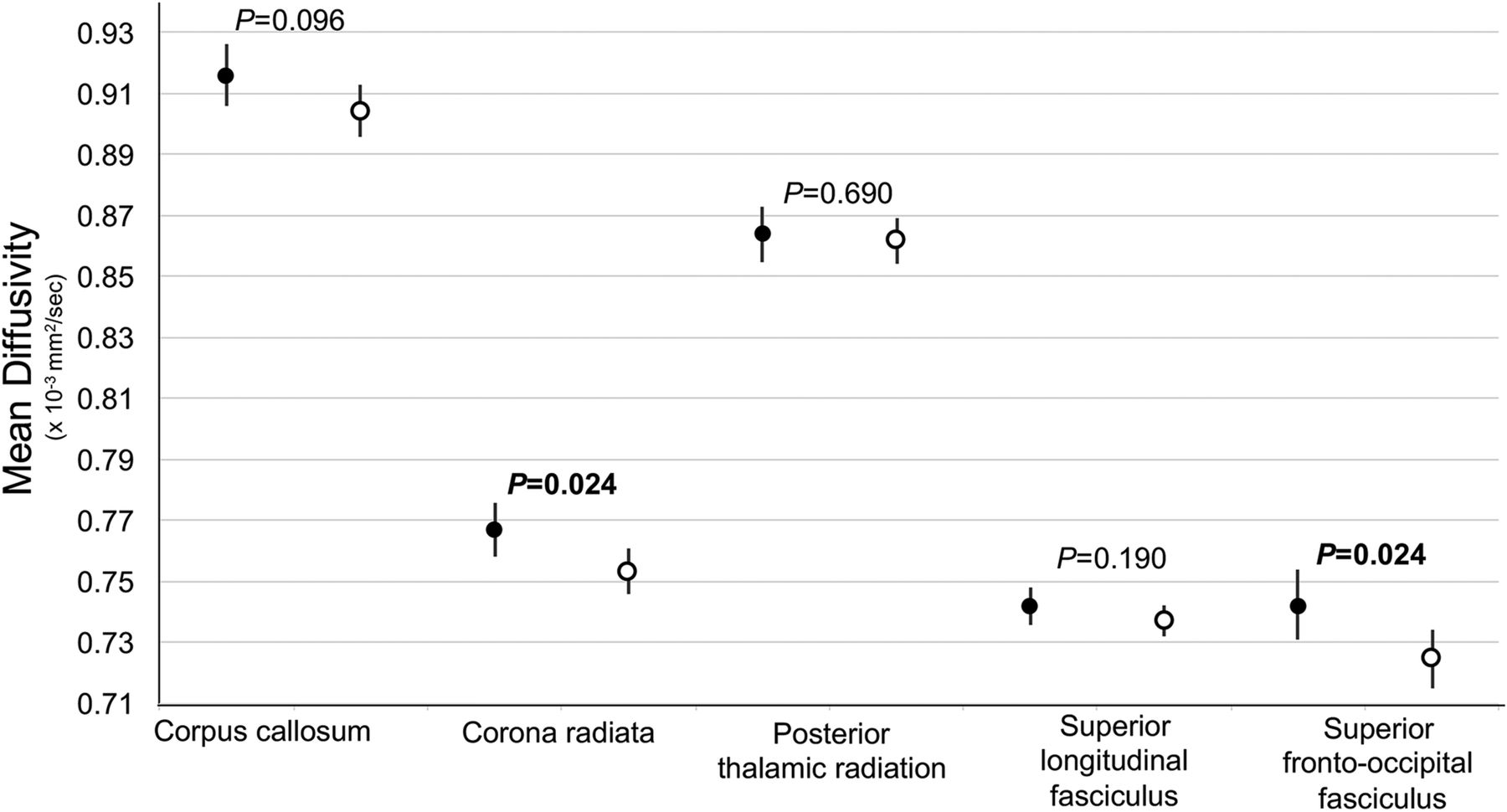
  • Fig 2.
    • Download figure
    • Open in new tab
    • Download powerpoint
    Fig 2.

    Leukoencephalopathy and white matter integrity. MD indicates mean diffusivity. Higher MD is indicative of worse white matter integrity. Point estimates represent the differences in white matter integrity between survivors with persistent leukoencephalopathy and survivors without a history of leukoencephalopathy within the global tracts listed on the x-axis. Error bars represent the 95% confidence intervals. The P values compare the MD between survivors with and without acute leukoencephalopathy using general linear modeling, adjusted for current age. All models have been corrected for the false discovery rate within the global tracts. The image shows that survivors with persistent leukoencephalopathy had reduced white matter integrity, demonstrated by higher global MD, in the corpus callosum, corona radiata, posterior thalamic radiations, superior longitudinal fasciculi, and superior fronto-occipital fasciculi. Details on the associations between leukoencephalopathy and lateralized diffusion tensor imaging measures within the specific tracts can be found in On-line Table 2.

  • Fig 3.
    • Download figure
    • Open in new tab
    • Download powerpoint
    Fig 3.

    Association between white matter integrity and global neurocognitive impairment. Black circles represent survivors with global neurocognitive impairment. White circles represent survivors without global neurocognitive impairment. Global neurocognitive impairment is defined as having ≥2 neurocognitive tests (listed in On-line Table 3) that fall >1.5 SDs or 1 test that falls >2 SDs below the age-adjusted population normative data. The y-axis represents mean diffusivity. Higher mean diffusivity is indicative of worse white matter integrity. Error bars represent the 95% confidence intervals. P values compare the mean diffusivity between survivors with neurocognitive impairment (closed circle, n = 63) and without neurocognitive impairment (open circle, n = 87) using general linear modeling, adjusted for current age. The image shows that survivors with neurocognitive impairment had reduced white matter integrity demonstrated by higher global mean diffusivity in the corona radiata and superior fronto-occipital fasciculus. Details on the associations between global neurocognitive impairment and lateralized diffusion tensor imaging measures within the specific tracts can be found in On-line Table 4.

Tables

  • Figures
    • View popup
    Table 1:

    Demographics and treatment characteristics (N = 173)

    No. (%)Mean (SD)Median (IQR)
    Demographics
        Sex
            Male89 (51)
            Female84 (49)
        Race/ethnicity
            White124 (72)
            Asian3 (2)
            Black21 (12)
            Hispanic18 (10)
            Others7 (4)
        Current age (yr)14.4 (4.6)13.5 (10.7–17.6)
        Patient's highest education (yr)7.7 (3.9)7.0 (4.0–11.0)
        Maternal education (yr)13.6 (2.5)13.0 (12.0–16.0)
        Paternal education (yr)13.6 (3.1)12.0 (12.0–16.0)
    Treatment characteristics
        Age at diagnosis (yr)6.7 (4.3)5.3 (3.5–8.6)
        Time since diagnosis (yr)7.7 (1.8)7.5 (6.3–9.1)
    Treatment risk stratum
            Low102 (59)
            Standard71 (41)
    Chemotherapy dosesa
        Oral dexamethasone (mg/m2)1096.4 (303.2)1099.9 (985.3–1246.1)
        IV high-dose cytarabine (g/m2)8.5 (3.5)8.0 (8.0–8.0)
        IV leucovorin (mg/m2)343.5 (207.1)300.0 (220.0–390.0)
        IV high-dose methotrexateb (g/m2)15.4 (6.7)14.2 (11.4–19.0)
        IT MHA (No. of counts)14.4 (4.0)13.0 (12.0–16.0)
    • Note:—IQR indicates interquartile range; IT MHA, intrathecal injection of methotrexate plus hydrocortisone plus cytarabine.

    • ↵a Except for IT MHA, all drug doses are presented as cumulative doses (g/m2 or mg/m2).

    • ↵b High-dose IV methotrexate was defined as a daily dose of >1 g/m2 of IV methotrexate.

    • View popup
    Table 2:

    Association between methotrexate and white matter integritya

    TractsIntrathecal MHAbHigh-Dose Methotrexateb
    Mean DiffusivityFractional AnisotropyMean DiffusivityFractional Anisotropy
    Est.SEPEst.SEPEst.SEPEst.SEP
    Corpus callosum0.00170.0008.04c−0.00090.0007.21−0.00030.0005.600.00060.0004.17
        Genu0.00160.0009.07−0.00150.0012.21−0.00010.0005.82−0.00020.0007.76
        Body0.00080.0010.450.00000.0009.99−0.00080.0006.210.00110.0005.05
        Splenium0.00220.0011.04c−0.00110.0007.130.00000.0006.950.00070.0004.09
    Corona radiata0.00140.0007.04c−0.00040.0005.36−0.00030.0004.440.00020.0003.57
        Anterior (left)0.00170.0009.06−0.00100.0005.06−0.00020.0005.73−0.00010.0003.75
        Anterior (right)0.00110.0009.19−0.00050.0006.41−0.00050.0005.310.00010.0003.78
        Superior (left)0.00130.0005.02c0.00030.0007.70−0.00010.0003.690.00040.0004.33
        Superior (right)0.00150.0005.005c−0.00030.0007.66−0.00020.0003.580.00020.0004.53
        Posterior (left)0.00090.0007.230.00030.0007.65−0.00070.0004.080.00070.0004.12
        Posterior (right)0.00160.0007.02c−0.00020.0007.79−0.00030.0004.420.00020.0004.58
    Posterior thalamic radiation0.00150.0007.03c−0.00030.0007.630.00000.0004.940.00070.0004.11
        Left0.00110.0010.290.00000.0008.98−0.00030.0006.650.00090.0005.05
        Right0.00180.0006.003c−0.00060.0007.420.00010.0004.690.00050.0004.24
    Superior longitudinal fasciculus0.00070.0005.160.00060.0005.25−0.00010.0003.750.00050.0003.14
        Left0.00060.0005.220.00080.0006.16−0.00010.0003.610.00040.0003.30
        Right0.00070.0005.130.00050.0006.410.00000.0003.870.00060.0003.10
    Superior fronto-occipital fasciculus0.00200.0009.02c−0.00040.0009.65−0.00030.0005.600.00090.0005.09
        Left0.00210.0010.04c−0.00040.0009.63−0.00050.0006.410.00090.0005.10
        Right0.00190.0007.01c−0.00030.0010.730.00000.0005.970.00080.0006.14
    • Note:—Est. indicates parameter estimate; SE, standard error; MHA, methotrexate plus hydrocortisone plus cytarabine.

    • ↵a General linear modeling was applied for the test of strength of association between treatment variables with mean diffusivity and fractional anisotropy for each tract, adjusted for age at evaluation. Higher mean diffusivity and lower fractional anisotropy are indicative of worse white matter integrity.

    • ↵b Intrathecal MHA was defined as the number of intrathecal injections of methotrexate plus hydrocortisone plus cytarabine; high-dose IV methotrexate was defined as a daily dose of >1 g/m2, presented as cumulative doses (g/m2).

    • ↵c Significant.

PreviousNext
Back to top

In this issue

American Journal of Neuroradiology: 39 (10)
American Journal of Neuroradiology
Vol. 39, Issue 10
1 Oct 2018
  • Table of Contents
  • Index by author
  • Complete Issue (PDF)
Advertisement
Print
Download PDF
Email Article

Thank you for your interest in spreading the word on American Journal of Neuroradiology.

NOTE: We only request your email address so that the person you are recommending the page to knows that you wanted them to see it, and that it is not junk mail. We do not capture any email address.

Enter multiple addresses on separate lines or separate them with commas.
The Impact of Persistent Leukoencephalopathy on Brain White Matter Microstructure in Long-Term Survivors of Acute Lymphoblastic Leukemia Treated with Chemotherapy Only
(Your Name) has sent you a message from American Journal of Neuroradiology
(Your Name) thought you would like to see the American Journal of Neuroradiology web site.
CAPTCHA
This question is for testing whether or not you are a human visitor and to prevent automated spam submissions.
Cite this article
N.D. Sabin, Y.T. Cheung, W.E. Reddick, D. Bhojwani, W. Liu, J.O. Glass, T.M. Brinkman, S.N. Hwang, D. Srivastava, C.-H. Pui, L.L. Robison, M.M. Hudson, K.R. Krull
The Impact of Persistent Leukoencephalopathy on Brain White Matter Microstructure in Long-Term Survivors of Acute Lymphoblastic Leukemia Treated with Chemotherapy Only
American Journal of Neuroradiology Oct 2018, 39 (10) 1919-1925; DOI: 10.3174/ajnr.A5791

Citation Manager Formats

  • BibTeX
  • Bookends
  • EasyBib
  • EndNote (tagged)
  • EndNote 8 (xml)
  • Medlars
  • Mendeley
  • Papers
  • RefWorks Tagged
  • Ref Manager
  • RIS
  • Zotero
0 Responses
Respond to this article
Share
Bookmark this article
The Impact of Persistent Leukoencephalopathy on Brain White Matter Microstructure in Long-Term Survivors of Acute Lymphoblastic Leukemia Treated with Chemotherapy Only
N.D. Sabin, Y.T. Cheung, W.E. Reddick, D. Bhojwani, W. Liu, J.O. Glass, T.M. Brinkman, S.N. Hwang, D. Srivastava, C.-H. Pui, L.L. Robison, M.M. Hudson, K.R. Krull
American Journal of Neuroradiology Oct 2018, 39 (10) 1919-1925; DOI: 10.3174/ajnr.A5791
del.icio.us logo Twitter logo Facebook logo Mendeley logo
  • Tweet Widget
  • Facebook Like
  • Google Plus One
Purchase

Jump to section

  • Article
    • Abstract
    • ABBREVIATION:
    • Materials and Methods
    • Results
    • Discussion
    • Conclusions
    • Acknowledgments
    • Footnotes
    • References
  • Figures & Data
  • Supplemental
  • Info & Metrics
  • Responses
  • References
  • PDF

Related Articles

  • PubMed
  • Google Scholar

Cited By...

  • No citing articles found.
  • Crossref
  • Google Scholar

This article has not yet been cited by articles in journals that are participating in Crossref Cited-by Linking.

More in this TOC Section

  • Clinical SVR of Fetal Brain MRI
  • FRACTURE MR in Congenital Vertebral Anomalies
  • Comparing MRI Perfusion in Pediatric Brain Tumors
Show more Pediatric Neuroimaging

Similar Articles

Advertisement

Indexed Content

  • Current Issue
  • Accepted Manuscripts
  • Article Preview
  • Past Issues
  • Editorials
  • Editor's Choice
  • Fellows' Journal Club
  • Letters to the Editor
  • Video Articles

Cases

  • Case Collection
  • Archive - Case of the Week
  • Archive - Case of the Month
  • Archive - Classic Case

More from AJNR

  • Trainee Corner
  • Imaging Protocols
  • MRI Safety Corner
  • Book Reviews

Multimedia

  • AJNR Podcasts
  • AJNR Scantastics

Resources

  • Turnaround Time
  • Submit a Manuscript
  • Submit a Video Article
  • Submit an eLetter to the Editor/Response
  • Manuscript Submission Guidelines
  • Statistical Tips
  • Fast Publishing of Accepted Manuscripts
  • Graphical Abstract Preparation
  • Imaging Protocol Submission
  • Evidence-Based Medicine Level Guide
  • Publishing Checklists
  • Author Policies
  • Become a Reviewer/Academy of Reviewers
  • News and Updates

About Us

  • About AJNR
  • Editorial Board
  • Editorial Board Alumni
  • Alerts
  • Permissions
  • Not an AJNR Subscriber? Join Now
  • Advertise with Us
  • Librarian Resources
  • Feedback
  • Terms and Conditions
  • AJNR Editorial Board Alumni

American Society of Neuroradiology

  • Not an ASNR Member? Join Now

© 2025 by the American Society of Neuroradiology All rights, including for text and data mining, AI training, and similar technologies, are reserved.
Print ISSN: 0195-6108 Online ISSN: 1936-959X

Powered by HighWire