
Article Figures & Data
Figures
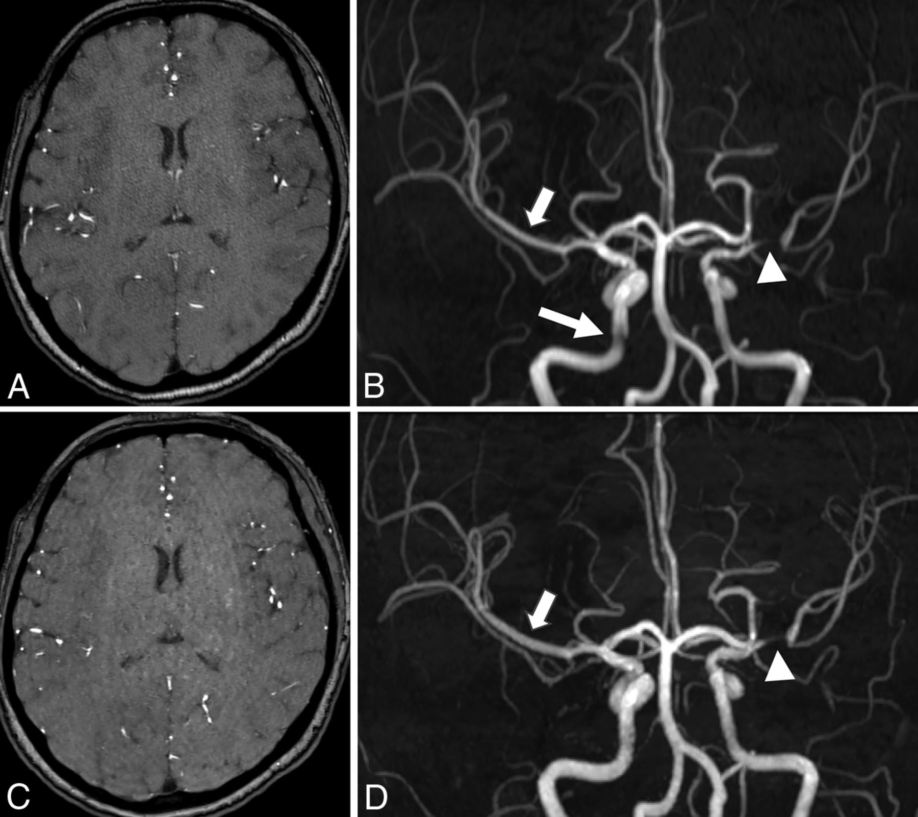
- Fig 1.
Source images and coronal view of MIP images in a 42-year-old patient. The speckled noise in the center can be seen on the source image of conventional PI-TOF (A), whereas some artifacts with a curved stripe pattern can be seen on the source image of CS-TOF (C). These artifacts are eliminated on the MIP images and have little effect on the visualization of the stenosis. An obvious stenosis located in the M1 segment of the left middle cerebral artery is sufficiently visualized on both PI-TOF (B) and CS-TOF (D) (arrowheads). The edge sharpness of vessels on CS-TOF (D) is higher than that on PI-TOF (B) (short arrows). The image quality of the right intracranial internal carotid artery (long arrow, B) is improved on CS-TOF (D).
- Fig 2.
MIP images of a 68-year-old patient. Mild stenosis located in the proximal M1 segment of left middle cerebral artery can be sufficiently visualized on both PI-TOF (A) and CS-TOF (B) (arrowheads). The edge sharpness of vessels on CS-TOF is higher than that on PI-TOF (arrows).
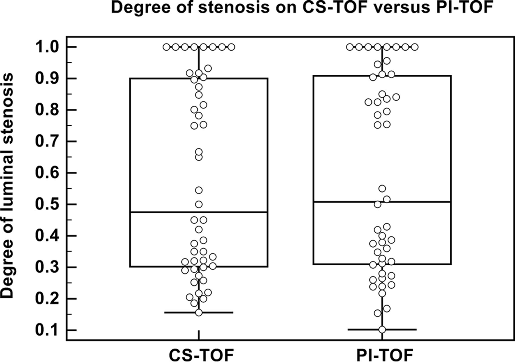
- Fig 3.
The degree of each luminal stenosis measured on CS-TOF and PI-TOF, respectively.
- Fig 4.
Bar plots of the 2 readers' preferences. CS-TOF is considered not inferior to PI-TOF in all cases. In 50.0% and 45.5% of patients, the diagnostic quality of CS-TOF is considered better than that of PI-TOF by each of the 2 readers.


Tables
Parameters CS-TOF PI-TOF FOV (mm2) 220 × 200 220 × 200 TR/TE (ms) 21/3.49 21/3.49 Flip angle 18° 18° Matrix 368 × 334 368 × 334 Slice thickness (mm) 0.4 0.6 No. of slabs 4 4 Slices per slab 60 40 Slice oversampling 20% 20% Phase partial Fourier factor None 6/8 Slice partial Fourier factor None None Acceleration factor 10.3 GRAPPA 2 Reconstructed voxel size (mm3) 0.4 × 0.4 × 0.4 0.4 × 0.4 × 0.6 Characteristics Mean ± SD or Number (%) Male 11 (50.0%) Age (yr) 61.8 ± 16.8 Stenosis location (R/L) 48 Intracranial internal carotid artery 6/5 Middle cerebral artery 6/13 Anterior cerebral artery 5/2 Posterior cerebral artery 3/4 Basilar artery 1 Intracranial vertebral artery 2/1 Note:—R indicates right; L, left; SD, standard deviation.
- Table 3:
Comparison between CS-TOF and PI-TOF for evaluating intracranial arterial stenosis
Variables CS-TOF PI-TOF P Value Diagnostic qualitya .046 Grade 3 20 (90.9%) 17 (77.3%) Grade 2 2 (9.1%) 4 (18.2%) Grade 1 0 (0.0%) 1 (4.5%) Grade 0 0 (0.0%) 0 (0.0%) Stenosis visualizationb .025 Grade 2 48 (100.0%) 43 (89.6%) Grade 1 0 (0.0%) 5 (10.4%) Grade 0 0 (0.0%) 0 (0.0%) Luminal stenosis ratio (mean ± SD) 57.9% ± 30.5% 58.9% ± 31.0% .241 Edge sharpness of artery (mean ± SD) 0.358 ± 0.038 0.267 ± 0.042 <.001 ↵a The diagnostic quality of the CS-TOF and PI-TOF images was graded on an ordinal scale from 0 to 3, with 0 indicating completely blurred arteries and severe artifacts; 1 indicating partially obscured arteries and moderate artifacts; 2 indicating good and clear arteries and slight artifacts; and 3 indicating excellent arteries and no artifacts.
↵b The visualization of arterial stenosis was graded as follows: grade 2, definite stenosis and sufficiently recognized, high confidence; grade 1, probable stenosis, moderately confident; grade 0, low confidence.


