
Article Figures & Data
Figures
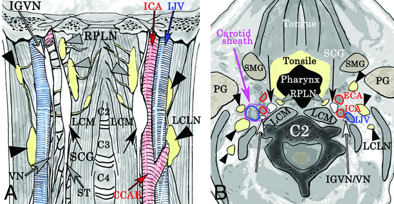
- Fig 1.
The coronal (A) and axial (B) views around the superior cervical ganglion according to anatomic reports show the superior cervical ganglion (black arrow), the inferior ganglion of the vagus nerve (white arrow), and the retropharyngeal lymph node (gray arrowheads). IJV indicates inferior jugular vein; ECA, external carotid artery; LCLN, lateral cervical lymph node (black arrowheads); LCM, longus capitis muscle; PG, parotid gland; SMG, submandibular gland; ST, sympathetic trunk; VN, vagus nerve. The magenta circle indicates the right carotid sheath.
- Fig 2.
We assessed bilateral superior cervical ganglia (black arrows), the inferior ganglion of the vagus nerve (white arrows), and the retropharyngeal lymph nodes (gray arrowheads) using 3D-STIR (A, maximum-intensity-projection image; B, a section of original coronal images). The artery and vein clearly demonstrate flow voids. IJV indicates inferior jugular vein; LCLN, lateral cervical lymph node (black arrowhead); LCM, longus capitis muscle.
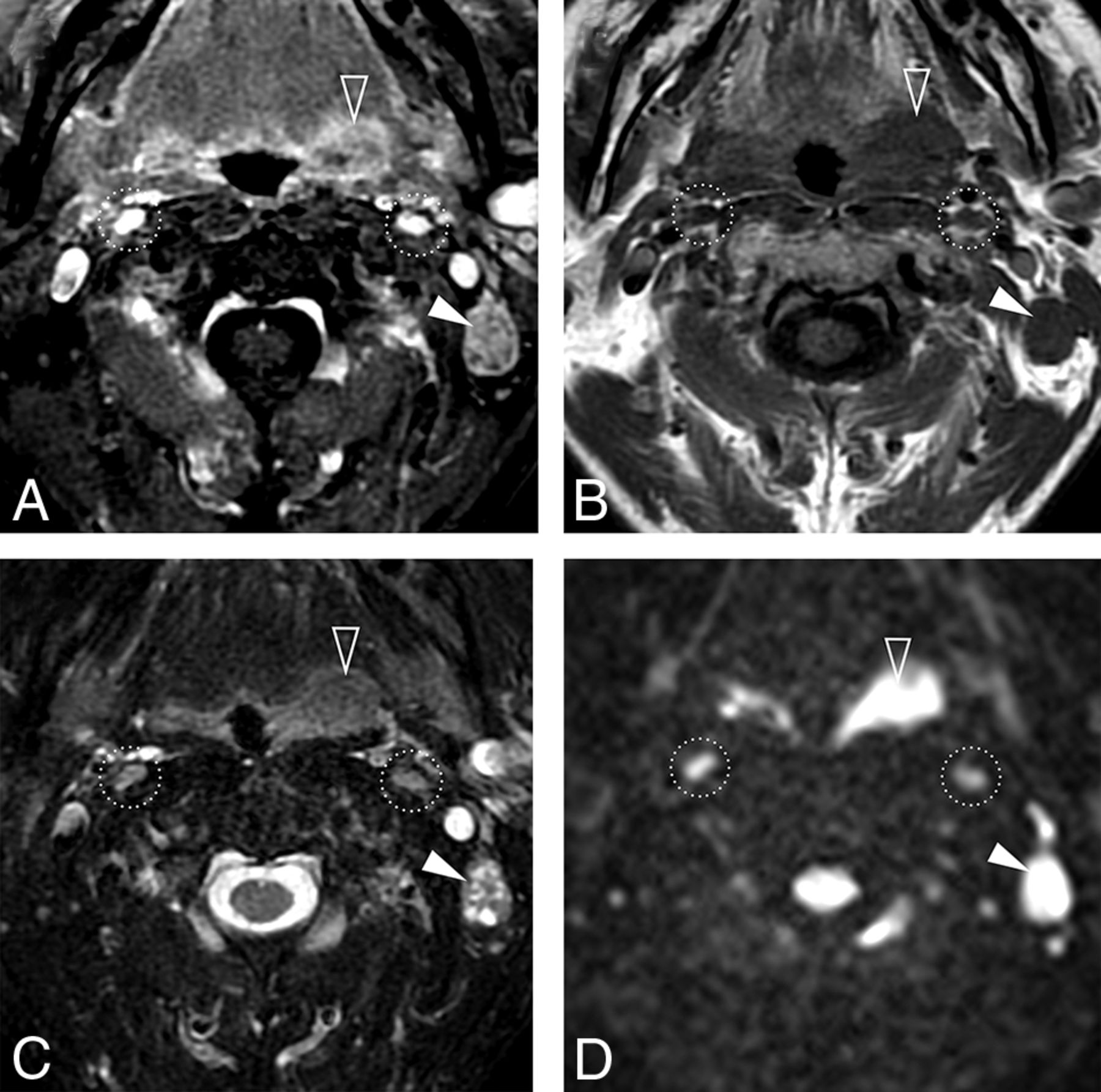
- Fig 3.
A 62-year-old man with oropharyngeal cancer with left lymph node metastasis. Bilateral superior cervical ganglia (circles), metastatic lymph node (arrowhead), and cancer (open arrowhead) are noted. Contrast-enhanced fat-saturated T1-wighted image (A) shows stronger enhancement of the SCG than the metastatic lymph node. The T1-weighted image (B) shows almost the same signals. Fat-saturated T2-weighted image (C) shows heterogeneous signal of the metastatic lymph node, whereas the SCG is homogeneous. Diffusion-weighted image shows that the signals of the SCGs are lower than those of the metastatic lymph node. ADC values of the right SCG, left SCG, and metastatic lymph nodes were 1.32, 1.54, and 0.90 × 10⁻³mm2/s, respectively.
- Fig 4.
A 58-year-old woman with an intraganglionic hypointense spot (arrow) on the right on T2-weighted image (A). This spot is less evident (arrow) on the gadolinium-enhanced fat-saturated T1-weighted image (B). T1-weighted images in a 65-year-old man show slit-shaped hyperintense areas in the bilateral superior cervical ganglia (C, circles). Signals of these regions are suppressed on the gadolinium-enhanced fat-saturated T1-weighted image (D).
- Fig 5.
A, A 14-year-old adolescent with a prominent but normal RPLN. Coronal maximum-intensity-projection image shows the superior cervical ganglia (arrows) and the retropharyngeal lymph nodes (arrowheads). Each SCG was inferior to the RPLN. B, Reconstructed axial MIP image of 15-mm thickness shows that each SCG (arrows) is posterolateral to the RPLN (arrowheads). C, Coronal MIP image shows the SCG (arrows), inferior ganglia of vagus nerve (open arrows), and RPLN (arrowheads) in a 38-year-old man. The SCG is inferior to the IGVN. D, Reconstructed axial MIP image shows that the RPLN (arrowhead), SCG (arrow), internal carotid artery, IGVN (open arrow), and the internal jugular vein (IJV) form a line from anteromedial to posterolateral. B and D, Blurring on the images is because the MIP was used to superimpose the SCG, IGVN, and RPLN. IJV indicates inferior jugular vein.
- Fig 6.
Probability maps of the superior cervical ganglion against the C2 transverse process (A, asterisk) and common carotid artery bifurcation (B, asterisk). The SCG was located at the anterior side against the C2 transverse process and superior and posterolateral to the CCAB. The location of bilateral SCGs is similar. R indicates right; L, left; Cor, coronal; Sag, sagittal; Axi, axial.


Jump to section
Related Articles
Cited By...
- No citing articles found.