Advertisement

Main menu

  • Home
  • Content
    • Current Issue
    • Accepted Manuscripts
    • Article Preview
    • Past Issue Archive
    • Video Articles
    • AJNR Case Collection
    • Case of the Week Archive
    • Case of the Month Archive
    • Classic Case Archive
  • Special Collections
    • AJNR Awards
    • Low-Field MRI
    • Alzheimer Disease
    • ASNR Foundation Special Collection
    • Photon-Counting CT
    • View All
  • Multimedia
    • AJNR Podcasts
    • AJNR SCANtastic
    • Trainee Corner
    • MRI Safety Corner
    • Imaging Protocols
  • For Authors
    • Submit a Manuscript
    • Submit a Video Article
    • Submit an eLetter to the Editor/Response
    • Manuscript Submission Guidelines
    • Statistical Tips
    • Fast Publishing of Accepted Manuscripts
    • Graphical Abstract Preparation
    • Imaging Protocol Submission
    • Author Policies
  • About Us
    • About AJNR
    • Editorial Board
    • Editorial Board Alumni
  • More
    • Become a Reviewer/Academy of Reviewers
    • Subscribers
    • Permissions
    • Alerts
    • Feedback
    • Advertisers
    • ASNR Home

User menu

  • Alerts
  • Log in

Search

  • Advanced search
American Journal of Neuroradiology

American Journal of Neuroradiology

ASHNR American Society of Functional Neuroradiology ASHNR American Society of Pediatric Neuroradiology ASSR
  • Alerts
  • Log in

Advanced Search

  • Home
  • Content
    • Current Issue
    • Accepted Manuscripts
    • Article Preview
    • Past Issue Archive
    • Video Articles
    • AJNR Case Collection
    • Case of the Week Archive
    • Case of the Month Archive
    • Classic Case Archive
  • Special Collections
    • AJNR Awards
    • Low-Field MRI
    • Alzheimer Disease
    • ASNR Foundation Special Collection
    • Photon-Counting CT
    • View All
  • Multimedia
    • AJNR Podcasts
    • AJNR SCANtastic
    • Trainee Corner
    • MRI Safety Corner
    • Imaging Protocols
  • For Authors
    • Submit a Manuscript
    • Submit a Video Article
    • Submit an eLetter to the Editor/Response
    • Manuscript Submission Guidelines
    • Statistical Tips
    • Fast Publishing of Accepted Manuscripts
    • Graphical Abstract Preparation
    • Imaging Protocol Submission
    • Author Policies
  • About Us
    • About AJNR
    • Editorial Board
    • Editorial Board Alumni
  • More
    • Become a Reviewer/Academy of Reviewers
    • Subscribers
    • Permissions
    • Alerts
    • Feedback
    • Advertisers
    • ASNR Home
  • Follow AJNR on Twitter
  • Visit AJNR on Facebook
  • Follow AJNR on Instagram
  • Join AJNR on LinkedIn
  • RSS Feeds

AJNR Awards, New Junior Editors, and more. Read the latest AJNR updates

Research ArticleHead and Neck Imaging

Patterns of Sonographically Detectable Echogenic Foci in Pediatric Thyroid Carcinoma with Corresponding Histopathology: An Observational Study

I. Erdem Toslak, B. Martin, G.A. Barkan, A.I. Kılıç and J.E. Lim-Dunham
American Journal of Neuroradiology January 2018, 39 (1) 156-161; DOI: https://doi.org/10.3174/ajnr.A5419
I. Erdem Toslak
aFrom the Departments of Radiology (I.E.T., J.E.L.-D.)
  • Find this author on Google Scholar
  • Find this author on PubMed
  • Search for this author on this site
  • ORCID record for I. Erdem Toslak
B. Martin
cClinical Research Office (B.M.), Loyola University Chicago Health Sciences Division, Loyola University Chicago Stritch School of Medicine, Maywood, Illinois.
  • Find this author on Google Scholar
  • Find this author on PubMed
  • Search for this author on this site
  • ORCID record for B. Martin
G.A. Barkan
bPathology (G.A.B., A.I.K.), Loyola University Medical Center, Maywood, Illinois
  • Find this author on Google Scholar
  • Find this author on PubMed
  • Search for this author on this site
  • ORCID record for G.A. Barkan
A.I. Kılıç
bPathology (G.A.B., A.I.K.), Loyola University Medical Center, Maywood, Illinois
  • Find this author on Google Scholar
  • Find this author on PubMed
  • Search for this author on this site
  • ORCID record for A.I. Kılıç
J.E. Lim-Dunham
aFrom the Departments of Radiology (I.E.T., J.E.L.-D.)
  • Find this author on Google Scholar
  • Find this author on PubMed
  • Search for this author on this site
  • ORCID record for J.E. Lim-Dunham
  • Article
  • Figures & Data
  • Info & Metrics
  • Responses
  • References
  • PDF
Loading

Article Figures & Data

Figures

  • Tables
  • Fig 1.
    • Download figure
    • Open in new tab
    • Download powerpoint
    Fig 1.

    Typical sonography samples of categories of ACR TI-RADS echogenic foci and types of distribution. A, Macrocalcifications with posterior acoustic shadowing (arrow) in a left-sided 4.7-cm thyroid nodule in an 18-year-old girl with histologically proved papillary thyroid carcinoma. The distribution is classified as sparse. B, Punctate echogenic foci with small, 0.7-mm comet-tail artifacts in the same patient as in A. The distribution is diffuse. Pathology revealed stromal calcifications and sticky colloid, both sparsely distributed, and the absence of psammomatous calcifications. C, Punctate echogenic foci with no posterior artifacts (arrows) in a left-sided 3.5-cm thyroid nodule in a 12-year-old girl with a histologically proved follicular variant of papillary thyroid carcinoma. The distribution is sparse. Histology revealed no corresponding calcifications or sticky colloid.
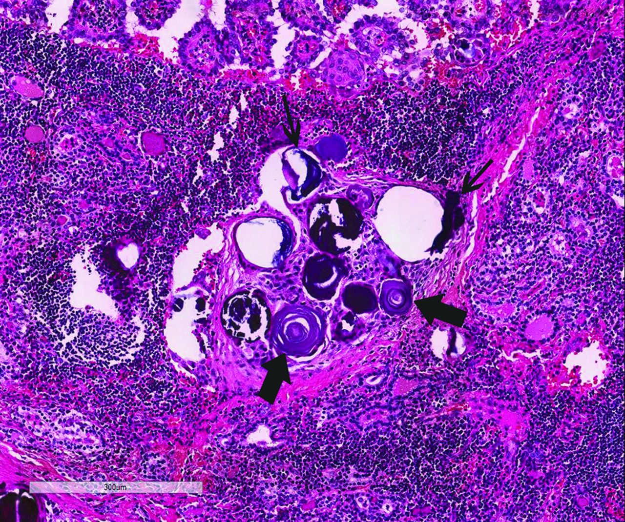
  • Fig 2.
    • Download figure
    • Open in new tab
    • Download powerpoint
    Fig 2.

    Examples of psammomatous (thick arrows) and stromal calcifications (thin arrows) from a thyroidectomy specimen of a 16-year-old girl showing papillary thyroid carcinoma in a background of lymphocytic thyroiditis (hematoxylin-eosin stain, 20×).

Tables

  • Figures
    • View popup
    Table 1:

    Observed agreement between radiologists for presence and distribution of thyroid nodule echogenic focia

    US ClassificationAgreement for Presence or Absence (No.) (%)Agreement for Distribution (No.) (%)
    Punctate echogenic foci15/15 (100%)14/15 (93%)
    Echogenic foci with large comet-tail artifacts11/15 (73%)11/15 (73%)
    Macrocalcifications8/15 (53%)8/15 (53%)
    Peripheral rim calcifications15/15 (100%)15/15 (100%)
    • Note:—No. indicates number of nodules.

    • ↵a The valid number of observations is 15.

    • View popup
    Table 2:

    Thyroid nodule pathology as a function of US patternsa

    Pathology Finding (No.)Punctate Echogenic Foci (No.)Macrocalcifications (No.)
    Present (n = 15)Absent (n = 0)Present (n = 4)Absent (n = 11)
    Stromal calcification present (9)9 (60%)0 (0%)4 (100%)5 (45%)
    Stromal calcification absent (6)6 (40%)0 (0%)0 (0%)6 (55%)
    Psammomatous calcification present (4)4 (27%)0 (0%)1 (25%)3 (27%)
    Psammomatous calcification absent (11)11 (73%)0 (0%)3 (75%)8 (73%)
    Sticky colloid present (8)8 (53%)0 (0%)3 (75%)5 (45%)
    Sticky colloid absent (7)7 (47%)0 (0%)1 (25%)6 (55%)
    • Note:—No. indicates number of nodules.

    • ↵a The valid number of observations is 15.

PreviousNext
Back to top

In this issue

American Journal of Neuroradiology: 39 (1)
American Journal of Neuroradiology
Vol. 39, Issue 1
1 Jan 2018
  • Table of Contents
  • Index by author
  • Complete Issue (PDF)
Advertisement
Print
Download PDF
Email Article

Thank you for your interest in spreading the word on American Journal of Neuroradiology.

NOTE: We only request your email address so that the person you are recommending the page to knows that you wanted them to see it, and that it is not junk mail. We do not capture any email address.

Enter multiple addresses on separate lines or separate them with commas.
Patterns of Sonographically Detectable Echogenic Foci in Pediatric Thyroid Carcinoma with Corresponding Histopathology: An Observational Study
(Your Name) has sent you a message from American Journal of Neuroradiology
(Your Name) thought you would like to see the American Journal of Neuroradiology web site.
CAPTCHA
This question is for testing whether or not you are a human visitor and to prevent automated spam submissions.
Cite this article
I. Erdem Toslak, B. Martin, G.A. Barkan, A.I. Kılıç, J.E. Lim-Dunham
Patterns of Sonographically Detectable Echogenic Foci in Pediatric Thyroid Carcinoma with Corresponding Histopathology: An Observational Study
American Journal of Neuroradiology Jan 2018, 39 (1) 156-161; DOI: 10.3174/ajnr.A5419

Citation Manager Formats

  • BibTeX
  • Bookends
  • EasyBib
  • EndNote (tagged)
  • EndNote 8 (xml)
  • Medlars
  • Mendeley
  • Papers
  • RefWorks Tagged
  • Ref Manager
  • RIS
  • Zotero
0 Responses
Respond to this article
Share
Bookmark this article
Patterns of Sonographically Detectable Echogenic Foci in Pediatric Thyroid Carcinoma with Corresponding Histopathology: An Observational Study
I. Erdem Toslak, B. Martin, G.A. Barkan, A.I. Kılıç, J.E. Lim-Dunham
American Journal of Neuroradiology Jan 2018, 39 (1) 156-161; DOI: 10.3174/ajnr.A5419
del.icio.us logo Twitter logo Facebook logo Mendeley logo
  • Tweet Widget
  • Facebook Like
  • Google Plus One
Purchase

Jump to section

  • Article
    • Abstract
    • ABBREVIATIONS:
    • Materials and Methods
    • Results
    • Discussion
    • Conclusions
    • References
  • Figures & Data
  • Info & Metrics
  • Responses
  • References
  • PDF

Related Articles

  • No related articles found.
  • PubMed
  • Google Scholar

Cited By...

  • No citing articles found.
  • Crossref
  • Google Scholar

This article has not yet been cited by articles in journals that are participating in Crossref Cited-by Linking.

More in this TOC Section

  • Diagnostic Performance of US in Neck Node NIRADS 2
  • Hydrops Herniation into the Semicircular Canals
  • ASL Sensitivity for Head and Neck Paraganglioma
Show more HEAD AND NECK IMAGING

Similar Articles

Advertisement

Indexed Content

  • Current Issue
  • Accepted Manuscripts
  • Article Preview
  • Past Issues
  • Editorials
  • Editor's Choice
  • Fellows' Journal Club
  • Letters to the Editor
  • Video Articles

Cases

  • Case Collection
  • Archive - Case of the Week
  • Archive - Case of the Month
  • Archive - Classic Case

More from AJNR

  • Trainee Corner
  • Imaging Protocols
  • MRI Safety Corner
  • Book Reviews

Multimedia

  • AJNR Podcasts
  • AJNR Scantastics

Resources

  • Turnaround Time
  • Submit a Manuscript
  • Submit a Video Article
  • Submit an eLetter to the Editor/Response
  • Manuscript Submission Guidelines
  • Statistical Tips
  • Fast Publishing of Accepted Manuscripts
  • Graphical Abstract Preparation
  • Imaging Protocol Submission
  • Evidence-Based Medicine Level Guide
  • Publishing Checklists
  • Author Policies
  • Become a Reviewer/Academy of Reviewers
  • News and Updates

About Us

  • About AJNR
  • Editorial Board
  • Editorial Board Alumni
  • Alerts
  • Permissions
  • Not an AJNR Subscriber? Join Now
  • Advertise with Us
  • Librarian Resources
  • Feedback
  • Terms and Conditions
  • AJNR Editorial Board Alumni

American Society of Neuroradiology

  • Not an ASNR Member? Join Now

© 2025 by the American Society of Neuroradiology All rights, including for text and data mining, AI training, and similar technologies, are reserved.
Print ISSN: 0195-6108 Online ISSN: 1936-959X

Powered by HighWire