
Article Figures & Data
Figures
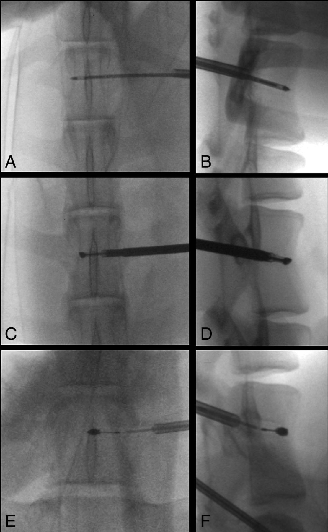
- Fig 1.
Fluoroscopic documentation of ablation probe placement in sheep. Anteroposterior (A, C, and E) and lateral (B, D, and F) fluoroscopic images show transpedicular placement of the cryoablation probe within the T14 vertebra (A and B), the radiofrequency ablation probe within the L3 vertebra (C and D), and the microwave ablation probe within the L6 vertebra (E and F).
- Fig 2.
Anteroposterior (A) and lateral (B) CT images of a normal lumbar vertebra. The thick black line represents the unilateral transpedicular trajectory of the ablation probe. By definition, the transverse and anteroposterior dimensions of the ablation zone are parallel and perpendicular to the ablation probe in the axial plane, respectively, and the craniocaudal dimension of the ablation zone is perpendicular to the ablation probe in the sagittal plane.
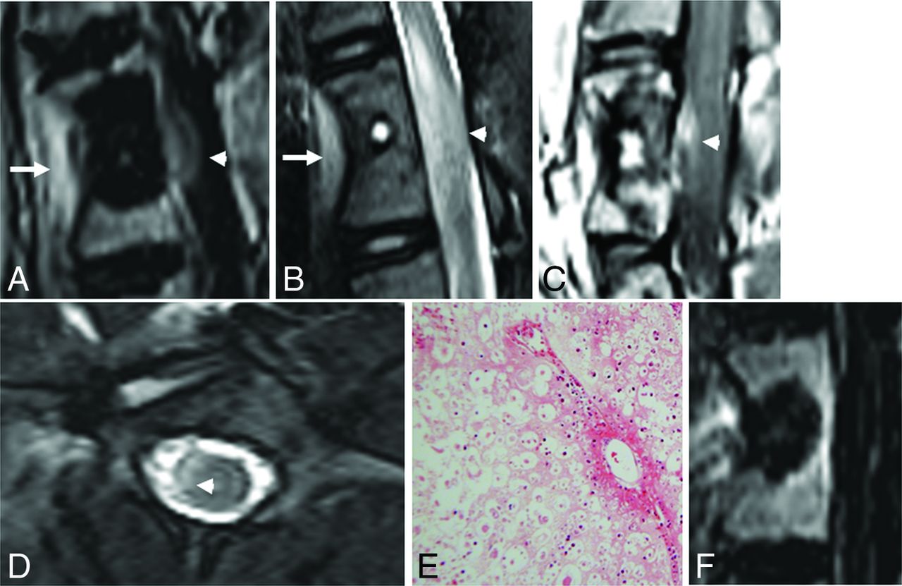
- Fig 3.
Sagittal postcontrast T1-weighted image with fat suppression (A) and a T2-weighted image (B) of the vertebral level treated with cryoablation show the ablation zone extending beyond the posterior vertebral body into the spinal canal. There is enhancement and intramedullary T2 signal hyperintensity in the ventral aspect of the edematous spinal cord (white arrowheads). There is also inflammation in the soft tissues ventral to the spine where the ablation zone extended beyond the anterior vertebral body cortex (white arrows). Sagittal postcontrast T1-weighted image (C) and axial T2-weighted image (D) of the vertebral level treated with microwave ablation similarly show extension of the ablation zone into the spinal canal (white arrowheads). E, Hematoxylin-eosin staining of the spinal cord at the level of the cryoablated vertebra at ×100 total magnification shows axonal necrosis and edema. Similar findings were seen at the microwave-ablated level (not shown). F, Sagittal postcontrast T1-weighted image with fat suppression of the radiofrequency-ablated vertebra shows the posterior margin of the ablation zone confined to the vertebral body.
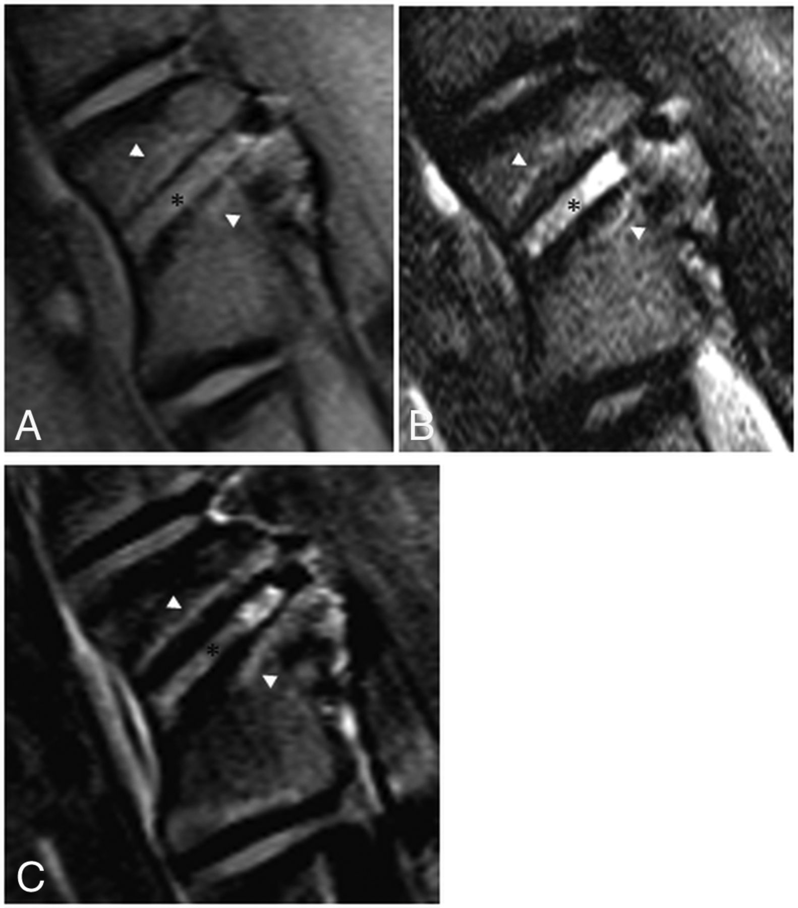
- Fig 4.
MR imaging findings 7 days after RFA of an L2 vertebral body in a sheep model. A, T1-weighted oblique sagittal MR imaging (left, anterior; right, posterior) shows T1 hyperintense soft tissue within the probe tract (black asterisk) outlined by a signal void. The surrounding ablation zone is isointense to normal marrow and outlined by a thin hyperintense rim (white arrowheads). B, T2-weighted oblique sagittal MR imaging with fat suppression shows the hyperintense probe tract (black asterisk) and slightly hypointense ablation zone surrounded by a hyperintense rim (white arrowheads). C, T1-weighted, postcontrast subtraction images show enhancement along the probe tract (black asterisk) and the nonenhancing ablation zone surrounded by a rim of enhancement (white arrowheads).
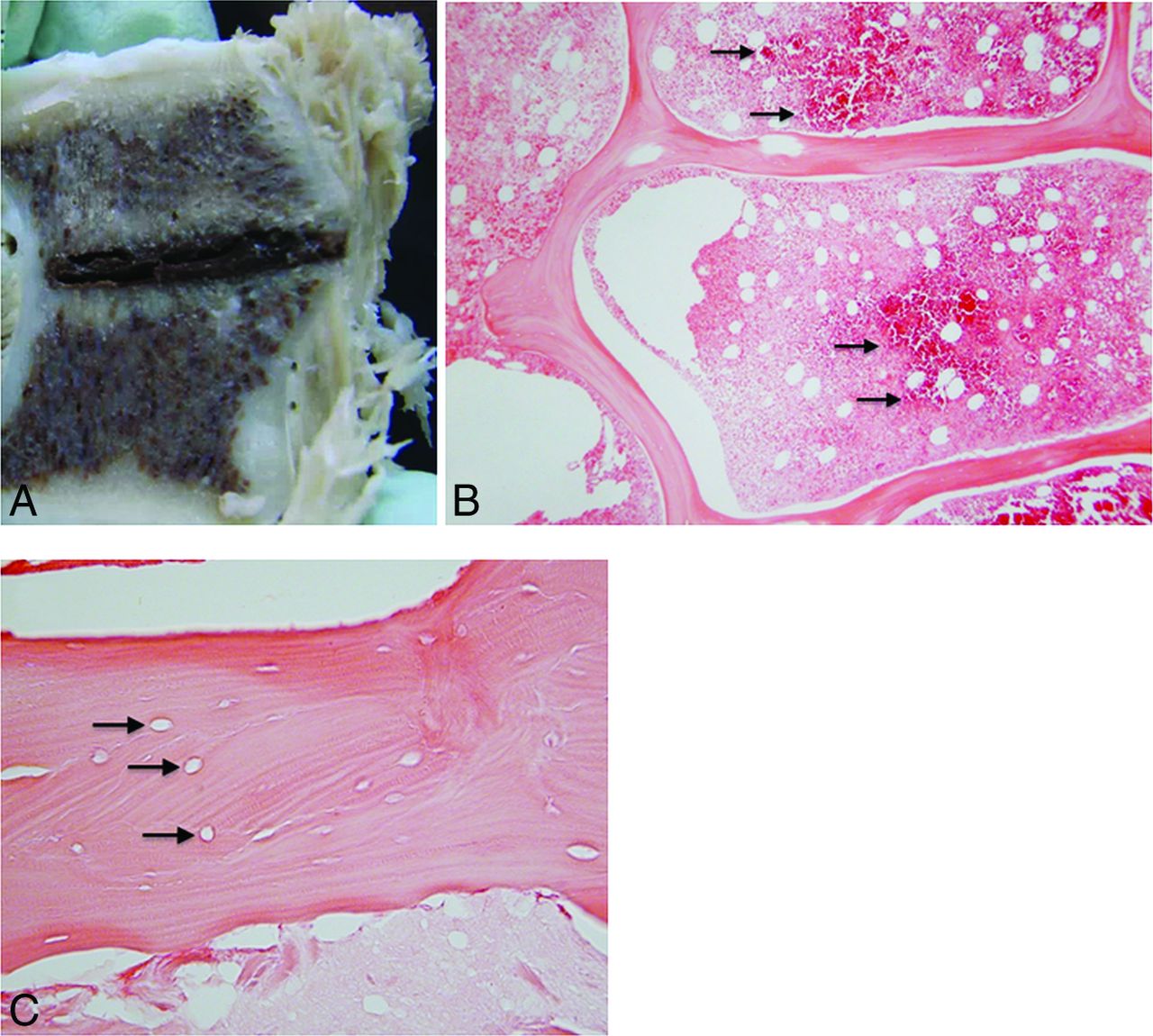
- Fig 5.
A, Gross pathology of the vertebral body shown in Fig 2 cut along the plane of the radiofrequency ablation probe tract. The tract is filled with hemorrhagic debris and is surrounded by a pale, tan zone of necrosis. B, Hematoxylin-eosin staining of the margin of the ablation zone at ×40 total magnification shows a dense band of red blood cells (black arrows), which demarcate necrotic marrow on the left from the intact region on the right. C, At ×200 magnification, the ablation zone shows empty lacunae (black arrows), representing a loss of osteocytes, within an intact trabecula.


Tables
- Table 1:
Technical parameters used for radiofrequency ablation, expected minor-axis diameter of the ablation zones based on nonanimal models, and measured ablation zone diameters on MRI and gross pathology
Watts Thermocouple Temperatures (°C) Ablation Time (sec) Diameter of Ablation Zone Orthogonal to Ablation Probe (mm) Proximal Distal Expected MRI Gross Pathology 10 72 51 92 20.0 13.4 13.8 5 58 42 107 16.6 9.2 10.0 5 51 40 42 13.0 7.3 8.0 - Table 2:
Technical parameters used for cryoablation, expected minor-axis diameter of the ablation zones based on nonanimal models, and measured ablation zone diameters on MRI and gross pathology
Probe Intensity (%) Freeze Cycles (min) Thaw (min) Diameter of Ablation Zone Orthogonal to Ablation Probe (mm) Expected MRI Gross Pathology IceSphere 60 6.0 7.0 20.0 19.2 20.1 IceSeed 50 4.0 5.0 6.0 6.6 7.4 IceSeed 50 3.5 5.0 6.0 6.4 7.0 IceSeed 50 3.0 5.0 5.0 5.5 6.8 - Table 3:
Technical parameters used for microwave ablation, expected minor-axis diameter of the ablation zones based on nonanimal models, and measured ablation zone diameters on MRI and gross pathology
Watts Ablation Time (sec) Diameter of Ablation Zone Orthogonal to Ablation Probe (mm) Expected MRI Gross Pathology 100 50 20.0 17.4 20.0 100 15 9.5 7.9 9.0 100 10 6.0 5.9 6.5 - Table 4:
Mean differences among the anteroposterior, transverse, and craniocaudal ablation zone dimensions expected on the basis of manufacturers' preclinical data and those measured on MRI and gross pathologya
Expected vs MRI (mm) Expected vs Pathology (mm) MRI vs Pathology (mm) AP TV CC TV CC TV CC Radiofrequency ablation 6.7 ± 0.9 11.0 ± 2.5 6.6 ± 0.7 13.4 ± 2.1 5.9 ± 0.7 −1.2 ± 0.7 −0.8 ± 0.1 Cryoablation −0.5 ± 0.3 0.1 ± 0.7 −0.1 ± 0.7 −0.4 ± 0.6 −1.0 ± 0.5 −3.4 ± 0.2 −0.9 ± 0.3 Microwave ablation 0.9 ± 0.4 0.3 ± 0.5 1.4 ± 1.0 0.1 ± 0.5 0 ± 0.4 −0.8 ± 0.2 −0.9 ± 0.3 Note:—AP indicates anteroposterior; TV, transverse; CC, craniocaudal.
↵a A positive value indicates that the value listed first in the upper row was larger than the value listed second. For example, a positive value under the “Expected vs MRI” column indicates that the expected ablation zone dimension was larger than that measured on MRI. Orientations of the ablation zone dimensions with respect to the ablation probe are defined in Fig 2.
T1WI T2WI CE-T1WI Histopathology Probe track Hyperintense Hyperintense Enhancement Hemorrhage, trabecular fragments, serum exudate, granulation tissue Center Slightly hyperintense Slightly hypointense No enhancement Marrow necrosis, granulation tissue, intact trabeculae Rim Hyperintense Hyperintense Enhancement Hemorrhagic congestion Note:—CE indicates contrast-enhanced.


Jump to section
Related Articles
Cited By...
- Evaluation of Conditions for the Development of Cryogenic Spinal Cord Injury Using a Canine Model: An Experimental Study on the Safety of Cryoablation for Metastatic Spinal Tumors
- Transforaminal Insertion of a Thermocouple on the Posterior Vertebral Wall Combined with Hydrodissection during Lumbar Spinal Radiofrequency Ablation
- Bipolar Radiofrequency Ablation of Spinal Tumors: The Effect of the Posterior Vertebral Cortex Defect on Temperature Distribution in the Spinal Canal
- Is an Intact Posterior Vertebral Body Cortex Protective for Percutaneous Ablation?