
Article Figures & Data
Figures
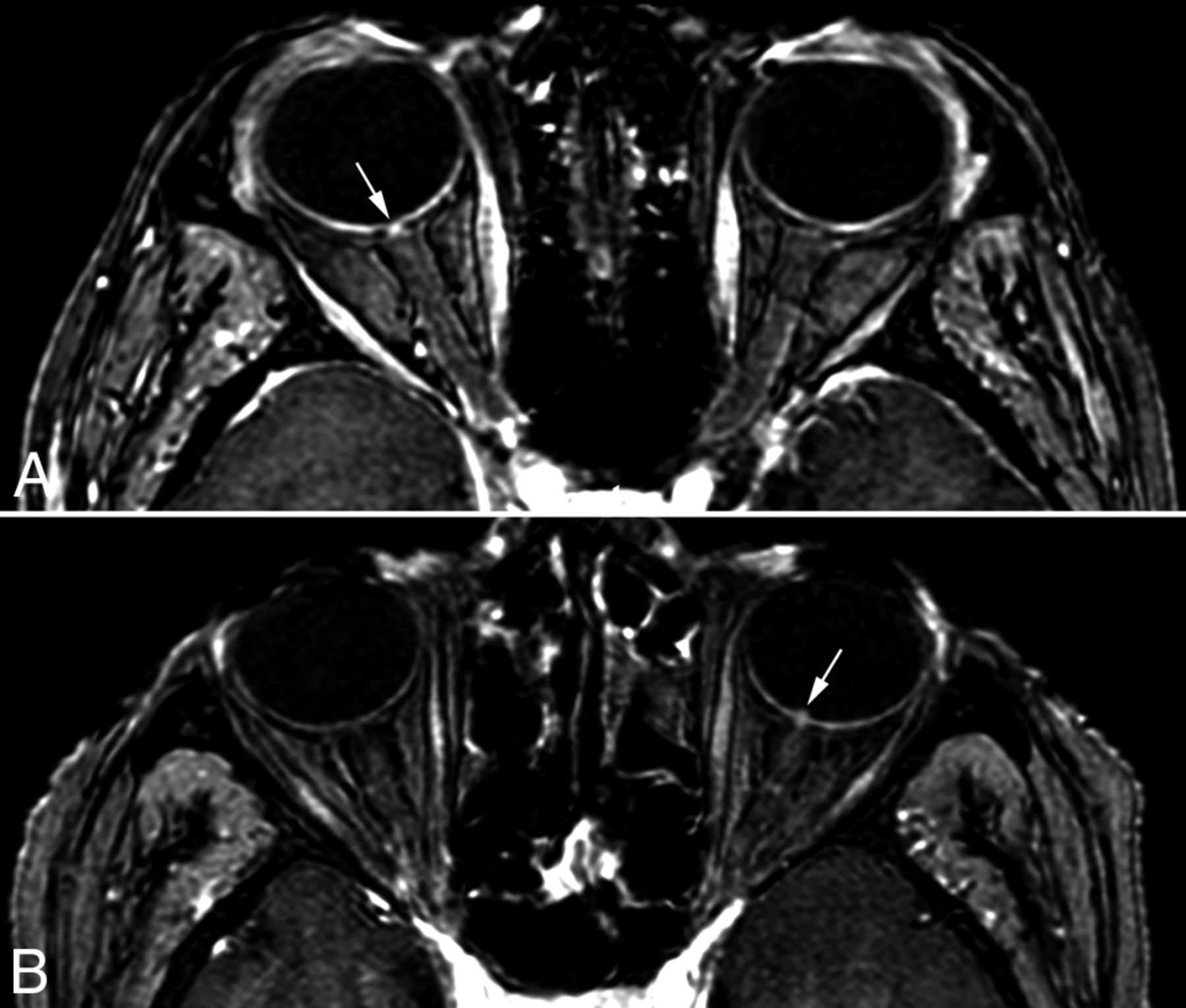
- Fig 1.
High-resolution T1 sequences in the axial plane showing the bright spot sign ranking score. A, The first patient was referred with right nonarteritic AION and a score of 1, with no optic nerve head intravitreal protrusion but local enhancement (white arrow). B, The second patient was referred with left GCA-related AION and both protrusion and local enhancement, defined as score 2 (white arrow).

Tables
Reader Healthy Subjects (n = 15) NA-AION (n = 15) GCA-AION (n = 15) Reader 1 0/15 7/15 15/15 Reader 2 0/15 6/15 15/15 Reader 3 0/15 6/15 15/15 - Table 2:
Sensitivity, specificity, positive/negative predictive values, and positive/negative likelihood ratios of the MR imaging bright spot sign in healthy subjects and patients with GCA-AION or NA-AION
No. of Participants TP Results TN Results FP Results FN Results Sensitivity (%) Specificity (%) PPV (%) NPV (%) PLR NLR NA-AION vs GCA-AION Observer 1 30 15 8 7 0 100 53.3 68.2 100 2.1 0 Observer 2 30 15 9 6 0 100 60 71.4 100 3.5 0 Observer 3 30 15 9 6 0 100 60 71.4 100 3.5 0 NA-AION vs healthy subjects Observer 1 30 7 15 0 8 46.7 100 100 65.2 0.5 Observer 2 30 6 15 0 9 40 100 100 62.5 0.6 Observer 3 30 6 15 0 9 40 100 100 62.5 0.6 GCA-AION vs healthy subjects Observer 1 30 15 15 0 0 100 100 100 100 0 Observer 2 30 15 15 0 0 100 100 100 100 0 Observer 3 30 15 15 0 0 100 100 100 100 0 Note:—FN indicates false-negative; FP, false-positive; PLR, positive likelihood ratio; NLR, negative likelihood ratio; NPV, negative predictive value; PPV, positive predictive value; TN, true-negative; TP, true-positive.


