
Article Figures & Data
Figures
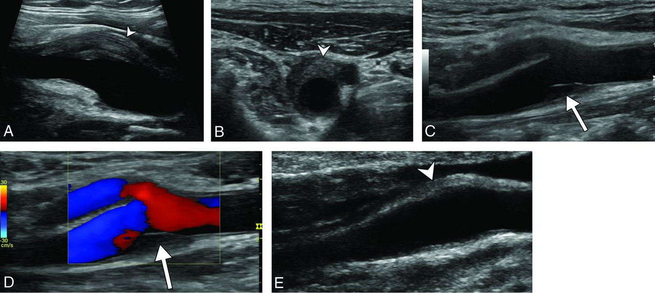
- Fig 1.
Diagnostic ultrasonography (A–D) shows an eccentric perivascular infiltration (arrowhead) at the level of bifurcation, with a soft intimal plaque (arrow) and a mild lumen narrowing without a hemodynamic change in Doppler mode. Follow-up ultrasonography (E) shows a marked decrease in the perivascular infiltration (arrowhead) and complete disappearance of the soft intimal plaque.
- Fig 2.
CTA shows a left posterolateral eccentric perivascular infiltration (arrowhead) surrounding the carotid artery, with a distinct low-density soft intimal plaque (arrow).
- Fig 3.
Initial diagnostic ultrasonography (A) and follow-up ultrasonography at 14 days (B) and 6 months (C) show a perivascular infiltration (arrowheads) at the level of the internal carotid artery just at the level of bifurcation, with a quick decrease at 14 days and the persistence of a thin abnormality at 6 months.
- Fig 4.
Pre (A) and post (B) contrast fat-suppressed 3D T1 and 3D T2-weighted (C) MR imaging in an axial plane shows T1 hypointense and T2 hyperintense perivascular infiltration (arrowhead) at the level of the carotid artery bifurcation, enhanced after gadolinium injection. A distinct soft intimal plaque (arrow) is visible at the posterior part of the carotid artery. A sagittal curvilinear reconstruction of the right internal carotid artery on the postcontrast T1-weighted imaging (D) shows the PVI (arrowhead) centered at the level of the right carotid artery bifurcation and extended to both the distal common carotid artery and proximal internal carotid artery. Note that there is no vascular or perivascular abnormality involving other parts of the common or the internal carotid arteries.


Tables
Characteristics Patients % No. of patients 47 No. of carotid arteries 49 Sex ratio (women/men) 27/18; 1.5:1 Median age (yr) (IQR) 48 (39–56) At least 1 vascular risk factor 22 47% High blood pressure 8 17% Dyslipidemia 3 6% Mellitus diabetes 0 0% Smoking 19 40% History of autoimmune disease 8 17% History of vascular event 2 4% Recent history of viral episode 2 4% Recent history of cervical chiropractic manipulation 0 0% Recent history of cervical trauma 1 2% Acute neck tenderness or cervical pain 47 100% Affected side (left/right) 28/21 Bilateral involvement 2 4% Median pain intensity (10-point scale) (IQR) 5 (3–6) Cervical swelling or palpable abnormality over the carotid bifurcation 6 13% Enlarged lymph nodes at palpation 8 17% Fever 2 4% Flu-like symptoms 1 2% Neurologic-associated symptoms 8 17% No abnormal biologic test findings 42 89% Biologic inflammatory syndrome (elevated ESR or CRP) 3 6% Positive serum IgM 2/18 11% Treatments 37 79% Anti-inflammatory treatment 34 72% Steroids 3 6% Follow-up 47 100% Median follow-up duration (days) (IQR) 163 (88–354) Full clinical recovery (SD) 47 100% Mean recovery delay (days) (IQR) 13 (10–15) Relapse 9 19% Median delay before relapse (mo) 6 Persistence of abnormal laboratory test findings 0 0% Note:—ESR indicates erythrocyte sedimentation rate; CRP, C-reactive protein; IgM, immunoglobulin-M; IQR, interquartile range.
Characteristics US % CT % MRI % No. of patients 23 49% 13 28% 43 91% No. of carotid arteries 24 49% 13 27% 45 92% Median delay between onset of symptoms and diagnostic exam (days) (IQR) 5 (3–7.5) High confidence in diagnosis 24 100% 9 69% 45 100% Preferential side of PVI: lateral/medial 39/10: 3.9 Preferential side of PVI: posterior/anterior 38/11: 3.5 Localization of PVI: carotid bifurcation 23 96% 12 92% 41 91% Circumferential 0 0% 0 0% 2 4% Percentage of carotid circumference involved (%) (IQR) 40 (30–40) 30 (20–40) 40 (30–57.5) PVI 24 100% 13 100% 45 100% Pericarotid fat stranding 24 100% 13 100% 45 100% Lumen caliber narrowing 9 38% 4 31% 12 27% Mean percentage of carotid stenosis (%) (IQR) 30 (20–30) 22.5 (18–26) 20 (14–30) Mean PVI span (mm) (IQR) 15 (10–22) 15 (10–26) 28 (17–34) Median PVI largest diameter (mm) 4 (2.4–6) 4 (3.6–4.8) 5 (4–7) Soft intimal plaque 14 58% 6 46% 12 27% Enlarged lymph nodes 8 33% 0 0% 8 18% PVI enhancement NA 11 85% 45 100% Laryngeal or pharyngeal inflammation 0 0% 0 0% 1 2% Note:—IQR indicates interquartile range; NA, not applicable.
Characteristics US % MRI % No. of patients 11 44% 23 51% Median delay between onset of symptoms and follow-up exam (days) (IQR) 89 (71.5–171) PVI 11 100% 15 65% Complete PVI disappearance 0 0% 8 35% PVI decrease 11 100% 15 65% PVI stability 0 0% 0 0% PVI increase 0 0% 0 0% Lumen caliber narrowing 2 18% 1 4% Median PVI largest diameter (mm) (IQR) 1.5 (0.9–2.4) 2.9 (2.0–3.5) Median decrease of the PVI largest diameter 61% 55% Median PVI span (mm) (IQR) 9 (5–10) 13 (9.5–16) Median decrease of the PVI span 62% 50% Persistence of a soft intimal plaque 4 50% 3 33% Disappearance of a soft intimal plaque 4 50% 6 67% PVI enhancement NA 12 52%


Jump to section
Related Articles
Cited By...
- Transient perivascular inflammation of the carotid artery syndrome
- Images of the month 3: Transient perivascular inflammation of the carotid artery syndrome
- Brain and Lung Imaging Correlation in Patients with COVID-19: Could the Severity of Lung Disease Reflect the Prevalence of Acute Abnormalities on Neuroimaging? A Global Multicenter Observational Study
- Secondary Otalgia: Referred Pain Pathways and Pathologies
- Teaching NeuroImages: An aTIPICal cause of acute neck pain
- TIPIC syndrome