Advertisement

Main menu

  • Home
  • Content
    • Current Issue
    • Accepted Manuscripts
    • Article Preview
    • Past Issue Archive
    • Video Articles
    • AJNR Case Collection
    • Case of the Week Archive
    • Case of the Month Archive
    • Classic Case Archive
  • Special Collections
    • AJNR Awards
    • Low-Field MRI
    • Alzheimer Disease
    • ASNR Foundation Special Collection
    • Photon-Counting CT
    • View All
  • Multimedia
    • AJNR Podcasts
    • AJNR SCANtastic
    • Trainee Corner
    • MRI Safety Corner
    • Imaging Protocols
  • For Authors
    • Submit a Manuscript
    • Submit a Video Article
    • Submit an eLetter to the Editor/Response
    • Manuscript Submission Guidelines
    • Statistical Tips
    • Fast Publishing of Accepted Manuscripts
    • Graphical Abstract Preparation
    • Imaging Protocol Submission
    • Author Policies
  • About Us
    • About AJNR
    • Editorial Board
    • Editorial Board Alumni
  • More
    • Become a Reviewer/Academy of Reviewers
    • Subscribers
    • Permissions
    • Alerts
    • Feedback
    • Advertisers
    • ASNR Home

User menu

  • Alerts
  • Log in

Search

  • Advanced search
American Journal of Neuroradiology

American Journal of Neuroradiology

ASHNR American Society of Functional Neuroradiology ASHNR American Society of Pediatric Neuroradiology ASSR
  • Alerts
  • Log in

Advanced Search

  • Home
  • Content
    • Current Issue
    • Accepted Manuscripts
    • Article Preview
    • Past Issue Archive
    • Video Articles
    • AJNR Case Collection
    • Case of the Week Archive
    • Case of the Month Archive
    • Classic Case Archive
  • Special Collections
    • AJNR Awards
    • Low-Field MRI
    • Alzheimer Disease
    • ASNR Foundation Special Collection
    • Photon-Counting CT
    • View All
  • Multimedia
    • AJNR Podcasts
    • AJNR SCANtastic
    • Trainee Corner
    • MRI Safety Corner
    • Imaging Protocols
  • For Authors
    • Submit a Manuscript
    • Submit a Video Article
    • Submit an eLetter to the Editor/Response
    • Manuscript Submission Guidelines
    • Statistical Tips
    • Fast Publishing of Accepted Manuscripts
    • Graphical Abstract Preparation
    • Imaging Protocol Submission
    • Author Policies
  • About Us
    • About AJNR
    • Editorial Board
    • Editorial Board Alumni
  • More
    • Become a Reviewer/Academy of Reviewers
    • Subscribers
    • Permissions
    • Alerts
    • Feedback
    • Advertisers
    • ASNR Home
  • Follow AJNR on Twitter
  • Visit AJNR on Facebook
  • Follow AJNR on Instagram
  • Join AJNR on LinkedIn
  • RSS Feeds

AJNR Awards, New Junior Editors, and more. Read the latest AJNR updates

Research ArticleSpine Imaging and Spine Image-Guided Interventions
Open Access

Clinically Feasible Microstructural MRI to Quantify Cervical Spinal Cord Tissue Injury Using DTI, MT, and T2*-Weighted Imaging: Assessment of Normative Data and Reliability

A.R. Martin, B. De Leener, J. Cohen-Adad, D.W. Cadotte, S. Kalsi-Ryan, S.F. Lange, L. Tetreault, A. Nouri, A. Crawley, D.J. Mikulis, H. Ginsberg and M.G. Fehlings
American Journal of Neuroradiology June 2017, 38 (6) 1257-1265; DOI: https://doi.org/10.3174/ajnr.A5163
A.R. Martin
aFrom the Division of Neurosurgery, Department of Surgery (A.R.M., D.W.C., S.K.-R., L.T., A.N., H.G., M.G.F.)
  • Find this author on Google Scholar
  • Find this author on PubMed
  • Search for this author on this site
  • ORCID record for A.R. Martin
B. De Leener
cPolytechnique Montreal (B.D.L., J.C.-A.), Montréal, Quebec, Canada
  • Find this author on Google Scholar
  • Find this author on PubMed
  • Search for this author on this site
  • ORCID record for B. De Leener
J. Cohen-Adad
cPolytechnique Montreal (B.D.L., J.C.-A.), Montréal, Quebec, Canada
dFunctional Neuroimaging Unit (J.C.-A.), Centre de recherche de l'Institut universitaire de gériatrie de Montréal, Université de Montréal, Montréal, Quebec, Canada
  • Find this author on Google Scholar
  • Find this author on PubMed
  • Search for this author on this site
  • ORCID record for J. Cohen-Adad
D.W. Cadotte
aFrom the Division of Neurosurgery, Department of Surgery (A.R.M., D.W.C., S.K.-R., L.T., A.N., H.G., M.G.F.)
  • Find this author on Google Scholar
  • Find this author on PubMed
  • Search for this author on this site
  • ORCID record for D.W. Cadotte
S. Kalsi-Ryan
aFrom the Division of Neurosurgery, Department of Surgery (A.R.M., D.W.C., S.K.-R., L.T., A.N., H.G., M.G.F.)
  • Find this author on Google Scholar
  • Find this author on PubMed
  • Search for this author on this site
  • ORCID record for S. Kalsi-Ryan
S.F. Lange
eUniversity of Groningen (S.F.L.), Groningen, the Netherlands.
  • Find this author on Google Scholar
  • Find this author on PubMed
  • Search for this author on this site
  • ORCID record for S.F. Lange
L. Tetreault
aFrom the Division of Neurosurgery, Department of Surgery (A.R.M., D.W.C., S.K.-R., L.T., A.N., H.G., M.G.F.)
  • Find this author on Google Scholar
  • Find this author on PubMed
  • Search for this author on this site
  • ORCID record for L. Tetreault
A. Nouri
aFrom the Division of Neurosurgery, Department of Surgery (A.R.M., D.W.C., S.K.-R., L.T., A.N., H.G., M.G.F.)
  • Find this author on Google Scholar
  • Find this author on PubMed
  • Search for this author on this site
  • ORCID record for A. Nouri
A. Crawley
bDepartment of Medical Imaging (A.C., D.J.M.), University of Toronto and the University Health Network, Toronto Western Hospital, Toronto, Ontario, Canada
  • Find this author on Google Scholar
  • Find this author on PubMed
  • Search for this author on this site
  • ORCID record for A. Crawley
D.J. Mikulis
bDepartment of Medical Imaging (A.C., D.J.M.), University of Toronto and the University Health Network, Toronto Western Hospital, Toronto, Ontario, Canada
  • Find this author on Google Scholar
  • Find this author on PubMed
  • Search for this author on this site
  • ORCID record for D.J. Mikulis
H. Ginsberg
aFrom the Division of Neurosurgery, Department of Surgery (A.R.M., D.W.C., S.K.-R., L.T., A.N., H.G., M.G.F.)
  • Find this author on Google Scholar
  • Find this author on PubMed
  • Search for this author on this site
  • ORCID record for H. Ginsberg
M.G. Fehlings
aFrom the Division of Neurosurgery, Department of Surgery (A.R.M., D.W.C., S.K.-R., L.T., A.N., H.G., M.G.F.)
  • Find this author on Google Scholar
  • Find this author on PubMed
  • Search for this author on this site
  • ORCID record for M.G. Fehlings
  • Article
  • Figures & Data
  • Info & Metrics
  • Responses
  • References
  • PDF
Loading

Article Figures & Data

Figures

  • Tables
  • Fig 1.
    • Download figure
    • Open in new tab
    • Download powerpoint
    Fig 1.

    Representative images showing FA maps (A), MTR maps (B), and T2*WI (C) with probabilistic maps of the lateral corticospinal tracts (blue) and dorsal columns (red-yellow) overlaid (D–F) following registration to the SCT atlas.
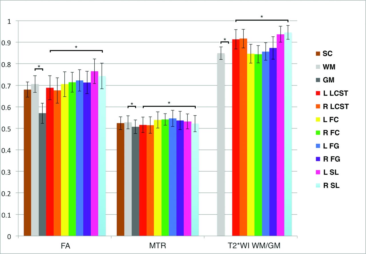
  • Fig 2.
    • Download figure
    • Open in new tab
    • Download powerpoint
    Fig 2.

    Normative data in the rostral cervical cord for FA, MTR, and T2*WI WM/GM ratios. Metrics are extracted from SC, WM, GM, and key WM tracts averaged over rostral sections (C1–C3). Values are displayed as mean ± intersubject SD (error bars). The asterisk denotes P < .05 with paired t tests between WM and GM and ANOVA among WM tracts. L indicates left; R, right; FC, fasciculus cuneatus; FG, fasciculus gracilis; SL, spinal lemniscus; LCST, lateral corticospinal tract.

  • Fig 3.
    • Download figure
    • Open in new tab
    • Download powerpoint
    Fig 3.

    Variations by rostrocaudal level. MR imaging metrics displayed for each vertebral and intervertebral level from C1 to C7. FA, MTR, and T2*WI WM/GM ratios are extracted from WM. ANOVA shows significant differences by level for all metrics. Monotonic variations are present for CSA, FA, and MTR.

  • Fig 4.
    • Download figure
    • Open in new tab
    • Download powerpoint
    Fig 4.

    Test-retest coefficients of variation of FA, MTR, and T2*WI WM/GM extracted from SC, WM, GM, and key WM tracts in rostral sections (C1–C3) are displayed. T2*WI WM/GM ratio shows better reliability than FA and MTR. Metrics derived from the SC and WM show TRCOV < 3%, while GM and key WM tracts show TRCOV < 5% except for FA of the spinal lemniscus. FC indicates fasciculus cuneatus; FG, fasciculus gracilis; SL, spinal lemniscus; LCST, lateral corticospinal tract; R, right; L, left.

Tables

  • Figures
    • View popup
    Table 1:

    Acquisition protocola

    Imaging TypePulse Sequence; OrientationTechnical DetailsAcquisition TimeMetric
    T2WI3D FIESTA-C; sagittalTR/TE = 5.4/2.6 s, FOV = 200 × 200 mm2, matrix = 256 × 256, resolution = 0.8 × 0.8 × 0.8 mm3, NEX = 2, flip angle = 35°6 min 56 sCSA
    DTISpin-echo ssEPI with OVS; axialTR/TE = 4050/91.2 ms, FOV = 80 × 80 mm2, matrix = 64 × 64, resolution = 1.25 × 1.25 × 5 mm3, 25 directions (b=800 s/mm2), 5 b=0 s/mm2 images, AP saturation bands, phase encoding = AP, 2nd-order shimming3 × 2 min 6 s, 1 min 30 s for shimmingFA
    MT2D SPGR with/without prepulse; axialTR/TE = 32/5.9 ms, FOV = 190 × 190 mm2, matrix = 192 × 192, resolution = 1 × 1 × 5 mm3, NEX = 3, flip angle = 6°, flow compensation, phase encoding = AP, prepulse: Gaussian, duration = 9984 μs, offset = 1200 Hz3 min 45 s each, with and without prepulseMTR
    T2*WI2D MERGE; axialTR/TE = 650/5, 10, 15 ms, FOV = 200 × 200 mm2, matrix = 320 × 320, resolution = 0.6 × 0.6 × 4 mm3, NEX = 1, flip angle = 20°, BW = 62 kHz per line3 min 33 sWM/GM ratio
    • Note:—AP indicates anteroposterior; BW, bandwidth; FIESTA-C, FIESTA-cycled phases; MERGE, multiecho recombined gradient echo; OVS, outer volume suppression; SPGR, echo-spoiled gradient echo; ssEPI, single-shot echo-planar imaging.

    • ↵a Technical specifications of our multiparametric cervical SC MRI protocol, with an acquisition time of 25 minutes (30–35 minutes, including positioning, section prescription, shimming, and prescans).

    • View popup
    Table 2:

    Subject characteristicsa

    CharacteristicHealthy Subjects (n = 40)Subjects with DCM (n = 18)
    Age (yr)47.1 ± 15.3 (range, 19–79)56.4 ± 11.0 (range, 36–76)
    Sex21 men, 19 women11 men, 7 women
    Height (cm)171.4 ± 8.6172.8 ± 8.9
    Weight (kg)74.6 ± 11.579.0 ± 15.1
    Cervical cord length (cm)10.6 ± 1.011.1 ± 0.9
    • ↵a Demographics and characteristics of 40 healthy subjects and 18 with DCM are shown. Data (other than sex) are reported as mean ± SD.

    • View popup
    Table 3:

    Univariate relationships of MRI metrics with healthy subject characteristicsa

    MetricAgeSex (M vs F)HeightWeightCervical Cord Length
    CSA (mm2)r = −0.25 (P = .12)80.0 ± 11.2 vs 73.5 ± 8.5 (P = .03b)r = 0.31 (P = .06c)r = 0.34 (P = .03b)r = 0.51 (P < .001b)
    FAr = −0.43 (P = .009b)0.658 ± 0.037 vs 0.663 ± 0.034 (P = .75)r = −0.02 (P = .89)r = −0.26 (P = .12)r = 0.11 (P = .53)
    MTRr = −0.25 (P = .11)48.8 ± 2.5 vs 51.4 ± 2.7 (P = .006b)r = −0.41 (P = .008b)r = −0.40 (P = .01)r = −0.18 (P = .26)
    T2*WI WM/GMr = 0.31 (P = .06)0.863 ± 0.034 vs 0.858 ± 0.031 (P = .64)r = −0.12 (P = .48)r = 0.31 (P = .06c)r = −0.09 (P = .55)
    • ↵a Values for sex are reported as mean ± SD, and other values are Pearson correlation coefficient. FA, MTR, and T2*WI WM/GM ratios are extracted from WM, while CSA of the spinal cord is measured, averaged across C1–C7.

    • ↵b Significance (P < .05).

    • ↵c Trends (P < .10).

    • View popup
    Table 4:

    Test-retest reliability across rostrocaudal levelsa

    LevelMetricHealthyDCMP ValuePooled
    Rostral (C1–C3)FA2.5 ± 2.0%2.8 ± 1.8%.712.6 ± 1.9%
    MTR2.7 ± 1.9%1.3 ± 0.5%.172.4 ± 1.9%
    T2*WI WM/GM0.9 ± 0.6%1.0 ± 0.7%.770.9 ± 0.7%b
    Midcervical (C4–C5) or MCLFA3.0 ± 2.2%5.0 ± 5.7%.213.6 ± 3.6%
    MTR3.2 ± 3.0%6.1 ± 0.9%.08c3.7 ± 3.2%
    T2*WI WM/GM1.4 ± 1.1%3.5 ± 2.2%.112.9 ± 2.2%
    Caudal (C6–C7)FA2.2 ± 1.6%4.6 ± 4.7%.07c3.2 ± 3.5%
    MTR4.4 ± 3.8%3.1 ± 3.9%.564.2 ± 3.7%
    T2*WI WM/GM3.4 ± 3.0%2.2 ± 2.1%.372.6 ± 2.4%
    • ↵a TRCOV ± SD is displayed for healthy subjects and those with DCM at rostral (C1–C3), midcervical (C4–5), or maximally compressed levels in subjects with DCM, and caudal (C6–C7) levels. Sample size was 26 subjects (17 healthy, 9 with DCM) for DTI, 17 subjects (13 healthy, 4 with DCM) for MT, and 16 subjects (5 healthy, 11 with DCM) for T2*WI.

    • ↵b Significant differences (P < .05) between pooled TRCOV of metrics at each level.

    • ↵c Trends (P < .10) in reliability between healthy subjects and those with DCM for each level/metric, and pooled reliability was calculated if no significant differences were found.

    • View popup
    Table 5:

    DTI with and without cardiac triggeringa

    MeasureLevelNo TriggeringTriggeringP Value
    FARostral0.651 ± 0.0540.664 ± 0.064.41
    Mid/MCL0.514 ± 0.0680.558 ± 0.081.06b
    Caudal0.534 ± 0.0570.562 ± 0.044.07b
    TRCOVRostral2.6 ± 1.9%1.5 ± 1.2%.11
    Mid/MCL3.6 ± 3.6%2.2 ± 2.3%.27
    Caudal3.2 ± 3.5%2.4 ± 2.3%.52
    • ↵a Paired t tests were used to compare FA values extracted from WM at rostral (C1–C3), midcervical (C4–5, healthy subjects), or MCL (subjects with DCM), and caudal (C6–C7) levels between no triggering vs triggering in 10 subjects (4 healthy, 6 with DCM). Welch t tests were used to compare test-retest coefficient of variation between no triggering (n = 26) and triggering (n = 10).

    • ↵b Trends (P < .10).

PreviousNext
Back to top

In this issue

American Journal of Neuroradiology: 38 (6)
American Journal of Neuroradiology
Vol. 38, Issue 6
1 Jun 2017
  • Table of Contents
  • Index by author
  • Complete Issue (PDF)
Advertisement
Print
Download PDF
Email Article

Thank you for your interest in spreading the word on American Journal of Neuroradiology.

NOTE: We only request your email address so that the person you are recommending the page to knows that you wanted them to see it, and that it is not junk mail. We do not capture any email address.

Enter multiple addresses on separate lines or separate them with commas.
Clinically Feasible Microstructural MRI to Quantify Cervical Spinal Cord Tissue Injury Using DTI, MT, and T2*-Weighted Imaging: Assessment of Normative Data and Reliability
(Your Name) has sent you a message from American Journal of Neuroradiology
(Your Name) thought you would like to see the American Journal of Neuroradiology web site.
CAPTCHA
This question is for testing whether or not you are a human visitor and to prevent automated spam submissions.
Cite this article
A.R. Martin, B. De Leener, J. Cohen-Adad, D.W. Cadotte, S. Kalsi-Ryan, S.F. Lange, L. Tetreault, A. Nouri, A. Crawley, D.J. Mikulis, H. Ginsberg, M.G. Fehlings
Clinically Feasible Microstructural MRI to Quantify Cervical Spinal Cord Tissue Injury Using DTI, MT, and T2*-Weighted Imaging: Assessment of Normative Data and Reliability
American Journal of Neuroradiology Jun 2017, 38 (6) 1257-1265; DOI: 10.3174/ajnr.A5163

Citation Manager Formats

  • BibTeX
  • Bookends
  • EasyBib
  • EndNote (tagged)
  • EndNote 8 (xml)
  • Medlars
  • Mendeley
  • Papers
  • RefWorks Tagged
  • Ref Manager
  • RIS
  • Zotero
0 Responses
Respond to this article
Share
Bookmark this article
Clinically Feasible Microstructural MRI to Quantify Cervical Spinal Cord Tissue Injury Using DTI, MT, and T2*-Weighted Imaging: Assessment of Normative Data and Reliability
A.R. Martin, B. De Leener, J. Cohen-Adad, D.W. Cadotte, S. Kalsi-Ryan, S.F. Lange, L. Tetreault, A. Nouri, A. Crawley, D.J. Mikulis, H. Ginsberg, M.G. Fehlings
American Journal of Neuroradiology Jun 2017, 38 (6) 1257-1265; DOI: 10.3174/ajnr.A5163
del.icio.us logo Twitter logo Facebook logo Mendeley logo
  • Tweet Widget
  • Facebook Like
  • Google Plus One
Purchase

Jump to section

  • Article
    • Abstract
    • ABBREVIATIONS:
    • Materials and Methods
    • Results
    • Discussion
    • Conclusions
    • ACKNOWLEDGMENTS
    • Footnotes
    • References
  • Figures & Data
  • Info & Metrics
  • Responses
  • References
  • PDF

Related Articles

  • No related articles found.
  • PubMed
  • Google Scholar

Cited By...

  • Extent of cord pathology in the lumbosacral enlargement in non-traumatic versus traumatic spinal cord injury
  • Tracking white and grey matter degeneration along the spinal cord axis in degenerative cervical myelopathy
  • The Restless Spinal Cord in Degenerative Cervical Myelopathy
  • Quantification of DTI in the Pediatric Spinal Cord: Application to Clinical Evaluation in a Healthy Patient Population
  • Convolutional Neural Network-Based Automated Segmentation of the Spinal Cord and Contusion Injury: Deep Learning Biomarker Correlates of Motor Impairment in Acute Spinal Cord Injury
  • In vivo evidence of remote neural degeneration in the lumbar enlargement after cervical injury
  • Can microstructural MRI detect subclinical tissue injury in subjects with asymptomatic cervical spinal cord compression? A prospective cohort study
  • Crossref
  • Google Scholar

This article has not yet been cited by articles in journals that are participating in Crossref Cited-by Linking.

More in this TOC Section

  • Dynamic CT Myelography: Patient Positioning
  • Evaluation of SIH MR Scoring Systems in Normals
  • Management Outcomes For VO Spine Biopsy
Show more SPINE IMAGING AND SPINE IMAGE-GUIDED INTERVENTIONS

Similar Articles

Advertisement

Indexed Content

  • Current Issue
  • Accepted Manuscripts
  • Article Preview
  • Past Issues
  • Editorials
  • Editor's Choice
  • Fellows' Journal Club
  • Letters to the Editor
  • Video Articles

Cases

  • Case Collection
  • Archive - Case of the Week
  • Archive - Case of the Month
  • Archive - Classic Case

More from AJNR

  • Trainee Corner
  • Imaging Protocols
  • MRI Safety Corner
  • Book Reviews

Multimedia

  • AJNR Podcasts
  • AJNR Scantastics

Resources

  • Turnaround Time
  • Submit a Manuscript
  • Submit a Video Article
  • Submit an eLetter to the Editor/Response
  • Manuscript Submission Guidelines
  • Statistical Tips
  • Fast Publishing of Accepted Manuscripts
  • Graphical Abstract Preparation
  • Imaging Protocol Submission
  • Evidence-Based Medicine Level Guide
  • Publishing Checklists
  • Author Policies
  • Become a Reviewer/Academy of Reviewers
  • News and Updates

About Us

  • About AJNR
  • Editorial Board
  • Editorial Board Alumni
  • Alerts
  • Permissions
  • Not an AJNR Subscriber? Join Now
  • Advertise with Us
  • Librarian Resources
  • Feedback
  • Terms and Conditions
  • AJNR Editorial Board Alumni

American Society of Neuroradiology

  • Not an ASNR Member? Join Now

© 2025 by the American Society of Neuroradiology All rights, including for text and data mining, AI training, and similar technologies, are reserved.
Print ISSN: 0195-6108 Online ISSN: 1936-959X

Powered by HighWire