Advertisement

Main menu

  • Home
  • Content
    • Current Issue
    • Accepted Manuscripts
    • Article Preview
    • Past Issue Archive
    • Video Articles
    • AJNR Case Collection
    • Case of the Week Archive
    • Case of the Month Archive
    • Classic Case Archive
  • Special Collections
    • AJNR Awards
    • Low-Field MRI
    • Alzheimer Disease
    • ASNR Foundation Special Collection
    • Photon-Counting CT
    • View All
  • Multimedia
    • AJNR Podcasts
    • AJNR SCANtastic
    • Trainee Corner
    • MRI Safety Corner
    • Imaging Protocols
  • For Authors
    • Submit a Manuscript
    • Submit a Video Article
    • Submit an eLetter to the Editor/Response
    • Manuscript Submission Guidelines
    • Statistical Tips
    • Fast Publishing of Accepted Manuscripts
    • Graphical Abstract Preparation
    • Imaging Protocol Submission
    • Author Policies
  • About Us
    • About AJNR
    • Editorial Board
    • Editorial Board Alumni
  • More
    • Become a Reviewer/Academy of Reviewers
    • Subscribers
    • Permissions
    • Alerts
    • Feedback
    • Advertisers
    • ASNR Home

User menu

  • Alerts
  • Log in

Search

  • Advanced search
American Journal of Neuroradiology

American Journal of Neuroradiology

ASHNR American Society of Functional Neuroradiology ASHNR American Society of Pediatric Neuroradiology ASSR
  • Alerts
  • Log in

Advanced Search

  • Home
  • Content
    • Current Issue
    • Accepted Manuscripts
    • Article Preview
    • Past Issue Archive
    • Video Articles
    • AJNR Case Collection
    • Case of the Week Archive
    • Case of the Month Archive
    • Classic Case Archive
  • Special Collections
    • AJNR Awards
    • Low-Field MRI
    • Alzheimer Disease
    • ASNR Foundation Special Collection
    • Photon-Counting CT
    • View All
  • Multimedia
    • AJNR Podcasts
    • AJNR SCANtastic
    • Trainee Corner
    • MRI Safety Corner
    • Imaging Protocols
  • For Authors
    • Submit a Manuscript
    • Submit a Video Article
    • Submit an eLetter to the Editor/Response
    • Manuscript Submission Guidelines
    • Statistical Tips
    • Fast Publishing of Accepted Manuscripts
    • Graphical Abstract Preparation
    • Imaging Protocol Submission
    • Author Policies
  • About Us
    • About AJNR
    • Editorial Board
    • Editorial Board Alumni
  • More
    • Become a Reviewer/Academy of Reviewers
    • Subscribers
    • Permissions
    • Alerts
    • Feedback
    • Advertisers
    • ASNR Home
  • Follow AJNR on Twitter
  • Visit AJNR on Facebook
  • Follow AJNR on Instagram
  • Join AJNR on LinkedIn
  • RSS Feeds

AJNR Awards, New Junior Editors, and more. Read the latest AJNR updates

Research ArticleHead and Neck Imaging

Imaging Manifestations of Pseudoprogression in Metastatic Melanoma Nodes Injected with Talimogene Laherparepvec: Initial Experience

C. Zamora, M. Lopez, F. Cunningham, F. Collichio and M. Castillo
American Journal of Neuroradiology June 2017, 38 (6) 1218-1222; DOI: https://doi.org/10.3174/ajnr.A5206
C. Zamora
aFrom the Division of Neuroradiology, Department of Radiology (C.Z., M.L., M.C.)
  • Find this author on Google Scholar
  • Find this author on PubMed
  • Search for this author on this site
  • ORCID record for C. Zamora
M. Lopez
aFrom the Division of Neuroradiology, Department of Radiology (C.Z., M.L., M.C.)
  • Find this author on Google Scholar
  • Find this author on PubMed
  • Search for this author on this site
  • ORCID record for M. Lopez
F. Cunningham
bthe Division of Hematology and Oncology, Department of Medicine (F. Cunningham, F. Collichio), University of North Carolina School of Medicine, Chapel Hill, North Carolina.
  • Find this author on Google Scholar
  • Find this author on PubMed
  • Search for this author on this site
  • ORCID record for F. Cunningham
F. Collichio
bthe Division of Hematology and Oncology, Department of Medicine (F. Cunningham, F. Collichio), University of North Carolina School of Medicine, Chapel Hill, North Carolina.
  • Find this author on Google Scholar
  • Find this author on PubMed
  • Search for this author on this site
  • ORCID record for F. Collichio
M. Castillo
aFrom the Division of Neuroradiology, Department of Radiology (C.Z., M.L., M.C.)
  • Find this author on Google Scholar
  • Find this author on PubMed
  • Search for this author on this site
  • ORCID record for M. Castillo
  • Article
  • Figures & Data
  • Info & Metrics
  • Responses
  • References
  • PDF
Loading

Article Figures & Data

Figures

  • Tables
  • Fig 1.
    • Download figure
    • Open in new tab
    • Download powerpoint
    Fig 1.

    Line graph showing change in size over time relative to baseline CT. A, Fifty percent of nodes showed initial growth followed by decreased size. One node (marked with an asterisk at last time point) demonstrated a continued increase in size, but could not be followed after 184 days. B, Forty-two percent of nodes showed a downward trend in size. The final size was smaller than baseline in all nodes except for the one indicated in A.
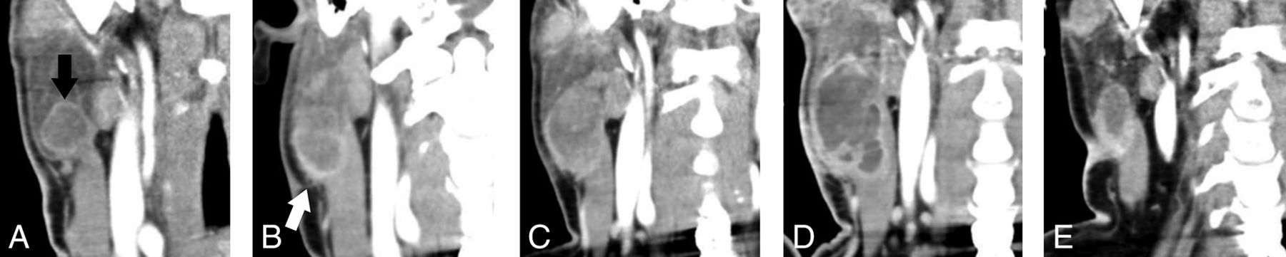
  • Fig 2.
    • Download figure
    • Open in new tab
    • Download powerpoint
    Fig 2.

    A, Coronal contrast-enhanced CT shows a round, well-circumscribed necrotic lesion (black arrow) at the inferior aspect of the right parotid gland. B, The lesion has increased in size and developed infiltrative margins (white arrow) 72 days after injection. Follow-up at 111 days (C) and 184 days (D) after injection demonstrates further growth and marked increase in central necrosis. E, The lesion shows decreased size after 337 days.
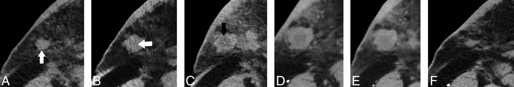
  • Fig 3.
    • Download figure
    • Open in new tab
    • Download powerpoint
    Fig 3.

    A, Axial contrast-enhanced CT shows a small nodule in the deep right breast/axillary region (white arrow). The lesion shows slight central hypoattenuation and increased size at 34 days after injection (white arrow, B) with the development of frank central necrosis after 72 days (black arrow, C). There is a mild further increase in size at 104 days (D), followed by decreased size at 125 days (E) and near complete resolution at 174 days (F).

Tables

  • Figures
  • Clinical features, disease stage, final nodal size (relative to baseline CT), and patient outcome

    Patient No.Lesion No.Age, yrSexStageBRAF StatusLocationFinal Lesion SizePatient Outcome
    1163FT3bN3M1aNegativeLeft supraclavicularDecreasedRemission.
    2266MTxNxM1cWild typeRight supraclavicularDecreasedT-VEC discontinued after disease progression in ribs. Placed on nivolumab with partial response.
    3Right supraclavicularDecreased
    3440MpT1bN3M1cPositiveRight preauricularDecreasedIL-2 followed by BRAF and MEK inhibitors after ipilimumab/T-VEC treatment. Deceased.
    5Right submandibularDecreased
    4644MT3bN3M1bPositiveRight axillaryDecreasedBRAF and MEK inhibitors after ipilimumab/T-VEC treatment, followed by disease progression. Deceased.
    7Right axillaryDecreased
    5858FT3bN2M1aWild typeLeft upper backDecreasedComplete response for 21 months, followed by axillary recurrence. Placed on nivolumab.
    9Left breastDecreased
    61032MT4bN3M1aNegativeLeft retroauricularDecreasedPartial response for 6 months. Placed on nivolumab.
    71163MT3aN3M1cNegativeRight chest wallIncreasedRapid deterioration after brain metastases. Deceased.
    12Right axillaDecreased
    • Note:—IL-2 indicates interleukin 2.

PreviousNext
Back to top

In this issue

American Journal of Neuroradiology: 38 (6)
American Journal of Neuroradiology
Vol. 38, Issue 6
1 Jun 2017
  • Table of Contents
  • Index by author
  • Complete Issue (PDF)
Advertisement
Print
Download PDF
Email Article

Thank you for your interest in spreading the word on American Journal of Neuroradiology.

NOTE: We only request your email address so that the person you are recommending the page to knows that you wanted them to see it, and that it is not junk mail. We do not capture any email address.

Enter multiple addresses on separate lines or separate them with commas.
Imaging Manifestations of Pseudoprogression in Metastatic Melanoma Nodes Injected with Talimogene Laherparepvec: Initial Experience
(Your Name) has sent you a message from American Journal of Neuroradiology
(Your Name) thought you would like to see the American Journal of Neuroradiology web site.
CAPTCHA
This question is for testing whether or not you are a human visitor and to prevent automated spam submissions.
Cite this article
C. Zamora, M. Lopez, F. Cunningham, F. Collichio, M. Castillo
Imaging Manifestations of Pseudoprogression in Metastatic Melanoma Nodes Injected with Talimogene Laherparepvec: Initial Experience
American Journal of Neuroradiology Jun 2017, 38 (6) 1218-1222; DOI: 10.3174/ajnr.A5206

Citation Manager Formats

  • BibTeX
  • Bookends
  • EasyBib
  • EndNote (tagged)
  • EndNote 8 (xml)
  • Medlars
  • Mendeley
  • Papers
  • RefWorks Tagged
  • Ref Manager
  • RIS
  • Zotero
0 Responses
Respond to this article
Share
Bookmark this article
Imaging Manifestations of Pseudoprogression in Metastatic Melanoma Nodes Injected with Talimogene Laherparepvec: Initial Experience
C. Zamora, M. Lopez, F. Cunningham, F. Collichio, M. Castillo
American Journal of Neuroradiology Jun 2017, 38 (6) 1218-1222; DOI: 10.3174/ajnr.A5206
del.icio.us logo Twitter logo Facebook logo Mendeley logo
  • Tweet Widget
  • Facebook Like
  • Google Plus One
Purchase

Jump to section

  • Article
    • Abstract
    • ABBREVIATION:
    • Discussion
    • CONCLUSIONS
    • Footnotes
    • References
  • Figures & Data
  • Info & Metrics
  • Responses
  • References
  • PDF

Related Articles

  • No related articles found.
  • PubMed
  • Google Scholar

Cited By...

  • No citing articles found.
  • Crossref
  • Google Scholar

This article has not yet been cited by articles in journals that are participating in Crossref Cited-by Linking.

More in this TOC Section

  • Diagnostic Performance of US in Neck Node NIRADS 2
  • Hydrops Herniation into the Semicircular Canals
  • ASL Sensitivity for Head and Neck Paraganglioma
Show more HEAD AND NECK IMAGING

Similar Articles

Advertisement

Indexed Content

  • Current Issue
  • Accepted Manuscripts
  • Article Preview
  • Past Issues
  • Editorials
  • Editor's Choice
  • Fellows' Journal Club
  • Letters to the Editor
  • Video Articles

Cases

  • Case Collection
  • Archive - Case of the Week
  • Archive - Case of the Month
  • Archive - Classic Case

More from AJNR

  • Trainee Corner
  • Imaging Protocols
  • MRI Safety Corner
  • Book Reviews

Multimedia

  • AJNR Podcasts
  • AJNR Scantastics

Resources

  • Turnaround Time
  • Submit a Manuscript
  • Submit a Video Article
  • Submit an eLetter to the Editor/Response
  • Manuscript Submission Guidelines
  • Statistical Tips
  • Fast Publishing of Accepted Manuscripts
  • Graphical Abstract Preparation
  • Imaging Protocol Submission
  • Evidence-Based Medicine Level Guide
  • Publishing Checklists
  • Author Policies
  • Become a Reviewer/Academy of Reviewers
  • News and Updates

About Us

  • About AJNR
  • Editorial Board
  • Editorial Board Alumni
  • Alerts
  • Permissions
  • Not an AJNR Subscriber? Join Now
  • Advertise with Us
  • Librarian Resources
  • Feedback
  • Terms and Conditions
  • AJNR Editorial Board Alumni

American Society of Neuroradiology

  • Not an ASNR Member? Join Now

© 2025 by the American Society of Neuroradiology All rights, including for text and data mining, AI training, and similar technologies, are reserved.
Print ISSN: 0195-6108 Online ISSN: 1936-959X

Powered by HighWire