Advertisement

Main menu

  • Home
  • Content
    • Current Issue
    • Accepted Manuscripts
    • Article Preview
    • Past Issue Archive
    • Video Articles
    • AJNR Case Collection
    • Case of the Week Archive
    • Case of the Month Archive
    • Classic Case Archive
  • Special Collections
    • AJNR Awards
    • Low-Field MRI
    • Alzheimer Disease
    • ASNR Foundation Special Collection
    • Photon-Counting CT
    • View All
  • Multimedia
    • AJNR Podcasts
    • AJNR SCANtastic
    • Trainee Corner
    • MRI Safety Corner
    • Imaging Protocols
  • For Authors
    • Submit a Manuscript
    • Submit a Video Article
    • Submit an eLetter to the Editor/Response
    • Manuscript Submission Guidelines
    • Statistical Tips
    • Fast Publishing of Accepted Manuscripts
    • Graphical Abstract Preparation
    • Imaging Protocol Submission
    • Author Policies
  • About Us
    • About AJNR
    • Editorial Board
    • Editorial Board Alumni
  • More
    • Become a Reviewer/Academy of Reviewers
    • Subscribers
    • Permissions
    • Alerts
    • Feedback
    • Advertisers
    • ASNR Home

User menu

  • Alerts
  • Log in

Search

  • Advanced search
American Journal of Neuroradiology

American Journal of Neuroradiology

ASHNR American Society of Functional Neuroradiology ASHNR American Society of Pediatric Neuroradiology ASSR
  • Alerts
  • Log in

Advanced Search

  • Home
  • Content
    • Current Issue
    • Accepted Manuscripts
    • Article Preview
    • Past Issue Archive
    • Video Articles
    • AJNR Case Collection
    • Case of the Week Archive
    • Case of the Month Archive
    • Classic Case Archive
  • Special Collections
    • AJNR Awards
    • Low-Field MRI
    • Alzheimer Disease
    • ASNR Foundation Special Collection
    • Photon-Counting CT
    • View All
  • Multimedia
    • AJNR Podcasts
    • AJNR SCANtastic
    • Trainee Corner
    • MRI Safety Corner
    • Imaging Protocols
  • For Authors
    • Submit a Manuscript
    • Submit a Video Article
    • Submit an eLetter to the Editor/Response
    • Manuscript Submission Guidelines
    • Statistical Tips
    • Fast Publishing of Accepted Manuscripts
    • Graphical Abstract Preparation
    • Imaging Protocol Submission
    • Author Policies
  • About Us
    • About AJNR
    • Editorial Board
    • Editorial Board Alumni
  • More
    • Become a Reviewer/Academy of Reviewers
    • Subscribers
    • Permissions
    • Alerts
    • Feedback
    • Advertisers
    • ASNR Home
  • Follow AJNR on Twitter
  • Visit AJNR on Facebook
  • Follow AJNR on Instagram
  • Join AJNR on LinkedIn
  • RSS Feeds

AJNR Awards, New Junior Editors, and more. Read the latest AJNR updates

Research ArticleFUNCTIONAL

Presurgical Brain Mapping of the Ventral Somatomotor Network in Patients with Brain Tumors Using Resting-State fMRI

N. Yahyavi-Firouz-Abadi, J.J. Pillai, M.A. Lindquist, V.D. Calhoun, S. Agarwal, R.D. Airan, B. Caffo, S.K. Gujar and H.I. Sair
American Journal of Neuroradiology May 2017, 38 (5) 1006-1012; DOI: https://doi.org/10.3174/ajnr.A5132
N. Yahyavi-Firouz-Abadi
aFrom the Department of Radiology (N.Y.-F.-A.), Mid-Atlantic Permanente Medical Group of Kaiser Permanente, Kensington, Maryland
bDivision of Neuroradiology, (N.Y.-F.-A., J.J.P., S.A., R.D.A., S.K.G., H.I.S.), The Russell H. Morgan Department of Radiology and Radiological Sciences, Johns Hopkins University School of Medicine, Baltimore, Maryland
  • Find this author on Google Scholar
  • Find this author on PubMed
  • Search for this author on this site
  • ORCID record for N. Yahyavi-Firouz-Abadi
J.J. Pillai
bDivision of Neuroradiology, (N.Y.-F.-A., J.J.P., S.A., R.D.A., S.K.G., H.I.S.), The Russell H. Morgan Department of Radiology and Radiological Sciences, Johns Hopkins University School of Medicine, Baltimore, Maryland
  • Find this author on Google Scholar
  • Find this author on PubMed
  • Search for this author on this site
  • ORCID record for J.J. Pillai
M.A. Lindquist
cDepartment of Biostatistics (M.A.L., B.C.), Johns Hopkins University, Baltimore, Maryland
  • Find this author on Google Scholar
  • Find this author on PubMed
  • Search for this author on this site
  • ORCID record for M.A. Lindquist
V.D. Calhoun
dThe Mind Research Network (S.A., V.D.C.), Departments of Electrical and Computer Engineering, University of New Mexico, Albuquerque, New Mexico.
  • Find this author on Google Scholar
  • Find this author on PubMed
  • Search for this author on this site
  • ORCID record for V.D. Calhoun
S. Agarwal
bDivision of Neuroradiology, (N.Y.-F.-A., J.J.P., S.A., R.D.A., S.K.G., H.I.S.), The Russell H. Morgan Department of Radiology and Radiological Sciences, Johns Hopkins University School of Medicine, Baltimore, Maryland
dThe Mind Research Network (S.A., V.D.C.), Departments of Electrical and Computer Engineering, University of New Mexico, Albuquerque, New Mexico.
  • Find this author on Google Scholar
  • Find this author on PubMed
  • Search for this author on this site
  • ORCID record for S. Agarwal
R.D. Airan
bDivision of Neuroradiology, (N.Y.-F.-A., J.J.P., S.A., R.D.A., S.K.G., H.I.S.), The Russell H. Morgan Department of Radiology and Radiological Sciences, Johns Hopkins University School of Medicine, Baltimore, Maryland
  • Find this author on Google Scholar
  • Find this author on PubMed
  • Search for this author on this site
  • ORCID record for R.D. Airan
B. Caffo
cDepartment of Biostatistics (M.A.L., B.C.), Johns Hopkins University, Baltimore, Maryland
  • Find this author on Google Scholar
  • Find this author on PubMed
  • Search for this author on this site
  • ORCID record for B. Caffo
S.K. Gujar
bDivision of Neuroradiology, (N.Y.-F.-A., J.J.P., S.A., R.D.A., S.K.G., H.I.S.), The Russell H. Morgan Department of Radiology and Radiological Sciences, Johns Hopkins University School of Medicine, Baltimore, Maryland
  • Find this author on Google Scholar
  • Find this author on PubMed
  • Search for this author on this site
  • ORCID record for S.K. Gujar
H.I. Sair
bDivision of Neuroradiology, (N.Y.-F.-A., J.J.P., S.A., R.D.A., S.K.G., H.I.S.), The Russell H. Morgan Department of Radiology and Radiological Sciences, Johns Hopkins University School of Medicine, Baltimore, Maryland
  • Find this author on Google Scholar
  • Find this author on PubMed
  • Search for this author on this site
  • ORCID record for H.I. Sair
  • Article
  • Figures & Data
  • Supplemental
  • Info & Metrics
  • Responses
  • References
  • PDF
Loading

Article Figures & Data

Figures

  • Tables
  • Fig 1.
    • Download figure
    • Open in new tab
    • Download powerpoint
    Fig 1.

    Task-based fMRI map demonstrating activation of the VSMN with the vertical tongue movement task in a single subject (A). Rs-fMRI maps with the best visual correlates to tb-fMRI at different ICA groups (B–E) were selected.

  • Fig 2.
    • Download figure
    • Open in new tab
    • Download powerpoint
    Fig 2.

    Rs-fMRI ICA maps (yellow) and Tb-fMRI T-maps (red) were thresholded, and 100 threshold maps were generated for each in a single subject. At a low threshold, there is artificially higher overlap (orange) between the maps due to noise. At a very high threshold, the overlap is smaller.

  • Fig 3.
    • Download figure
    • Open in new tab
    • Download powerpoint
    Fig 3.

    The Dice coefficient matrix at different thresholds at the subject level between the rs-fMRI (x-axis) at ICA 20 and tb-fMRI (y-axis). An artificially high Dice coefficient is seen in the top left corner of the left map due to overlap of noise. A noise matrix was generated (middle map) and was subtracted (right map). Noise-removed Dice coefficient maps were generated for all the subjects across 4 different ICA orders.

  • Fig 4.
    • Download figure
    • Open in new tab
    • Download powerpoint
    Fig 4.

    fMRI comparison among the patients. The number of patients with full, partial, and no match between tb- and rs-fMRI at different ICA levels is demonstrated.
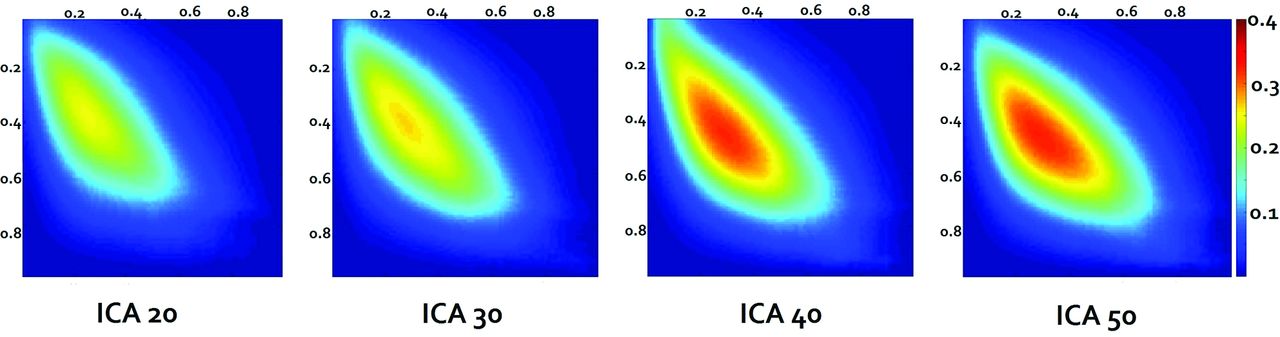
  • Fig 5.
    • Download figure
    • Open in new tab
    • Download powerpoint
    Fig 5.

    Group mean Dice coefficient maps across all the subjects at each ICA order calculated from subject-level noise-removed Dice coefficient maps. The x-axis depicts the rs-fMRI threshold levels, and the y-axis depicts the tb-fMRI threshold levels with the color bar demonstrating the Dice coefficient value.

  • Fig 6.
    • Download figure
    • Open in new tab
    • Download powerpoint
    Fig 6.

    Violin plots of Dice VUS across ICA orders. The mean Dice VUS for each ICA order is denoted by white diamonds. As the ICA order increases, there are larger numbers of subjects with higher Dice VUS values as reflected in the greater width of the violin plots corresponding to kernel densities.

  • Fig 7.
    • Download figure
    • Open in new tab
    • Download powerpoint
    Fig 7.

    Sample subject (patient 23 in the On-line Table) demonstrating an expansile mass lesion centered in the postcentral gyrus (arrows). Signal abnormality is extended to the subcortical white matter of the precentral gyrus. Red denotes tongue motor task activation, green denotes the VSMN network identified from rs-fMRI (ICA 50), and yellow denotes areas of overlap between tb- and rs-fMRI.

Tables

  • Figures
    • View popup
    Table 1:

    Probability of getting a full match between rs-fMRI and tb-fMRI maps as a function of number of ICA components

    ICA 20ICA 30ICA 40ICA 50
    Probability0.530.650.690.73
    • View popup
    Table 2:

    Dice coefficient values range, median, and mean at the subject level across different ICA orders

    MinimumMaximumMedianMean
    ICA 20−0.2140.5280.0090.062
    ICA 30−0.1340.5400.0150.071
    ICA 40−0.0560.5870.0240.085
    ICA 50−0.3680.6160.0240.086
PreviousNext
Back to top

In this issue

American Journal of Neuroradiology: 38 (5)
American Journal of Neuroradiology
Vol. 38, Issue 5
1 May 2017
  • Table of Contents
  • Index by author
  • Complete Issue (PDF)
Advertisement
Print
Download PDF
Email Article

Thank you for your interest in spreading the word on American Journal of Neuroradiology.

NOTE: We only request your email address so that the person you are recommending the page to knows that you wanted them to see it, and that it is not junk mail. We do not capture any email address.

Enter multiple addresses on separate lines or separate them with commas.
Presurgical Brain Mapping of the Ventral Somatomotor Network in Patients with Brain Tumors Using Resting-State fMRI
(Your Name) has sent you a message from American Journal of Neuroradiology
(Your Name) thought you would like to see the American Journal of Neuroradiology web site.
CAPTCHA
This question is for testing whether or not you are a human visitor and to prevent automated spam submissions.
Cite this article
N. Yahyavi-Firouz-Abadi, J.J. Pillai, M.A. Lindquist, V.D. Calhoun, S. Agarwal, R.D. Airan, B. Caffo, S.K. Gujar, H.I. Sair
Presurgical Brain Mapping of the Ventral Somatomotor Network in Patients with Brain Tumors Using Resting-State fMRI
American Journal of Neuroradiology May 2017, 38 (5) 1006-1012; DOI: 10.3174/ajnr.A5132

Citation Manager Formats

  • BibTeX
  • Bookends
  • EasyBib
  • EndNote (tagged)
  • EndNote 8 (xml)
  • Medlars
  • Mendeley
  • Papers
  • RefWorks Tagged
  • Ref Manager
  • RIS
  • Zotero
0 Responses
Respond to this article
Share
Bookmark this article
Presurgical Brain Mapping of the Ventral Somatomotor Network in Patients with Brain Tumors Using Resting-State fMRI
N. Yahyavi-Firouz-Abadi, J.J. Pillai, M.A. Lindquist, V.D. Calhoun, S. Agarwal, R.D. Airan, B. Caffo, S.K. Gujar, H.I. Sair
American Journal of Neuroradiology May 2017, 38 (5) 1006-1012; DOI: 10.3174/ajnr.A5132
del.icio.us logo Twitter logo Facebook logo Mendeley logo
  • Tweet Widget
  • Facebook Like
  • Google Plus One
Purchase

Jump to section

  • Article
    • Abstract
    • ABBREVIATIONS:
    • Materials and Methods
    • Results
    • Discussion
    • Conclusions
    • Footnotes
    • REFERENCES
  • Figures & Data
  • Supplemental
  • Info & Metrics
  • Responses
  • References
  • PDF

Related Articles

  • PubMed
  • Google Scholar

Cited By...

  • No citing articles found.
  • Crossref (20)
  • Google Scholar

This article has been cited by the following articles in journals that are participating in Crossref Cited-by Linking.

  • Rapid Precision Functional Mapping of Individuals Using Multi-Echo fMRI
    Charles J. Lynch, Jonathan D. Power, Matthew A. Scult, Marc Dubin, Faith M. Gunning, Conor Liston
    Cell Reports 2020 33 12
  • Functional MRI for Surgery of Gliomas
    Antonella Castellano, Sara Cirillo, Lorenzo Bello, Marco Riva, Andrea Falini
    Current Treatment Options in Neurology 2017 19 10
  • Functional Connectivity in Adult Brain Tumor Patients: A Systematic Review
    Michelle E. Fox, Tricia Z. King
    Brain Connectivity 2018 8 7
  • Improved resting state functional connectivity sensitivity and reproducibility using a multiband multi-echo acquisition
    Alexander D. Cohen, Baolian Yang, Brice Fernandez, Suchandrima Banerjee, Yang Wang
    NeuroImage 2021 225
  • The Problem of Neurovascular Uncoupling
    Shruti Agarwal, Haris I. Sair, Jay J. Pillai
    Neuroimaging Clinics of North America 2021 31 1
  • Decreased Hand Motor Resting-State Functional Connectivity in Patients with Glioma: Analysis of Factors including Neurovascular Uncoupling
    Herie Sun, Behroze Vachha, Maria E. Laino, Mehrnaz Jenabi, Jessica R. Flynn, Zhigang Zhang, Andrei I. Holodny, Kyung K. Peck
    Radiology 2020 294 3
  • Dynamic Brain Connectivity in Resting State Functional MR Imaging
    Rozita Jalilianhasanpour, Daniel Ryan, Shruti Agarwal, Elham Beheshtian, Sachin K. Gujar, Jay J. Pillai, Haris I. Sair
    Neuroimaging Clinics of North America 2021 31 1
  • Identification of the Somatomotor Network from Language Task–based fMRI Compared with Resting-State fMRI in Patients with Brain Lesions
    Elham Beheshtian, Rozita Jalilianhasanpour, Amirali Modir Shanechi, Varun Sethi, Guoqing Wang, Martin A. Lindquist, Brian S. Caffo, Shruti Agarwal, Jay J. Pillai, Sachin K. Gujar, Haris I. Sair
    Radiology 2021 301 1
  • Dynamic Functional Connectivity States Between the Dorsal and Ventral Sensorimotor Networks Revealed by Dynamic Conditional Correlation Analysis of Resting-State Functional Magnetic Resonance Imaging
    Maleeha F. Syed, Martin A. Lindquist, Jay J. Pillai, Shruti Agarwal, Sachin K. Gujar, Ann S. Choe, Brian Caffo, Haris I. Sair
    Brain Connectivity 2017 7 10
  • The utilisation of resting‐state fMRI as a pre‐operative mapping tool in patients with brain tumours in comparison to task‐based fMRI and intraoperative mapping: A systematic review
    Hanani Abdul Manan, Elizabeth A. Franz, Noorazrul Yahya
    European Journal of Cancer Care 2021 30 4

More in this TOC Section

  • Kurtosis and Epileptogenic Tubers: A Pilot Study
  • Glutaric Aciduria Type 1: DK vs. Conventional MRI
  • Multiparametric MRI in PEDS Pontine Glioma
Show more FUNCTIONAL

Similar Articles

Advertisement

Indexed Content

  • Current Issue
  • Accepted Manuscripts
  • Article Preview
  • Past Issues
  • Editorials
  • Editor's Choice
  • Fellows' Journal Club
  • Letters to the Editor
  • Video Articles

Cases

  • Case Collection
  • Archive - Case of the Week
  • Archive - Case of the Month
  • Archive - Classic Case

More from AJNR

  • Trainee Corner
  • Imaging Protocols
  • MRI Safety Corner
  • Book Reviews

Multimedia

  • AJNR Podcasts
  • AJNR Scantastics

Resources

  • Turnaround Time
  • Submit a Manuscript
  • Submit a Video Article
  • Submit an eLetter to the Editor/Response
  • Manuscript Submission Guidelines
  • Statistical Tips
  • Fast Publishing of Accepted Manuscripts
  • Graphical Abstract Preparation
  • Imaging Protocol Submission
  • Evidence-Based Medicine Level Guide
  • Publishing Checklists
  • Author Policies
  • Become a Reviewer/Academy of Reviewers
  • News and Updates

About Us

  • About AJNR
  • Editorial Board
  • Editorial Board Alumni
  • Alerts
  • Permissions
  • Not an AJNR Subscriber? Join Now
  • Advertise with Us
  • Librarian Resources
  • Feedback
  • Terms and Conditions
  • AJNR Editorial Board Alumni

American Society of Neuroradiology

  • Not an ASNR Member? Join Now

© 2025 by the American Society of Neuroradiology All rights, including for text and data mining, AI training, and similar technologies, are reserved.
Print ISSN: 0195-6108 Online ISSN: 1936-959X

Powered by HighWire