
Article Figures & Data
Figures
- Fig 1.
The cumulative dose-area product is significantly reduced with the dose-reduction technology platform. Bar graph and error bars represent least squares means and the associated 95% confidence intervals. Note a significant reduction in CPKA between the dose-reduction technology and reference platforms. Asterisk indicates P < .05; double asterisks, P < .0001.
- Fig 2.
Least squares means and 95% confidence intervals of total fluoroscopy duration (A) and total administered contrast volume (B) are plotted for the reference and dose-reduction technology platforms. Note significant differences in fluoroscopy duration or contrast volume between the dose-reduction technology and reference platforms. Asterisk indicates P < .05; double asterisks, P < .0001.
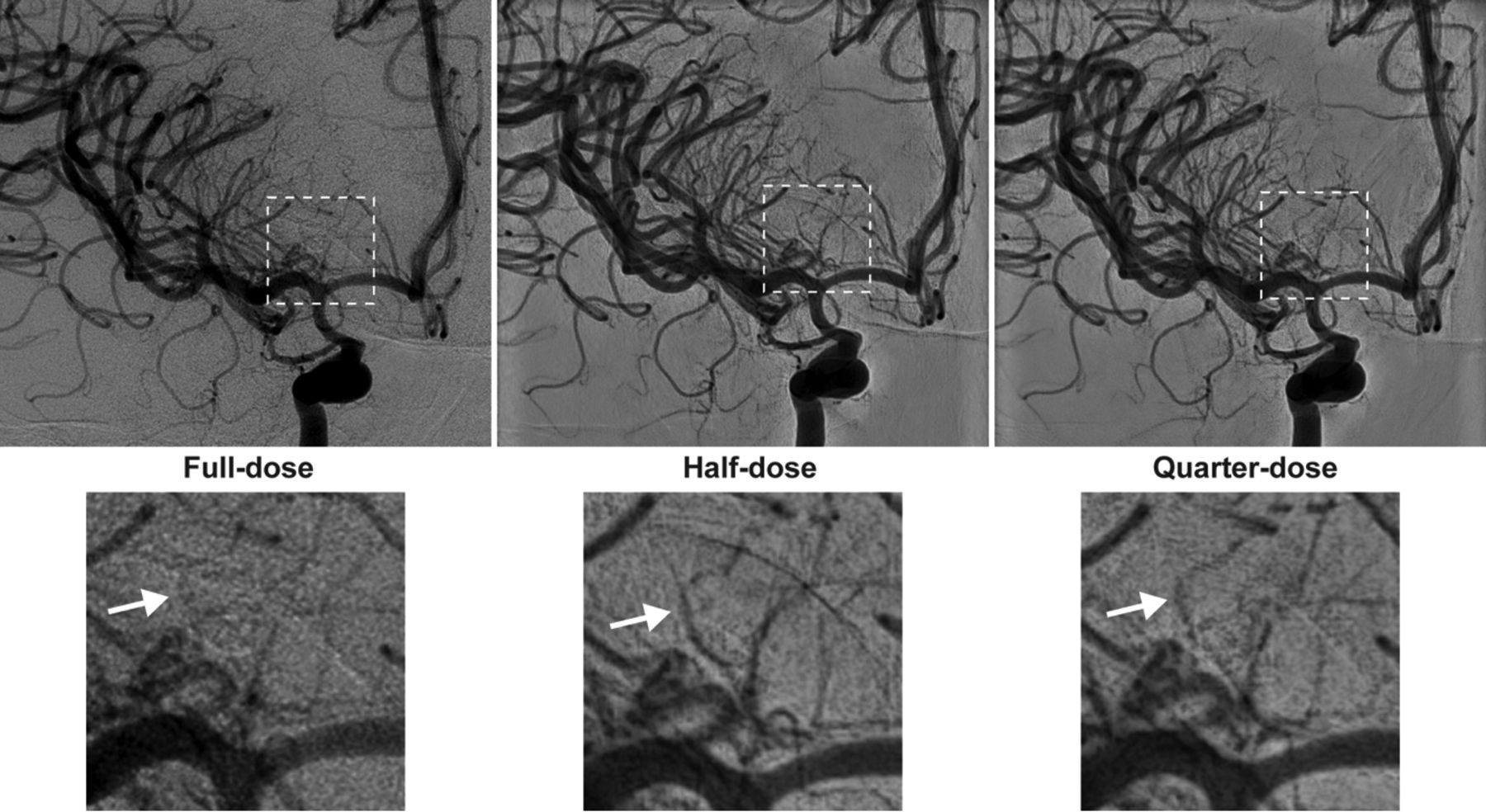
- Fig 3.
Diagnostic angiography was performed to assess the source of bleeding in a 43-year-old man who presented with diffuse SAH. During the examination, angiograms after contrast administration into the right ICA were obtained on IPDRT platform by using the “full-dose” protocol (left), which is identical to the reference platform in terms of hardware and software settings, “half-dose” protocol (middle), and “quarter-dose” protocol (right). Magnified views of the dashed area highlight improved visualization of small perforators (white arrows) with the “half-dose” and “quarter-dose” protocols (lower panels).


Tables
- Table 1:
Maximum entrance air kerma rates for the 3 fluoroscopic modes I, II, and III on the reference and dose-reduction platformsa
Platform, Mode Frames/Second Filtration Maximum EAKR (mGy/min) Reference I 6 0.4 mm Cu +1 mm Al 22 II 12.5 0.4 mm Cu +1 mm Al 44 III 12.5 0.1 mm Cu +1 mm Al 79 DRT I 15 0.4 mm Cu +1 mm Al 11 II 15 0.4 mm Cu +1 mm Al 26 III 15 0.1 mm Cu +1 mm Al 62 Note:—DRT indicates dose-reduction technology.
↵a The operator chooses the fluoroscopy mode on the dose-reduction platform independent of the DSA program and the targeted dose-reduction setting used for angiography.
- Table 2:
Measured entrance air kerma rates for a typical patient examination and for the largest FOV in fluoroscopic mode II preferred for clinical imaginga
System DRT Plane kV Focal Spot (mm) Measured EAKR (mGy/min) K̄a,r Ratio FD 20/20 No AP 68 ± 1 Small (0.4) 5.1 ± 0.2 1.03 ± 0.12 LAT 69 ± 1 Small (0.4) 6.1 ± 1.0 1.00 ± 0.04 FD 20/10 Before AP 68 ± 1 Small (0.4) 4.9 ± 1.0 1.07 ± 0.03 LAT 72 ± 4 Small (0.5) 6.3 ± 3.4 0.97 ± 0.004 After AP 68 ± 1 Small (0.4) 2.8 ± 0.4 0.96 ± 0.08 LAT 73 ± 2 Small (0.5) 4.4 ± 0.6 1.01 ± 0.1 Note:—AP indicates anteroposterior; DRT, dose-reduction technology.
↵a For each system, the selected kilovolt and x-ray focal spot along with its nominal size (millimeter) for each plane are summarized. For the FD 20/10 system, these values are reported before and after the installation of the DRT. The x-ray beam filtration is 0.4 mm Cu and 1 mm of Al for all systems, platforms, and planes.
- Table 3:
DSA programmed settings for the image-receptor (detector) entrance air kerma rate for a typical patient examination with the largest FOV on both platformsa
System DRT Acquisition Protocol kV Filtration Focal Spot (mm) Programmed RAKR (μGy/frame) FD 20/20 No Standard 80 0.1 mm Cu +1 mm Al Large (AP/LAT: 0.7 mm) 4.0 FD 20/10 Before Standard 80 0.1 mm Cu +1 mm Al Large (AP: 0.7 mm; LAT: 0.8 mm) 4.0 After Quarter 75 0.1 mm Cu +1 mm Al Small (AP: 0.4 mm; LAT: 0.5 mm) 0.7 Half 78 No added filtration Small (AP: 0.4 mm; LAT: 0.5 mm) 1.0 Full 80 0.1 mm Cu +1 mm Al Large (AP: 0.7 mm; LAT: 0.8 mm) 4.0 Note:—AP indicates anteroposterior; DRT, dose-reduction technology.
↵a The dose-reduction platform was equipped with 3 acquisition protocols in which the “full-dose” protocol reverts to the reference platform hardware and software settings (“standard” dose protocol). The programmed settings are identical for both planes in each system and for each acquisition protocol.
Reference Platform Dose-Reduction Platform No. of cases Diagnostic 654 (71.6%) 173 (53.4%) Coil embolization 45 (5.0%) 42 (12.2%) Flow diverter 58 (6.5%) 26 (7.6%) Vasospasm 34 (3.7%) 26 (8.0%) Thrombectomy 37 (4.1%) 19 (5.5%) Stent-assisted coiling 27 (3.0%) 15 (4.4%) Carotid stenting 25 (2.8%) 9 (2.6%) Epistaxis 17 (1.9%) 5 (1.5%) AVM 10 (1.1%) 6 (1.7%) AVF 7 (0.8%) 3 (0.9%) Total 914 (73.8%) 324 (26.2%) Patient characteristics Age (yr) 57.4 ± 14.7 56.6 ± 15.2 Weight (kg) 79.3 ± 19.9 77.6 ± 18.4 Male 370 (40.5%) 134 (41.4%) Female 544 (59.5%) 190 (58.6%) Hypertension 276 (30.2%) 89 (27.5%) Medical history Diabetes 113 (12.4%) 39 (12.0%) CAD 67 (7.3%) 21 (6.5%) COPD 61 (6.7%) 26 (8.0%) Hypertension 496 (54.3%) 177 (54.6%) Obesity 53 (5.8%) 17 (5.2%) Operator 1 59 (6.5%) 18 (5.6%) 2 433 (47.4%) 175 (54.0%) 3 249 (27.2%) 23 (7.1%) 4 79 (8.6%) 92 (28.4%) Multiple 94 (10.3%) 16 (4.9%) Note:—CAD indicates coronary artery disease; COPD, chronic obstructive pulmonary disorder.
↵a Data are presented as number (percentage) or mean ± SD. The number of patients with preprocedural hypertension is reported under “Patient characteristics,” while the number of patients with a documented history of hypertension is reported under “Medical history.”
- Table 5:
Reduction achieved with the dose-reduction platform in comparison with the reference platforma
Procedure CPKA (Gy × cm2) Fluoroscopy Duration (min) Contrast Volume (mL) Diagnostic 88.2 (63.3%)b −1.7 (−17.3%)c −12.6 (−8.3%) All interventions 171.8 (52.7%)b −5.9 (−16.6) −23.1 (−10.1%) Coil embolization 166.3 (50.3%)b −4.4 (−10.3%) −37.3 (−13.7%) Flow diverter 82.1 (30.5%)c −14.9 (−55.1%)c −34.6 (−15.5%) Vasospasm 124.9 (71.1%)b 1.7 (11.1%) 9.4 (6.2%) Thrombectomy 191.9 (60.2%)b −5.1 (−17.8%) 2.9 (1.4%) Stent-assisted coiling 112.1 (35.2%)c −11.1 (−26.2%) −72.6 (−27.1%)c Carotid stenting 122.2 (55.7%)b −1.7 (−6.7%) 17.6 (7.9%) Epistaxis 251.0 (73.8%b −7.4 (−22.7%) 73.7 (30.2%) AVM 165.4 (26.2%) −8.9 (−8.7%) −105.3 (−43.2%)c AVF 222.4 (37.6%) −24.9 (−36.2%) −51.3 (−15.6%) Overall 160.3 (53.2%)b −5.2 (−16.8%)c −15.3 (−6.7%)


Jump to section
Related Articles
Cited By...
- No citing articles found.