
Article Figures & Data
Figures
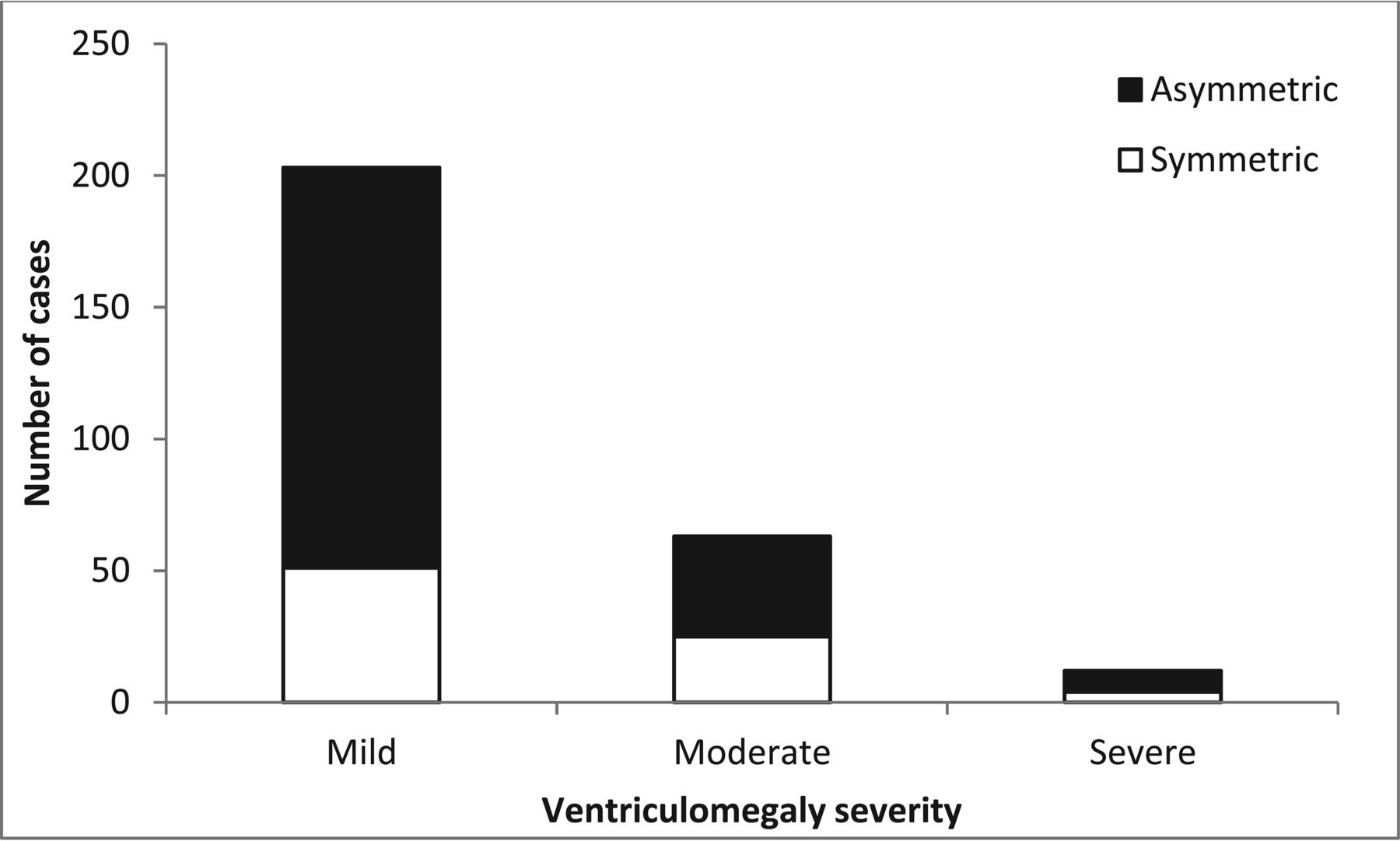
- Fig 1.
Distribution of ventriculomegaly cases according to severity and the presence or absence of asymmetry. Filled bars represent asymmetric ventriculomegaly; empty bars represent symmetric ventriculomegaly.
- Fig 2.
Distribution of additional CNS findings in cases of fetal ventriculomegaly according to maximal ventricle width. Filled bars represent major CNS findings, dotted bars represent minor CNS findings, and empty bars represent no additional CNS findings. Due to small numbers, cases with dilation of ≥16 mm were grouped together.
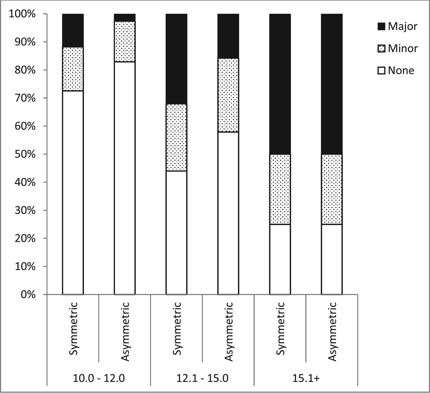
- Fig 3.
Distribution of additional CNS findings in cases of fetal ventriculomegaly according to ventriculomegaly severity and the presence or absence of asymmetry. Filled bars represent major CNS findings, dotted bars represent minor CNS findings, and empty bars represent no additional CNS findings.


Tables
Classification of MRI findings according to severity
Malformation Type Severity Normal Variant Minor Findings Major Findings NTD Meningocele Acrania/anencephaly encephalocele Myelocele Cystic lesions CPC Isolated PVPC Nonisolated PVPC Cavum verge Arachnoid cyst PVL CVI Porencephalic cyst Bleeding IVH grade 1 IVH grade 2–4 Ischemia Parenchymatic damage Cortical disorders Delayed sulcation (up to 2 weeks) Lissencephaly Schizencephaly Heterotopia Microcephaly White matter Isolated T2 hypersignal Nonisolated T2 hypersignal Midline CC Lipoma Short intact CC thick/thin Complete/partial CC agenesis Midline CSP Isolated septal agenesis SOD Posterior fossa MCM Blake pouch cyst Chiari 2 DWS Primary disruption of cerebellum/vermis/brain stem Secondary disruption of cerebellum/vermis/brain stem Vascular Vein of Galen aneurysm Note:—NTD indicates neural tube defect; CPC, choroid plexus cyst; CVI, cavum vellum interpositum; PVPC, periventricular pseudocyst; PVL, periventricular leukomalacia; IVH, intraventricular hemorrhage; CMV, cytomegalovirus; CC, corpus callosum; CSP, cavum septum pellucidum; SOD, septo-optic dysplasia; MCM, mega cysterna magna; DWS, Dandy-Walker syndrome; CSVT, cerebral sinovenous thrombosis.


Jump to section
Related Articles
Cited By...
- Correlation between 2D and 3D Fetal Brain MRI Biometry and Neurodevelopmental Outcomes in Fetuses with Suspected Microcephaly and Macrocephaly
- Fetal Exposure to MR Imaging: Long-Term Neurodevelopmental Outcome
- MR Imaging Diagnosis of Diencephalic-Mesencephalic Junction Dysplasia in Fetuses with Developmental Ventriculomegaly