
Article Figures & Data
Figures
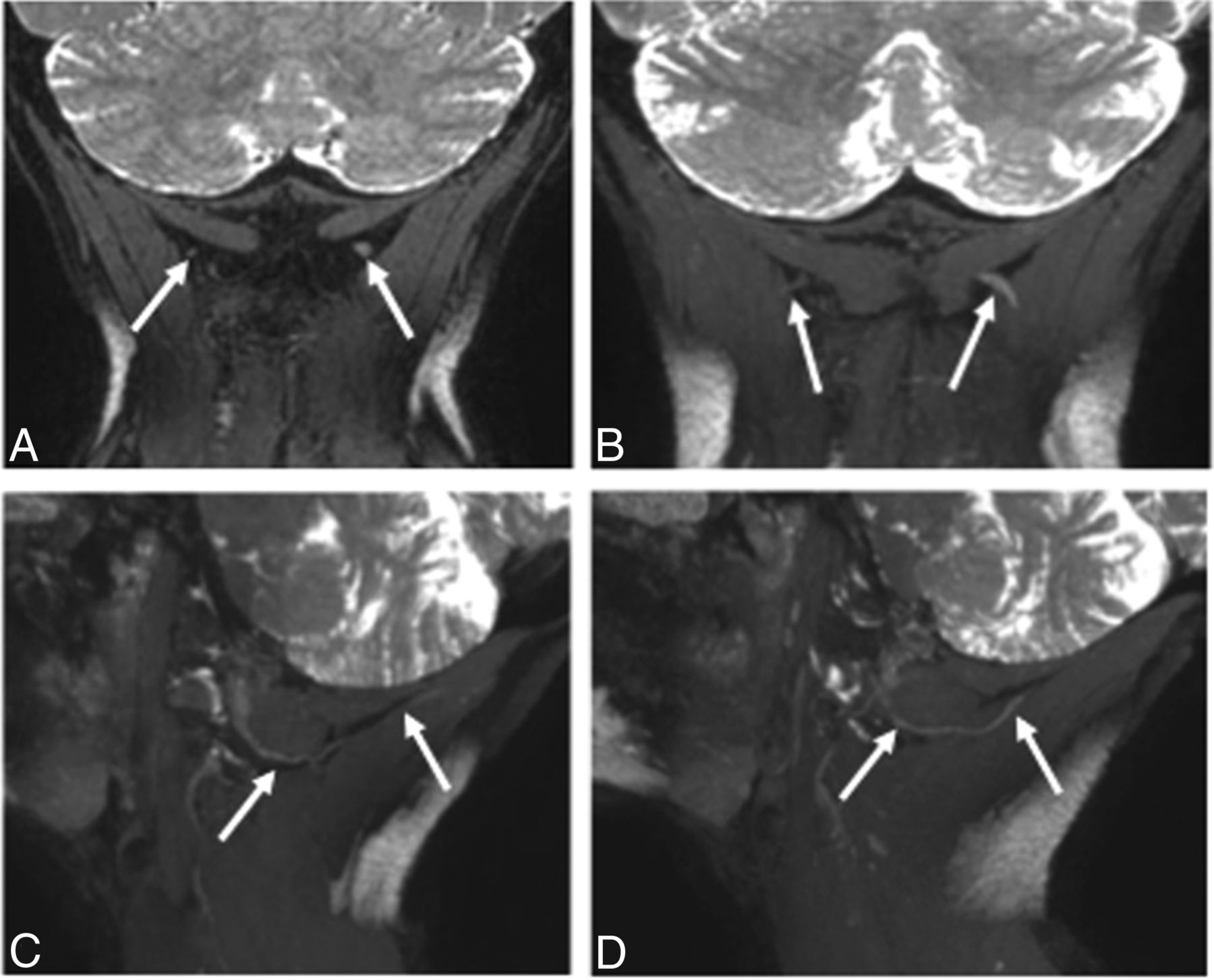
- Fig 1.
3T MRN demonstrating a normal GON. A, Coronal 3D PSIF shows bilateral GONs (arrows). B, Eight-millimeter-thick MIP reconstruction in the coronal plane shows the normal GONs (arrows). C and D, Eight-millimeter-thick isotropic MIP reconstruction in the sagittal plane shows right and left GONs (arrows).
- Fig 2.
3D Coronal PSIF images at 3T. A. Enlarged, hyperintense left greater occipital nerve (in comparison with the right). B, Caliber measurements show a larger left occipital nerve (1.6 mm) compared with the right (1.2 mm). C, Signal intensity measurements for both greater occipital nerves and semispinalis capitis muscles.
- Fig 3.
Coronal 3D PSIF images at 1.5T. A, Caliber measurements for the right and left GONs. B, Signal intensity measurements for the right GONs and semispinalis capitis muscles.
- Fig 4.
3T MRN demonstrating left GON neuropathy in a 62-year-old woman with left occipital neuralgia. A and B, Coronal 3D PSIF and 8-mm-thick MIP reconstruction show an asymmetrically thickened and hyperintense left GON (arrows). C and D, Eight-millimeter-thick isotropic MIP reconstruction in the sagittal planes. Note the normal right GON (arrows in C) and abnormal left GON (arrows in D) with increasing thickening proximal to the muscle entrapment site.
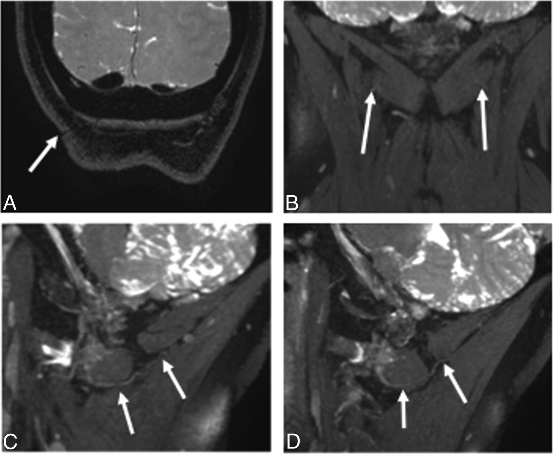
- Fig 5.
3T MRN demonstrating persistent right GON neuropathy in a 55-year-old woman with prior right occipital neurolysis and persistent right occipital neuralgia. A, Coronal 3D PSIF shows the surgical scar site (arrow). B, A more anterior coronal image shows minimal hyperintensity of the right GON (arrows). C and D, Eight-millimeter-thick isotropic MIP reconstruction in the sagittal planes. Note the normal persistently hyperintense right GON (arrows in C) and normal left GON (arrows in D).
- Fig 6.
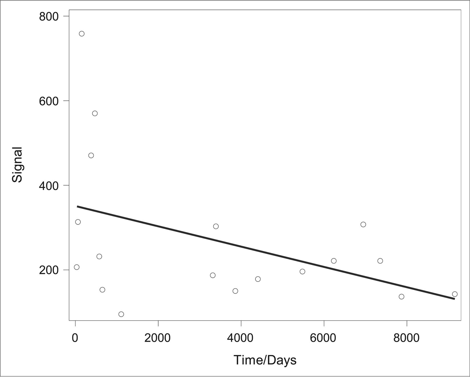
Evaluation of the correlation between the duration of migraine symptoms (defined as length of “time” in days from symptom onset to MRN acquisition) and the GON signal yielded a Spearman rank correlation coefficient of −0.499 with P = .351. When we compared the duration of symptoms and the GON diameter using the same statistical methodology, no significant correlation was found (Spearman rank correlation, 0.21; P = .4). The mean duration of migraine symptoms in this cohort of 18 patients was 3415 ± 3127 days (approximately 9 years).


Tables
- Table 1:
Patient demographic factors (n = 18) and migraine histories prior to MR neurography
Age (yr) Sex Migraine Frequencya Migraine Laterality Positive Family History Previous Head Trauma Younger than 40 Older than 40 Male Female <15 per mo >15 per mo Left Right Yes No Yes No 6 (33%) 12 (67%) 3 (17%) 15 (83%) 5 (38%) 8 (62%) 10 (56%) 8 (44%) 3 (17%) 15 (83%) 5 (28%) 13 (72%) ↵a Five patients were not reported.
- Table 3:
Statistical analysis of differences in GON diameter, signal intensity, calculated SNR, and calculated CNR comparing the symptomatic (subject group) versus asymptomatic (control group) side using a paired t test in patients with unilateral occipital migrainesa
MRN Characteristic Subject Group Control Group P Value Diameter 1.77 ± 0.4 1.29 ± 0.25 .001 Signal 269.06 ± 170.93 222.44 ± 170.46 .043 SNR 15.79 ± 4.59 14.02 ± 5.23 .009 CNR 2.57 ± 4.89 −1.26 ± 5.02 .004 ↵a All values are mean ± SD.
- Table 4:
Intra- and interobserver statistical analysis showing inter- and intraobserver performance among all 4 parameters using ICCs
ICC Reader 1 (initial) versus reader 1 (4 mo later) Nerve diameter (mm) 0.93 Nerve signal 0.79 SNR 0.74 CNR 0.68 Reader 1 versus reader 2 Nerve diameter (mm) 0.81 Nerve signal 0.71 SNR 0.67 CNR 0.54


