
Article Figures & Data
Figures
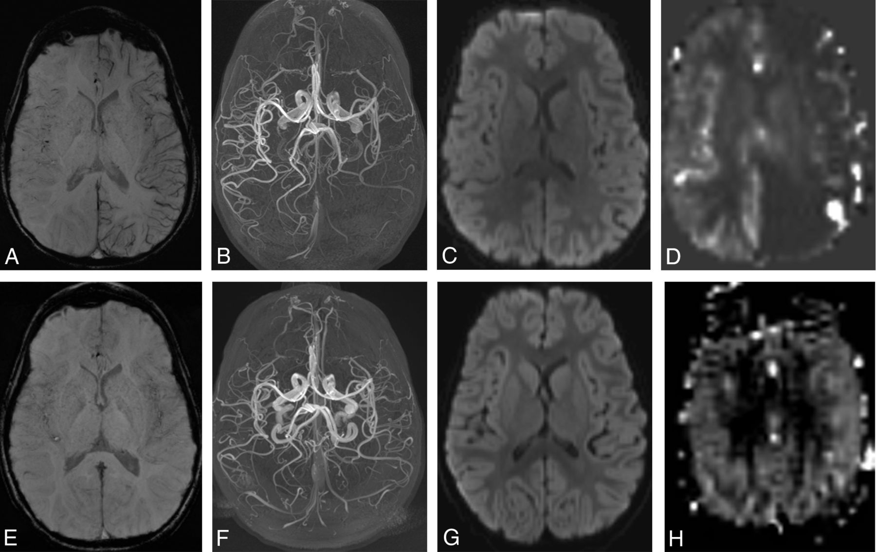
- Figure.
A 5-year-old boy presenting with right-sided weakness. Symptoms were improving at the time of the initial MR imaging performed 3 hours after presentation (A–D). A, Susceptibility-weighted imaging shows increased prominence of the cortical veins throughout the left cerebral hemisphere, indicating increased deoxyhemoglobin on the left. B, Collapsed maximum intensity projection from time-of-flight MR angiography shows reduced flow-related enhancement in the left anterior, middle, and posterior cerebral arteries. C, The average trace of the diffusion tensor image shows no abnormality of diffusion. D, Pulsed arterial spin-labeled perfusion-weighted imaging relative CBF map shows a marked decrease in perfusion throughout the left cerebral hemisphere. Follow-up imaging at 4 days after presentation (E–H). E, SWI shows resolution of venous asymmetry. F, MRA shows normal flow-related enhancement in the left anterior, middle, and posterior cerebral arteries. G, Findings of the average trace image continue to be negative. H, Pulsed arterial spin-labeling relative CBF map shows resolution of perfusion asymmetry.

Tables
Patient No. Age (yr) Sex Headache History of Migraine Headache Time from Presentation to Imaging Clinical Symptoms 1 15 Female Yes Yes 4.5 hr Right face and arm weakness, hypoesthesia, hemianopia, aphasia 2 13 Male Yes No 5.5 hr Left face and hand paresthesia 3 16 Female Yes No 6.75 hr Left hand and foot paresthesia and weakness 4 14 Male Yes Yes 4.5 hr Aphasia, right hemianopia 5 2 Male No No <12 hr Right hemiparesis and hemianopia 6 12 Female Yes Yes 6 hr Aphasia 7 13 Male Yes No 4.25 hr Aphasia and right face and arm weakness 8 8 Male Yes No 11 hr Aphasia, bilateral blurry vision, left hand paresthesia 9 10 Male Yes No 6 hr Right hand paresthesia, confusion 10 11 Female No Yes <1 hr Right hand paresthesia 11 5 Male No No 4 hr Right hemiparesis, dysarthria, and confusion 12 13 Female Yes No 7.5 hr Left hand weakness and paresthesia, aphasia 13 8 Male Yes Yes 16.25 hr Left facial weakness, hemiparesis 14 15 Male Yes Yes 10.5 hr Aphasia 15 10 Male Yes No 8.5 hr Aphasia and blurry vision 16 4 Female Yes No <16 hr Left facial weakness, dysarthria - Table 2:
Radiology findings of children with ASL and SWI reported in lobes and MRA in vessels involved
Patient No. Side pASL (Decrease) vASL (Decrease) SWI (Increase) MRA (Decrease) Resolution 1 Left P/T/O ND F/P/T/O MCA/PCA ND 2 Left P/T ND P/T MCA Yes 3 Right P ND P MCA ND 4 Left P/T/O ND P/T/O None Yes 5 Left F/P/T/O ND F/P/T/O ACA/MCA/PCA Yes 6 Left F/P/T/O Left F/P/O/T F/P/T/O ACA/MCA/PCA ND 7 Left F/T ND F/T MCA ND 8 Left P/T ND P/T None ND 9 Left F/P/T/O ND F/P/T/O ACA/MCA ND 10 Left ND ND P/T/O MCA/PCA ND 11 Left F/P/T/O ND F/P/T/O ACA/MCA/PCA Yes 12 Right F/P/T/O ND F/P/T/O ACA/MCA/PCA Yes 13 Right ND Right P/O/T P/T/O MCA/PCA Yes 14 Left ND ND F/P/T/O MCA/PCA ND 15 Left ND ND F/P MCA ND 16 Left ND ND P/T/O MCA ND Note:—pASL indicates pseudocontinuous arterial spin-labeling; vASL, velocity-selective arterial spin-labeling; ND, study not done; F, frontal; P, parietal; T, temporal; O, occipital; ACA, anterior cerebral artery; PCA, posterior cerebral artery.


