Advertisement

Main menu

  • Home
  • Content
    • Current Issue
    • Accepted Manuscripts
    • Article Preview
    • Past Issue Archive
    • Video Articles
    • AJNR Case Collection
    • Case of the Week Archive
    • Case of the Month Archive
    • Classic Case Archive
  • Special Collections
    • AJNR Awards
    • Low-Field MRI
    • Alzheimer Disease
    • ASNR Foundation Special Collection
    • Photon-Counting CT
    • View All
  • Multimedia
    • AJNR Podcasts
    • AJNR SCANtastic
    • Trainee Corner
    • MRI Safety Corner
    • Imaging Protocols
  • For Authors
    • Submit a Manuscript
    • Submit a Video Article
    • Submit an eLetter to the Editor/Response
    • Manuscript Submission Guidelines
    • Statistical Tips
    • Fast Publishing of Accepted Manuscripts
    • Graphical Abstract Preparation
    • Imaging Protocol Submission
    • Author Policies
  • About Us
    • About AJNR
    • Editorial Board
    • Editorial Board Alumni
  • More
    • Become a Reviewer/Academy of Reviewers
    • Subscribers
    • Permissions
    • Alerts
    • Feedback
    • Advertisers
    • ASNR Home

User menu

  • Alerts
  • Log in

Search

  • Advanced search
American Journal of Neuroradiology

American Journal of Neuroradiology

ASHNR American Society of Functional Neuroradiology ASHNR American Society of Pediatric Neuroradiology ASSR
  • Alerts
  • Log in

Advanced Search

  • Home
  • Content
    • Current Issue
    • Accepted Manuscripts
    • Article Preview
    • Past Issue Archive
    • Video Articles
    • AJNR Case Collection
    • Case of the Week Archive
    • Case of the Month Archive
    • Classic Case Archive
  • Special Collections
    • AJNR Awards
    • Low-Field MRI
    • Alzheimer Disease
    • ASNR Foundation Special Collection
    • Photon-Counting CT
    • View All
  • Multimedia
    • AJNR Podcasts
    • AJNR SCANtastic
    • Trainee Corner
    • MRI Safety Corner
    • Imaging Protocols
  • For Authors
    • Submit a Manuscript
    • Submit a Video Article
    • Submit an eLetter to the Editor/Response
    • Manuscript Submission Guidelines
    • Statistical Tips
    • Fast Publishing of Accepted Manuscripts
    • Graphical Abstract Preparation
    • Imaging Protocol Submission
    • Author Policies
  • About Us
    • About AJNR
    • Editorial Board
    • Editorial Board Alumni
  • More
    • Become a Reviewer/Academy of Reviewers
    • Subscribers
    • Permissions
    • Alerts
    • Feedback
    • Advertisers
    • ASNR Home
  • Follow AJNR on Twitter
  • Visit AJNR on Facebook
  • Follow AJNR on Instagram
  • Join AJNR on LinkedIn
  • RSS Feeds

AJNR Awards, New Junior Editors, and more. Read the latest AJNR updates

LetterLetter

Coregistration and Fusion: An Easy and Reliable Method for Identifying Cranial Nerve IV on MRI

A. Lecler, J. Savatovsky and F. Audren
American Journal of Neuroradiology October 2017, 38 (10) E81-E82; DOI: https://doi.org/10.3174/ajnr.A5286
A. Lecler
aDepartment of Radiology
  • Find this author on Google Scholar
  • Find this author on PubMed
  • Search for this author on this site
  • ORCID record for A. Lecler
J. Savatovsky
aDepartment of Radiology
  • Find this author on Google Scholar
  • Find this author on PubMed
  • Search for this author on this site
  • ORCID record for J. Savatovsky
F. Audren
bDepartment of Ophthalmology Fondation Ophtalmologique Adolphe de Rothschild Paris, France
  • Find this author on Google Scholar
  • Find this author on PubMed
  • Search for this author on this site
  • ORCID record for F. Audren
  • Article
  • Figures & Data
  • Info & Metrics
  • Responses
  • References
  • PDF
Loading

We read with much interest the article by Bunch et al1 in the March 2017 issue of the American Journal of Neuroradiology, in which they described the trochlear groove and trochlear cistern as reliable anatomic landmarks when searching for the tentorial segment of cranial nerve IV on MR imaging. The authors provided useful indications to recognize these 2 structures with a T2-weighted driven equilibrium radiofrequency reset pulse sequence. Confirming these 2 landmarks was significantly associated with correctly identifying the trochlear nerve.

We agree that short sequences are more practical for routine clinical use as opposed to those previously reported in the literature using high-resolution motion-sensitized driven equilibrium sequences with a 26-minute acquisition time.2 This sequence use is particularly true for cranial nerve IV imaging because a substantial number of patients being evaluated for suspected trochlear nerve pathology, including congenital palsy, are children, who are more prone to movements than adults. Therefore, a long scanning time may exacerbate motion artifacts on MR images.

However, we believe that this study has a few limitations that may impair its scope. The trochlear nerve remains the thinnest of all cranial nerves, with a mean diameter of approximately 0.5 mm (range, 0.3–0.9 mm). It has a complex course around the midbrain and is in close proximity to many arteries and veins with similar courses and calibers. The voxel size acquired in this study ranged from 0.3 × 0.35 × 3.0 to 0.4 × 0.44 × 3.0; thus, the section thickness (resolution of the z-axis) provided was 3.0 mm, which is too large to identify the trochlear nerve consistently. Moreover, the gap ranged from 0.3 to 1 mm, which is too high to observe a 0.5-mm structure.3 Much thinner sections are required to visualize the trochlear nerve, with a maximum of 0.25-mm section thickness and no gap.

The imaging findings of this study remain subjective and are not supported by surgical or pathologic correlations. Additionally, significant discrepancies between the MR imaging and anatomic findings were reported in cadaveric studies. Our colleagues' images are somewhat noisy, with CSF flow-related artifacts and are therefore not very convincing, even with annotations.

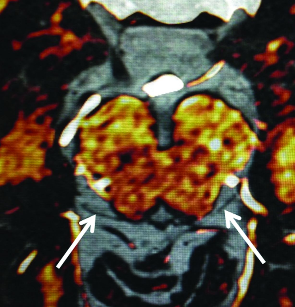
An interesting option for reliably identifying cranial nerve IV on MR imaging within a reasonable acquisition time may be the use of coregistration and fusion4 of an MR angiography sequence and of a “routine” T2-weighted driven equilibrium radiofrequency reset pulse sequence. Regrettably, even a very high resolution may still be insufficient due to the very complex course of the nerve and its close proximity to adjacent vessels. MRA enables the reader to clearly and reliably identify both arteries and veins, despite their highly variable anatomy. Therefore, this technique supports correct identification of the closely related trochlear nerve (Figure). This method is easy and is available in various software packages. A long posttreatment process is not necessary, and it can be performed during routine clinical practice in many institutions without involving highly specialized providers. With this method, all segments of the trochlear nerve are visible, especially its tentorial segment where the nerve is parallel to the vessels and runs along the tentorial edge. This approach may also be used successfully on larger cranial nerves such as the oculomotor or the abducens nerves.

Figure.
  • Download figure
  • Open in new tab
  • Download powerpoint
Figure.

Coregistration-fusion of an MR angiography and a T2-weighted driven equilibrium radiofrequency reset pulse sequence allowing to distinguish cisternal vessels colored in white from the trochlear nerves in black (arrows).

Finally, it is possible that the key for reliable and accurate identification of the trochlear nerve may be the use of a simple coregistration and fusion instead of long and complex high-resolution MR imaging acquisitions.

Footnotes

  • Disclosures: Julien Savatovsky—UNRELATED: Consultancy: Servier International Re-search Institute (IRIS), Madday Pharmaceutical, Bristol-Myers Squibb, GlaxoSmithKline, Comments: All except IRIS paid me around $5000 2 years ago*; Expert Testimony: Philips Healthcare, Bayer Pharmaceutical; Grants/Grants Pending: Association de la Recherche Sur le Syndrome Immuno-Déficitaire Acqui*; Payment for Lectures Including Service on Speakers Bureaus: Philips Healthcare, IRIS, Biogen; Travel/Accommodations/Meeting Expenses Unrelated to Activities Listed: GE Healthcare, Philips, Bayer. *Money paid to the institution.

References

  1. 1.↵
    1. Bunch PM,
    2. Kelly HR,
    3. Zander DA, et al
    . Trochlear groove and trochlear cistern: useful anatomic landmarks for identifying the tentorial segment of cranial nerve IV on MRI. AJNR Am J Neuroradiol 2017;38:1026–30 doi:10.3174/ajnr.A5117 pmid:28302606
    Abstract/FREE Full Text
  2. 2.↵
    1. Kanoto M,
    2. Toyoguchi Y,
    3. Hosoya T, et al
    . Visualization of the trochlear nerve in the cistern with use of high-resolution turbo spin-echo multisection motion-sensitized driven equilibrium. AJNR Am J Neuroradiol 2013;34:1434–37 doi:10.3174/ajnr.A3403 pmid:23391840
    Abstract/FREE Full Text
  3. 3.↵
    1. Choi BS,
    2. Kim JH,
    3. Jung C, et al
    . High-resolution 3D MR imaging of the trochlear nerve. AJNR Am J Neuroradiol 2010;31:1076–79 doi:10.3174/ajnr.A1992 pmid:20093316
    Abstract/FREE Full Text
  4. 4.↵
    1. Liu P,
    2. Eberhardt B,
    3. Wybranski C, et al
    . Nonrigid 3D medical image registration and fusion based on deformable models. Comput Math Methods Med 2013;2013:902470 doi:10.1155/2013/902470 pmid:23690883
    CrossRefPubMed
  • © 2017 by American Journal of Neuroradiology
PreviousNext
Back to top

In this issue

American Journal of Neuroradiology: 38 (10)
American Journal of Neuroradiology
Vol. 38, Issue 10
1 Oct 2017
  • Table of Contents
  • Index by author
  • Complete Issue (PDF)
Advertisement
Print
Download PDF
Email Article

Thank you for your interest in spreading the word on American Journal of Neuroradiology.

NOTE: We only request your email address so that the person you are recommending the page to knows that you wanted them to see it, and that it is not junk mail. We do not capture any email address.

Enter multiple addresses on separate lines or separate them with commas.
Coregistration and Fusion: An Easy and Reliable Method for Identifying Cranial Nerve IV on MRI
(Your Name) has sent you a message from American Journal of Neuroradiology
(Your Name) thought you would like to see the American Journal of Neuroradiology web site.
CAPTCHA
This question is for testing whether or not you are a human visitor and to prevent automated spam submissions.
Cite this article
A. Lecler, J. Savatovsky, F. Audren
Coregistration and Fusion: An Easy and Reliable Method for Identifying Cranial Nerve IV on MRI
American Journal of Neuroradiology Oct 2017, 38 (10) E81-E82; DOI: 10.3174/ajnr.A5286

Citation Manager Formats

  • BibTeX
  • Bookends
  • EasyBib
  • EndNote (tagged)
  • EndNote 8 (xml)
  • Medlars
  • Mendeley
  • Papers
  • RefWorks Tagged
  • Ref Manager
  • RIS
  • Zotero
0 Responses
Respond to this article
Share
Bookmark this article
Coregistration and Fusion: An Easy and Reliable Method for Identifying Cranial Nerve IV on MRI
A. Lecler, J. Savatovsky, F. Audren
American Journal of Neuroradiology Oct 2017, 38 (10) E81-E82; DOI: 10.3174/ajnr.A5286
del.icio.us logo Twitter logo Facebook logo Mendeley logo
  • Tweet Widget
  • Facebook Like
  • Google Plus One
Purchase

Jump to section

  • Article
    • Footnotes
    • References
  • Figures & Data
  • Info & Metrics
  • Responses
  • References
  • PDF

Related Articles

  • Reply:
  • PubMed
  • Google Scholar

Cited By...

  • No citing articles found.
  • Crossref
  • Google Scholar

This article has not yet been cited by articles in journals that are participating in Crossref Cited-by Linking.

More in this TOC Section

  • Letter to the Editor regarding “Automated Volumetric Software in Dementia: Help or Hindrance to the Neuroradiologist?”
  • Reply:
  • Brain AVM’s Nidus: What if We Hadn’t Understood Anything?
Show more LETTERS

Similar Articles

Advertisement

Indexed Content

  • Current Issue
  • Accepted Manuscripts
  • Article Preview
  • Past Issues
  • Editorials
  • Editor's Choice
  • Fellows' Journal Club
  • Letters to the Editor
  • Video Articles

Cases

  • Case Collection
  • Archive - Case of the Week
  • Archive - Case of the Month
  • Archive - Classic Case

More from AJNR

  • Trainee Corner
  • Imaging Protocols
  • MRI Safety Corner
  • Book Reviews

Multimedia

  • AJNR Podcasts
  • AJNR Scantastics

Resources

  • Turnaround Time
  • Submit a Manuscript
  • Submit a Video Article
  • Submit an eLetter to the Editor/Response
  • Manuscript Submission Guidelines
  • Statistical Tips
  • Fast Publishing of Accepted Manuscripts
  • Graphical Abstract Preparation
  • Imaging Protocol Submission
  • Evidence-Based Medicine Level Guide
  • Publishing Checklists
  • Author Policies
  • Become a Reviewer/Academy of Reviewers
  • News and Updates

About Us

  • About AJNR
  • Editorial Board
  • Editorial Board Alumni
  • Alerts
  • Permissions
  • Not an AJNR Subscriber? Join Now
  • Advertise with Us
  • Librarian Resources
  • Feedback
  • Terms and Conditions
  • AJNR Editorial Board Alumni

American Society of Neuroradiology

  • Not an ASNR Member? Join Now

© 2025 by the American Society of Neuroradiology All rights, including for text and data mining, AI training, and similar technologies, are reserved.
Print ISSN: 0195-6108 Online ISSN: 1936-959X

Powered by HighWire