
Article Figures & Data
Figures
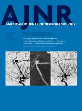
- Fig 1.
Axial 3D cisternographic (DRIVE) fast-recovery TSE MR imaging of the right temporal bone in a patient with normal hearing. A, Reformatted images parallel to the lateral semicircular canal (LSC) are created. Once created, the lateral semicircular canal is seen in its entirety on a single reformatted image. B, The osseous spiral lamina–basilar membrane complex of the upper basal turn (OSL-BM) and the first T2-hypointense linear band (arrow), in this case representing the interscalar septum, are demonstrated. C, A caliper is then placed to measure the distance between the OSL-BM and the linear band (distance X).
- Fig 2.
Axial 3D cisternographic (DRIVE) fast-recovery TSE MR image of the right temporal bone in a patient with a prospectively diagnosed IP-II anomaly. A, Reformatted images parallel to the lateral semicircular canal (LSC) are created. Once created, the lateral semicircular canal is seen in its entirety on a single reformatted image. Note the enlarged vestibular aqueduct. B, The osseous spiral lamina–basilar membrane complex of the upper basal turn (OSL-BM) and the first T2-hypointense linear band (arrow), in this case representing a band of dysplastic osseous spiral lamina–basilar membrane neural complex, are demonstrated. C, A caliper is then placed to measure the distance between the OSL-BM and the linear band (distance X).
- Fig 3.
Graph demonstrating the mean distance X measurements ± 2 SDs in patients with prospectively diagnosed IP-II anomalies and individuals with normal hearing.
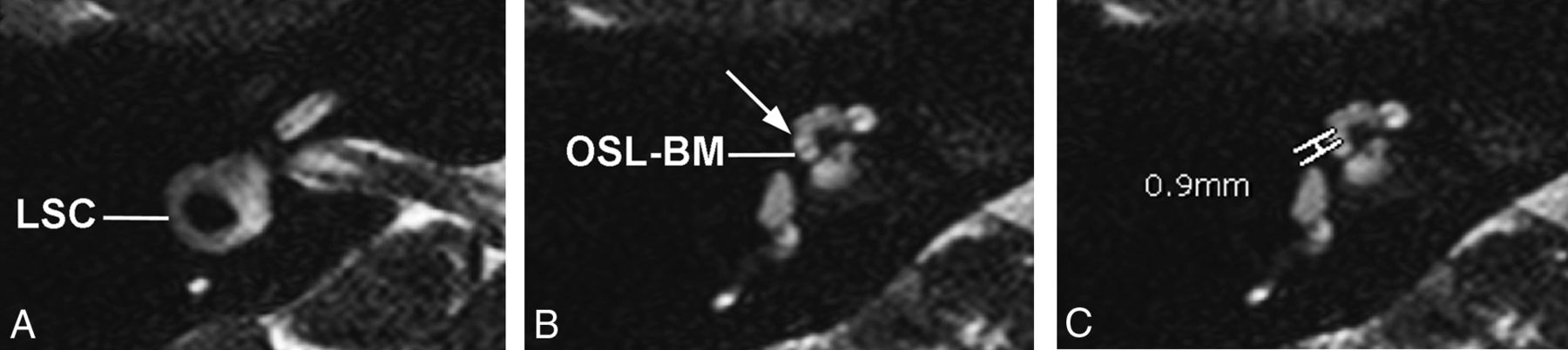
- Fig 4.
Axial CT and MR images of 1 of the 4 patients who was found, in retrospect, to have abnormal distance X measurements on MR imaging. A, On CT, the typical findings of IP-II including flattening of the interscalar ridge (anchor point) between the upper basal and upper middle turns (arrow) are demonstrated and confirm the presence of an IP-II anomaly. B, Corresponding MR image, on which distance X is found to be greater than 1.2 mm.


Tables
Patient demographics
Normal-Hearing Ears Patients with EVA Total No. of patients 28 (33 ears studied) 27 (54 ears studied) Mean patient age (yr) 16.7 24.9 SD of patient age (yr) 11.0 21.5 P value .08


