
Article Figures & Data
Figures
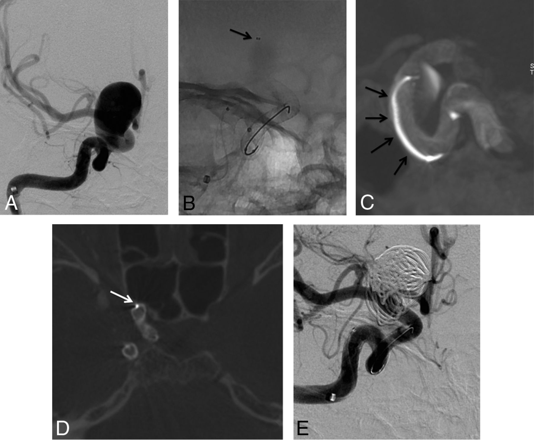
- Fig 1.
A 51-year-old woman presenting a recurrence of a left unruptured PComA aneurysm previously clipped. A, DSA in working projection showing the 5.2 × 5.0 mm recurrence with a 4.7 mm neck (arrowheads). B, Unsubtracted snapshot of the DSA in working projection displaying the clip (white arrow). C, Unsubtracted snapshot during the deployment of the FDS (Pipeline Embolization Device) from the left M1 segment to the carotid siphon. D and E, Snapshots from the FPV-CTA acquisition performed with 20% contrast media intra-arterial injection through the guiding catheter; MIP reconstruction. Satisfactory deployment of the stent is seen. Note the presence of the clip from the previous treatment (black arrows) and the microcatheter's tip, left in the FDS lumen during the acquisition (white arrows). Note that neither the clip nor the microcatheter's tip hamper the FDS visualization. F and G, Final DSA in working projection after FDS deployment (F, early phase; G, late phase). Stagnation of the contrast media within the aneurysm sac is seen at late phase (G). PComA indicates posterior communicating artery.
- Fig 2.
A 40-year-old woman treated for an incidental large right carotid ophthalmic aneurysm. A, Right ICA DSA in working projection showing the large paraclinoid aneurysm. B, Plain x-ray snapshot in working projection after the deployment of the FDS (Pipeline Embolization Device) in the parent artery, with a microcatheter jailed in the aneurysm sac (arrow). C, FPV-CTA acquisition, MIP reconstruction in sagittal view showing the satisfactory deployment of the FDS with the microcatheter jailed between the FDS and the wall of the parent artery (arrows). D, FPV-CTA, axial view. The jailed microcatheter is seen between the FDS and the wall of the parent artery (arrow). E, Final control DSA in working projection after loose coiling of the sac. Satisfactory exclusion of the sac is seen.
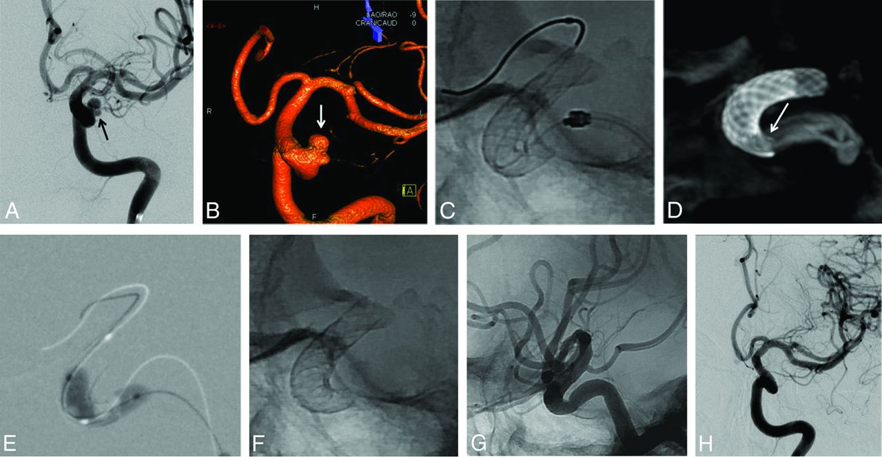
- Fig 3.
A 52-year-old woman treated for an unruptured left carotid ophthalmic large-neck aneurysm. A, Left ICA DSA in working projection (A) and 3D rotational angiography (B) showing the bi-lobed paraclinoid aneurysm with a large neck. Two overlapped FDSs (NeuroEndoGraft) were deployed in the carotid siphon to cover the aneurysm neck. C, Unsubtracted plain x-ray in lateral projection; the satisfactory opening of the distal FDS is demonstrated, but the proximal aspect of the proximal FDS is not clearly seen. D, FPV-CTA (sagittal view MIP reconstruction clearly separates the 2 FDSs and confirms an incomplete opening of the proximal aspect of the proximal FDS (arrow), which was subsequently treated by intrastent balloon angioplasty (E). F and G, Plain x-ray snapshots (F, without and G, with contrast media injection) in lateral projection showing a satisfactory opening of the FDS. H, One-year follow-up DSA in working projection showing the complete occlusion of the aneurysm.
- Fig 4.
A 30-year-old man treated for a traumatic left carotid cavernous fistula, 1 month after a severe traumatic brain injury. A, Left ICA DSA in lateral projection before stent placement. Note the filling of the ipsilateral cavernous sinus (arrows). Treatment with an FDS covering the ICA's arterial tear was chosen. One FDS (NeuroEndoGraft) was deployed in the carotid siphon to cover the arterial tear. B, Unsubtracted snapshot after the stent deployment. No obvious malapposition of the stent is seen. C and D, FPV-CTA after the stent deployment in sagittal MIP reconstruction (C) and thin section sagittal oblique reconstruction (D). This acquisition clearly shows a narrowing of the distal aspect of the FDS (arrow) that prompted the operator to perform an intrastent balloon angioplasty. E and F, Postangioplasty FPV-CTA (E, sagittal view, MIP reconstruction; F, thin-section sagittal oblique reconstruction) shows incomplete but satisfactory opening of the distal aspect of the FDS (arrow). G, One-year follow-up DSA showing the complete cure of the traumatic fistula.


Tables
Demographics/Characteristics Overall Population Patients Who Underwent FPV-CTA No. of patients 83 68 Age (mean ± SD) 51 ± 12 51 ± 12 Female (no., %) 62 (75) 51 (75) No. of aneurysms 87 70 Direct CC fistula (no.) 1 1 No. of procedures 84 70 Aneurysm locations Anterior circulation (no., %) 76 (87.4) 63 (90) Paraclinoid ICA (no., %) 36 (41.4) 28 (40) Cavernous ICA (no., %) 20 (23) 17 (24) ACA/AComA (no., %) 7 (8) 6 (8.6) ICA terminus (no., %) 4 (4.6) 4 (5.7) AChoA/PComA (no., %) 4 (4.6) 4 (5.7) MCA (no., %) 4 (4.6) 3 (4.3) Petrous ICA (no., %) 1 (1.1) 1 (1.4) Posterior circulation 11 (12.6) 7 (10) Vertebral artery (no., %) 5 (5.7) 3 (4.3) BA (no., %) 3 (3.4) 1 (1.4) PCA (no., %) 2 (2.3) 2 (2.9) SCA (no., %) 1 (1.1) 1 (1.45) Aneurysm maximum diameter (mean ± SD) 9.2 ± 6.5 9.8 ± 6.5 Aneurysm neck (mean ± SD) 5.4 ± 3 5.4 ± 3 Acutely ruptured aneurysms (no., %) 5 (5.7) 3 (4.3) Recanalized aneurysms (no., %) 16 (18.4) 13 (18.6) Previously clipped (no., %) 2 (2.3) 2 (2.9) Previously coiled (no., %) 14 (16.1) 11 (15.7) Note:—ACA indicates anterior cerebral artery; AChoA, anterior choroidal artery; AComA, anterior communicating artery; BA, basilar artery; CC, carotid cavernous; PCA, posterior cerebral artery; PComA, posterior communicating artery; SCA, superior cerebellar artery.
Interrater agreements Stent visualization quality κ = 0.38 Stent rate visualized κ = 0.49 Misdeployment κ = 0.57 Analysis in consensus Good visualization 83.8% Fair/poor visualization 16.2% Complete visualization 73.5% Partial visualization 27% Satisfactory opening 88.2% Misdeployment 11.8% Variables Poor/Fair FDS Visualization Good FDS Visualization Univariate Analysis (P Value) Multivariate Analysis (P Value) Age, yr (mean ± SD) 51.5 ± 12.7 51.5 ± 12.3 .12 .7 Female (no., %) 7 (64) 44 (77) .45 .61 Anterior location (no., %) 10 (90) 51 (89) 1 .94 Acute rupture (no., %) 0 (0) 3 (5) 1 .64 Recanalization/recurrence 4 (36) 7 (12) .73 .34 Aneurysm max. diameter, mm (mean ± SD) 15.5 ± 8.2 8.7 ± 5.2 <.001a .06 Aneurysm neck, mm (mean ± SD) 6.35 ± 3.65 5.1 ± 2.2 .13 .795 Stent type (PED; no., %) 36 (63) 8 (73) .73 .96 No. of stents (mean ± SD) 1.2 ± 0.5 1.1 ± 0.3 .25 .92 Additional coils (no., %) 8 (73) 14 (25) .004a .015a Microcatheter in place (no., %) 2 (18) 11 (19) 1 .28 Contrast media stagnation (no., %) 2 (18) 2 (3.5) .12 .02a Note:—max indicates maximum; PED, Pipeline Embolization Device.
↵a Statistically significant difference.


Jump to section
Related Articles
Cited By...
- SuperDyna: Unlocking the Potential of Post-Treatment Device Evaluation
- SuperDyna: Unlocking the Potential of Post-Treatment Device Evaluation
- Visualization of stent apposition after stent-assisted coiling of intracranial aneurysms using high resolution 3D fusion images acquired by C-arm CT
- A Multicenter Pilot Study on the Clinical Utility of Computational Modeling for Flow-Diverter Treatment Planning
- Flow-Diversion Effect of LEO Stents: Aneurysm Occlusion and Flow Remodeling of Covered Side Branches and Perforators