
Article Figures & Data
Figures
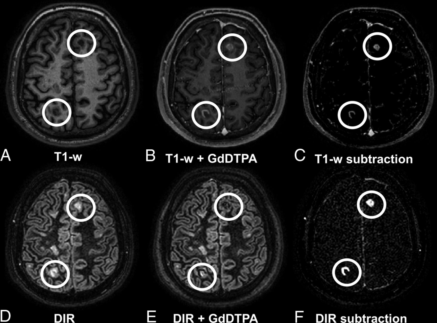
- Fig 1.
A 32-year-old male patient with relapsing-remitting MS with several lesions, including 2 contrast-enhancing juxta-/intracortical lesions in the left frontal and right parietal areas. Upper row (A–C): T1WI; lower row (D–F): DIR images with A and D being precontrast; B and E, postcontrast; and C and F, subtraction images. Enhancing lesions appear hypointense on postcontrast DIR and are visible in subtraction images. Note the high contrast of the lesions in the DIR subtraction image (F) compared with T1WI subtraction image (C).
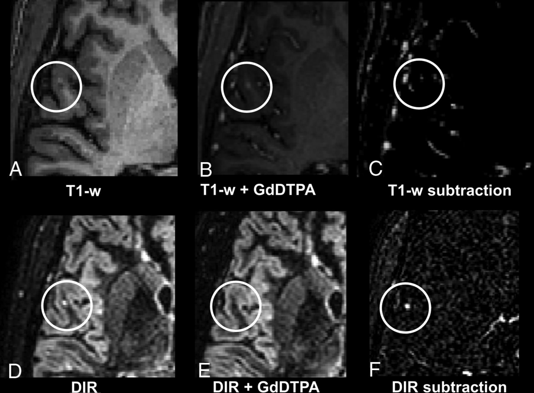
- Fig 2.
A 32-year-old male patient with relapsing-remitting MS. Upper row (A–C): T1WI; lower row (D–F): DIR images with A and D being precontrast; B and E, postcontrast; and C and F, subtraction images. Note the small juxtacortical lesion that is substantially more detectable in the DIR subtraction image (F) compared with the T1WI subtraction image (C). Contrary to DIR subtraction (F), in T1WI subtraction images (C), some contrast-enhancing vessels are visible near the lesion; thus, differentiation between an active contrast-enhancing lesion and surrounding enhancing vessels is difficult.
- Fig 3.
A 46-year-old female patient with relapsing-remitting MS. Upper row (A–C): T1WI; lower row (D–F): DIR images with A and D being precontrast; B and E, postcontrast; and C and F, subtraction images. Note that the small juxtacortical lesion is more detectable on the DIR subtraction image (F) compared with the T1-weighted subtraction image (C).
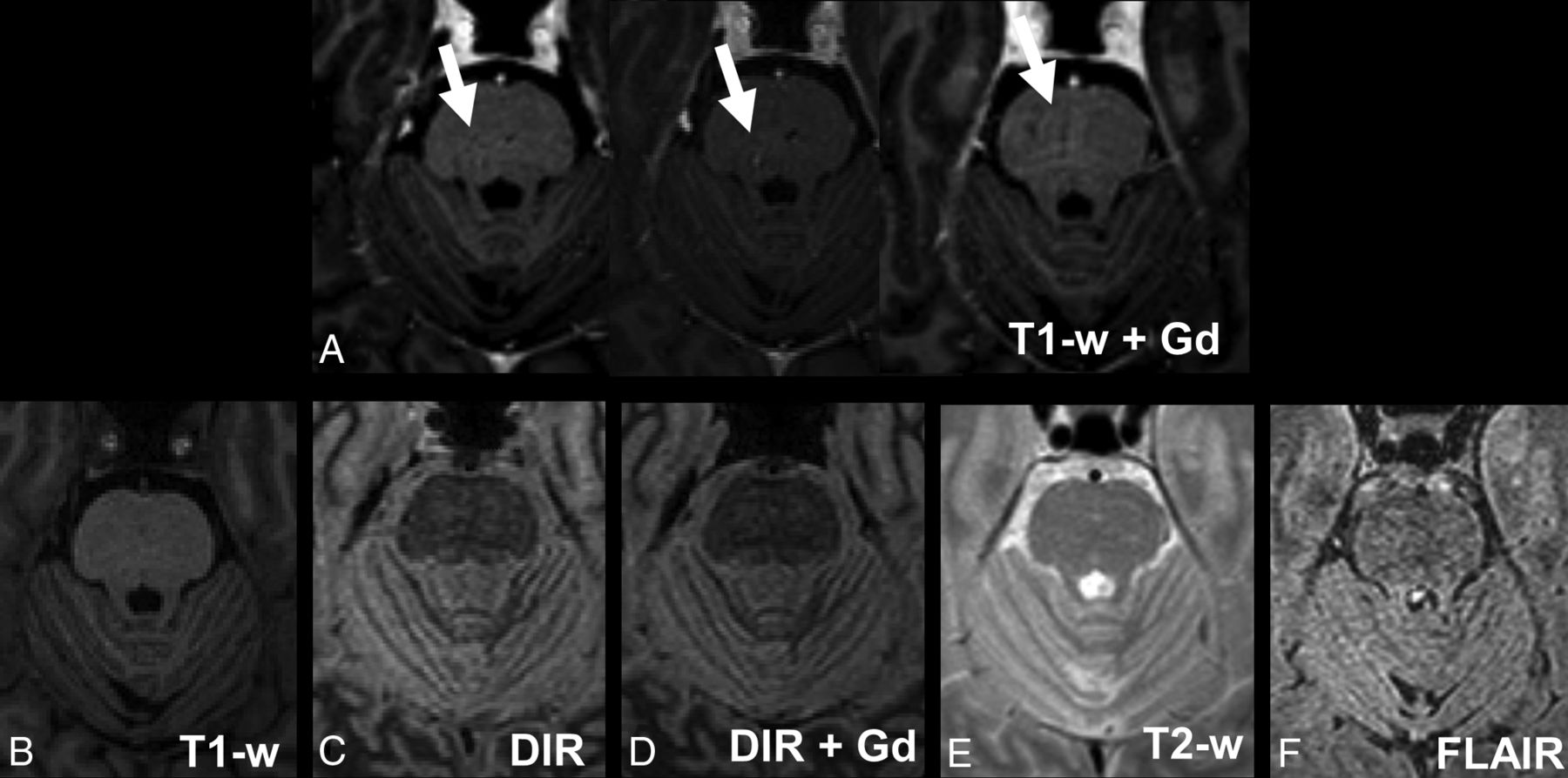
- Fig 4.
Pulsation artifacts detected in T1WI postcontrast. Images of a 52-year-old male patient with relapsing-remitting MS. Upper row: A, 3 consecutive postcontrast T1-weighted images; lower row: B, T1-weighted precontrast image. C, DIR precontrast image. D, DIR postcontrast image; E, T2WI; F, FLAIR. Note the small hyperintense signal alterations in the middle of the pons that are only visible on 3 consecutive T1-weighted postcontrast images and not on the other images.


