Advertisement

Main menu

  • Home
  • Content
    • Current Issue
    • Accepted Manuscripts
    • Article Preview
    • Past Issue Archive
    • Video Articles
    • AJNR Case Collection
    • Case of the Week Archive
    • Case of the Month Archive
    • Classic Case Archive
  • Special Collections
    • AJNR Awards
    • Low-Field MRI
    • Alzheimer Disease
    • ASNR Foundation Special Collection
    • Photon-Counting CT
    • View All
  • Multimedia
    • AJNR Podcasts
    • AJNR SCANtastic
    • Trainee Corner
    • MRI Safety Corner
    • Imaging Protocols
  • For Authors
    • Submit a Manuscript
    • Submit a Video Article
    • Submit an eLetter to the Editor/Response
    • Manuscript Submission Guidelines
    • Statistical Tips
    • Fast Publishing of Accepted Manuscripts
    • Graphical Abstract Preparation
    • Imaging Protocol Submission
    • Author Policies
  • About Us
    • About AJNR
    • Editorial Board
    • Editorial Board Alumni
  • More
    • Become a Reviewer/Academy of Reviewers
    • Subscribers
    • Permissions
    • Alerts
    • Feedback
    • Advertisers
    • ASNR Home

User menu

  • Alerts
  • Log in

Search

  • Advanced search
American Journal of Neuroradiology

American Journal of Neuroradiology

ASHNR American Society of Functional Neuroradiology ASHNR American Society of Pediatric Neuroradiology ASSR
  • Alerts
  • Log in

Advanced Search

  • Home
  • Content
    • Current Issue
    • Accepted Manuscripts
    • Article Preview
    • Past Issue Archive
    • Video Articles
    • AJNR Case Collection
    • Case of the Week Archive
    • Case of the Month Archive
    • Classic Case Archive
  • Special Collections
    • AJNR Awards
    • Low-Field MRI
    • Alzheimer Disease
    • ASNR Foundation Special Collection
    • Photon-Counting CT
    • View All
  • Multimedia
    • AJNR Podcasts
    • AJNR SCANtastic
    • Trainee Corner
    • MRI Safety Corner
    • Imaging Protocols
  • For Authors
    • Submit a Manuscript
    • Submit a Video Article
    • Submit an eLetter to the Editor/Response
    • Manuscript Submission Guidelines
    • Statistical Tips
    • Fast Publishing of Accepted Manuscripts
    • Graphical Abstract Preparation
    • Imaging Protocol Submission
    • Author Policies
  • About Us
    • About AJNR
    • Editorial Board
    • Editorial Board Alumni
  • More
    • Become a Reviewer/Academy of Reviewers
    • Subscribers
    • Permissions
    • Alerts
    • Feedback
    • Advertisers
    • ASNR Home
  • Follow AJNR on Twitter
  • Visit AJNR on Facebook
  • Follow AJNR on Instagram
  • Join AJNR on LinkedIn
  • RSS Feeds

AJNR Awards, New Junior Editors, and more. Read the latest AJNR updates

Research ArticleSpine Imaging and Spine Image-Guided Interventions

First-Pass Contrast-Enhanced MR Angiography in Evaluation of Treated Spinal Arteriovenous Fistulas: Is Catheter Angiography Necessary?

S. Mathur, S.P. Symons, T.J. Huynh, T.R. Marotta, R.I. Aviv and A. Bharatha
American Journal of Neuroradiology January 2017, 38 (1) 200-205; DOI: https://doi.org/10.3174/ajnr.A4971
S. Mathur
aFrom the Division of Diagnostic and Interventional Neuroradiology, Department of Medical Imaging (S.M., T.J.H., T.R.M., A.B.), St. Michael's Hospital, University of Toronto, Toronto, Ontario, Canada
bDivision of Neuroradiology, Department of Medical Imaging (S.M., T.J.H., R.I.A., S.P.S.)
  • Find this author on Google Scholar
  • Find this author on PubMed
  • Search for this author on this site
  • ORCID record for S. Mathur
S.P. Symons
bDivision of Neuroradiology, Department of Medical Imaging (S.M., T.J.H., R.I.A., S.P.S.)
cDepartment of Otolaryngology–Head and Neck Surgery (S.P.S.), Sunnybrook Health Sciences Centre, University of Toronto, Toronto, Ontario, Canada.
  • Find this author on Google Scholar
  • Find this author on PubMed
  • Search for this author on this site
  • ORCID record for S.P. Symons
T.J. Huynh
aFrom the Division of Diagnostic and Interventional Neuroradiology, Department of Medical Imaging (S.M., T.J.H., T.R.M., A.B.), St. Michael's Hospital, University of Toronto, Toronto, Ontario, Canada
bDivision of Neuroradiology, Department of Medical Imaging (S.M., T.J.H., R.I.A., S.P.S.)
  • Find this author on Google Scholar
  • Find this author on PubMed
  • Search for this author on this site
  • ORCID record for T.J. Huynh
T.R. Marotta
aFrom the Division of Diagnostic and Interventional Neuroradiology, Department of Medical Imaging (S.M., T.J.H., T.R.M., A.B.), St. Michael's Hospital, University of Toronto, Toronto, Ontario, Canada
  • Find this author on Google Scholar
  • Find this author on PubMed
  • Search for this author on this site
  • ORCID record for T.R. Marotta
R.I. Aviv
bDivision of Neuroradiology, Department of Medical Imaging (S.M., T.J.H., R.I.A., S.P.S.)
  • Find this author on Google Scholar
  • Find this author on PubMed
  • Search for this author on this site
  • ORCID record for R.I. Aviv
A. Bharatha
aFrom the Division of Diagnostic and Interventional Neuroradiology, Department of Medical Imaging (S.M., T.J.H., T.R.M., A.B.), St. Michael's Hospital, University of Toronto, Toronto, Ontario, Canada
  • Find this author on Google Scholar
  • Find this author on PubMed
  • Search for this author on this site
  • ORCID record for A. Bharatha
  • Article
  • Figures & Data
  • Supplemental
  • Info & Metrics
  • Responses
  • References
  • PDF
Loading

Article Figures & Data

Figures

  • Fig 1.
    • Download figure
    • Open in new tab
    • Download powerpoint
    Fig 1.

    Spinal epidural AVF with intradural venous drainage (patient 4). Sagittal T2, postgadolinium T1, MRA MIP, and frontal DSA images pretreatment (A1–4), after first attempted surgical disconnection (B1–4), and after second surgical disconnection (C1–4), respectively. Pretreatment study shows T2 cord hyperintensity and enhancement (A1–2, arrowheads) with surrounding enhancing perimedullary flow voids (A1–2, arrows). MRA shows arterial enhancement of intradural veins (A3, arrow). DSA confirmed the AVF (A4, arrow indicates the arterialized radicular vein). Postsurgical study after attempted AVF disconnection (B1–4) shows findings unchanged compared with the pretreatment study, consistent with residual AVF. Second postsurgical study shows persistent T2 cord hyperintensity and enhancement (C1–2, arrowheads) with decreased size of perimedullary veins (C1–2, arrows). MRA and DSA (C3–4) confirm absence of arterialized intradural veins, consistent with a successful surgical disconnection.

  • Fig 2.
    • Download figure
    • Open in new tab
    • Download powerpoint
    Fig 2.

    Spinal dural AVF in a 50-year-old man with 6 months of progressive lower extremity paraplegia. Pretreatment sagittal T2 (A) and postcontrast T1 (B) show abnormal perimedullary vessels (A, arrow), which enhance postcontrast (B, arrow), in addition to intramedullary T2 hyperintensity (A, arrowheads) with mild patchy intramedullary enhancement (B, arrowheads). Pretreatment sagittal MRA MIP (C) demonstrates arterially enhancing perimedullary veins (C, arrow). Right L1 segmental artery injection–frontal projection DSA demonstrates retrograde drainage into radicular vein (D, arrow). After surgical disconnection, there is reduction in intramedullary T2 hyperintensity (E, arrowheads) and perimedullary flow voids, with persistent cord enhancement (F, arrowheads) and lack of arterially enhancing perimedullary veins on MRA (G). Successful fistula disconnection was confirmed on repeat DSA (H).
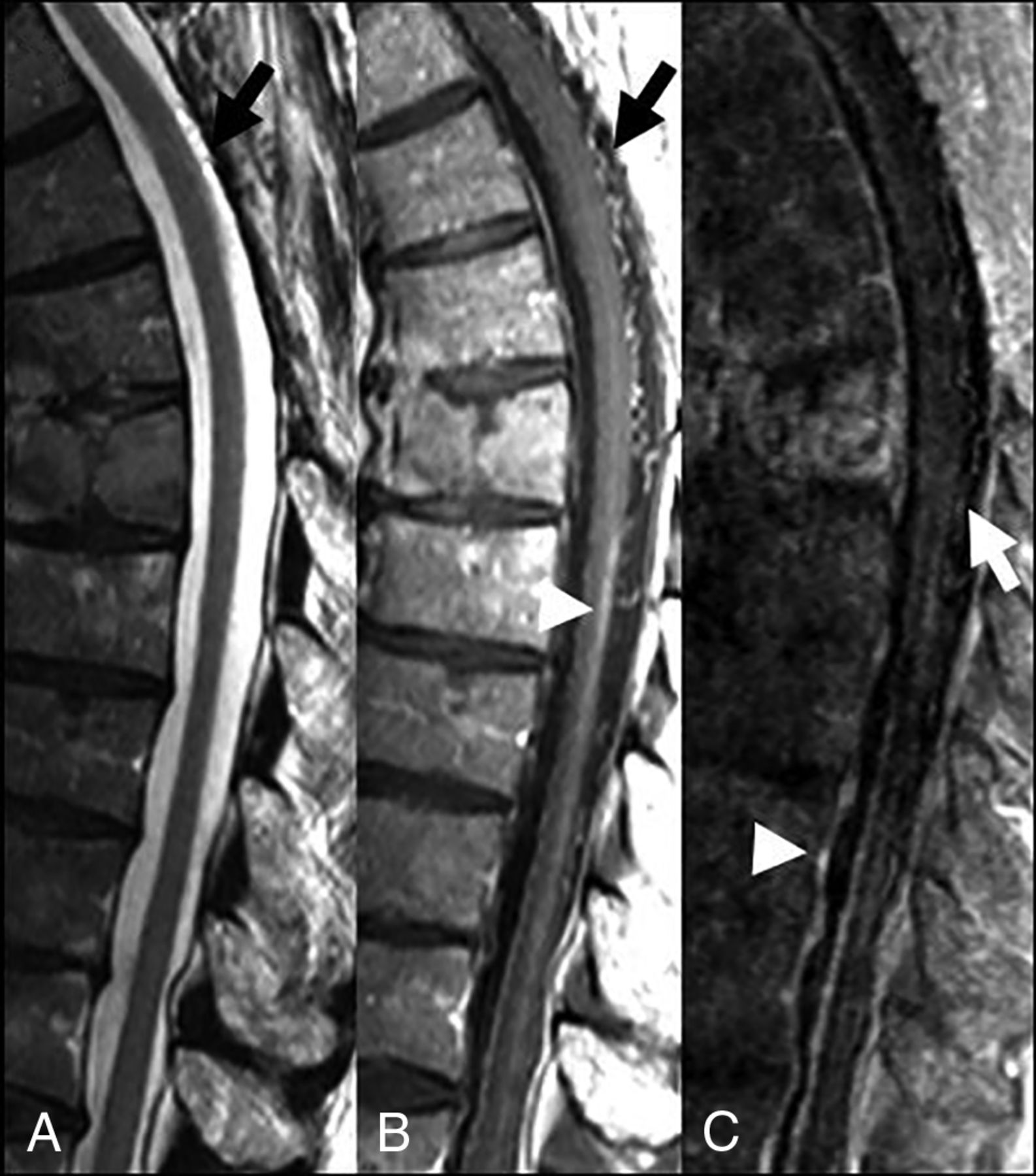
  • Fig 3.
    • Download figure
    • Open in new tab
    • Download powerpoint
    Fig 3.

    False-positive on MRA. 78-year-old man with prior surgical disconnection of a spinal dural AVF at left T5 level presented with worsening gait spasticity confounded by history of Parkinson disease. Few dorsal perimedullary flow voids were noted on the sagittal T2 (A, arrow), which enhanced on the postcontrast T1 study (B, arrow). Mild intramedullary enhancement was also noted on the postcontrast T1 (B, arrowhead). On MRA (C), there was faint enhancement of the perimedullary veins (C, arrow), which led to the suspicion of residual/recurrent fistula. No fistula was found at subsequent DSA (not shown). The findings on MR imaging and MRA were presumed to be residual changes from prior fistula. Mild enhancement of the perimedullary veins may be a result of venous contamination of contrast bolus, as suggested by enhancement of the basivertebral veins (C, arrowhead).

PreviousNext
Back to top

In this issue

American Journal of Neuroradiology: 38 (1)
American Journal of Neuroradiology
Vol. 38, Issue 1
1 Jan 2017
  • Table of Contents
  • Index by author
  • Complete Issue (PDF)
Advertisement
Print
Download PDF
Email Article

Thank you for your interest in spreading the word on American Journal of Neuroradiology.

NOTE: We only request your email address so that the person you are recommending the page to knows that you wanted them to see it, and that it is not junk mail. We do not capture any email address.

Enter multiple addresses on separate lines or separate them with commas.
First-Pass Contrast-Enhanced MR Angiography in Evaluation of Treated Spinal Arteriovenous Fistulas: Is Catheter Angiography Necessary?
(Your Name) has sent you a message from American Journal of Neuroradiology
(Your Name) thought you would like to see the American Journal of Neuroradiology web site.
CAPTCHA
This question is for testing whether or not you are a human visitor and to prevent automated spam submissions.
Cite this article
S. Mathur, S.P. Symons, T.J. Huynh, T.R. Marotta, R.I. Aviv, A. Bharatha
First-Pass Contrast-Enhanced MR Angiography in Evaluation of Treated Spinal Arteriovenous Fistulas: Is Catheter Angiography Necessary?
American Journal of Neuroradiology Jan 2017, 38 (1) 200-205; DOI: 10.3174/ajnr.A4971

Citation Manager Formats

  • BibTeX
  • Bookends
  • EasyBib
  • EndNote (tagged)
  • EndNote 8 (xml)
  • Medlars
  • Mendeley
  • Papers
  • RefWorks Tagged
  • Ref Manager
  • RIS
  • Zotero
0 Responses
Respond to this article
Share
Bookmark this article
First-Pass Contrast-Enhanced MR Angiography in Evaluation of Treated Spinal Arteriovenous Fistulas: Is Catheter Angiography Necessary?
S. Mathur, S.P. Symons, T.J. Huynh, T.R. Marotta, R.I. Aviv, A. Bharatha
American Journal of Neuroradiology Jan 2017, 38 (1) 200-205; DOI: 10.3174/ajnr.A4971
del.icio.us logo Twitter logo Facebook logo Mendeley logo
  • Tweet Widget
  • Facebook Like
  • Google Plus One
Purchase

Jump to section

  • Article
    • Abstract
    • ABBREVIATION:
    • Materials and Methods
    • Results
    • Discussion
    • Conclusions
    • Footnotes
    • References
  • Figures & Data
  • Supplemental
  • Info & Metrics
  • Responses
  • References
  • PDF

Related Articles

  • PubMed
  • Google Scholar

Cited By...

  • No citing articles found.
  • Crossref (3)
  • Google Scholar

This article has been cited by the following articles in journals that are participating in Crossref Cited-by Linking.

  • Time-Resolved Magnetic Resonance Angiography for Follow-Up of Treated Dural and Epidural Spinal Arteriovenous Fistula
    Giovanni Giulio Vercelli, Fabrizio Venturi, Massimiliano Minardi, Fabio Cofano, Francesco Zenga, Mauro Bergui, Diego Garbossa
    Journal of Neurological Surgery Part A: Central European Neurosurgery 2022 83 06
  • Diagnostic Accuracy of 4D-MRA for the Detection and Localization of Spinal Dural Arteriovenous Fistulas: A Systematic Review and Meta-Analysis
    Vincent Raymaekers, Ana Rodríguez-Hernández, Sjoert A.H. Pegge, Tomas Menovsky, Frederick J.A. Meijer, Jeroen H.D. Boogaarts
    World Neurosurgery 2025 193
  • Is Catheter Angiography Still Necessary for the Follow-Up of Spinal Malformations after Treatment?
    M.I. Vargas, J. Boto, Z. Kulcsar
    American Journal of Neuroradiology 2017 38 4

More in this TOC Section

Spine Imaging and Spine Image-Guided Interventions

  • Cone Beam CT Myelography
  • Post-Procedural Brachial Neuritis Features
  • Diagnostic Value of Brain WMH in SIH
Show more Spine Imaging and Spine Image-Guided Interventions

Neurointervention

  • Flow diversion for distal circulation aneurysms
  • Neuroform Atlas Stent for Intracranial Aneurysms
  • Transophthalmic Artery Embolization of Meningiomas
Show more Neurointervention

Similar Articles

Advertisement

Indexed Content

  • Current Issue
  • Accepted Manuscripts
  • Article Preview
  • Past Issues
  • Editorials
  • Editor's Choice
  • Fellows' Journal Club
  • Letters to the Editor
  • Video Articles

Cases

  • Case Collection
  • Archive - Case of the Week
  • Archive - Case of the Month
  • Archive - Classic Case

More from AJNR

  • Trainee Corner
  • Imaging Protocols
  • MRI Safety Corner
  • Book Reviews

Multimedia

  • AJNR Podcasts
  • AJNR Scantastics

Resources

  • Turnaround Time
  • Submit a Manuscript
  • Submit a Video Article
  • Submit an eLetter to the Editor/Response
  • Manuscript Submission Guidelines
  • Statistical Tips
  • Fast Publishing of Accepted Manuscripts
  • Graphical Abstract Preparation
  • Imaging Protocol Submission
  • Evidence-Based Medicine Level Guide
  • Publishing Checklists
  • Author Policies
  • Become a Reviewer/Academy of Reviewers
  • News and Updates

About Us

  • About AJNR
  • Editorial Board
  • Editorial Board Alumni
  • Alerts
  • Permissions
  • Not an AJNR Subscriber? Join Now
  • Advertise with Us
  • Librarian Resources
  • Feedback
  • Terms and Conditions
  • AJNR Editorial Board Alumni

American Society of Neuroradiology

  • Not an ASNR Member? Join Now

© 2025 by the American Society of Neuroradiology All rights, including for text and data mining, AI training, and similar technologies, are reserved.
Print ISSN: 0195-6108 Online ISSN: 1936-959X

Powered by HighWire