
Article Figures & Data
Figures
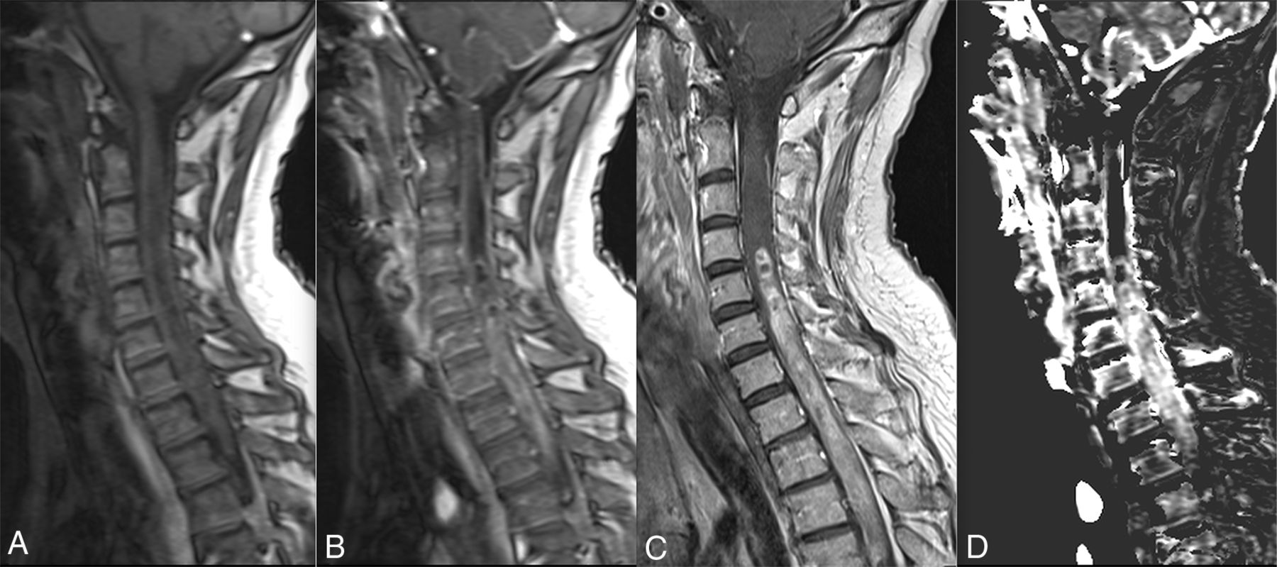
- Fig 1.
Spinal cord glioblastoma. Baseline (A) and timeframe after enhancement (B) of DCE perfusion show a spinal cord mass at the cervicothoracic junction. C, Gadolinium-enhanced sagittal T1 spin-echo image shows a contrast-enhanced mass with necrotic/cystic areas. D, Vp map based on the Tofts extended pharmacokinetic modeling shows increased plasmatic volume, suggesting increased neoangiogenesis.


