Advertisement
  • Main menu
  • User menu
  • Search
  • English ▼
    • English
    • Afrikaans
    • Albanian
    • Amharic
    • Arabic
    • Armenian
    • Azerbaijani
    • Basque
    • Belarusian
    • Bengali
    • Bosnian
    • Bulgarian
    • Catalan
    • Cebuano
    • Chichewa
    • Chinese (Simplified)
    • Chinese (Traditional)
    • Corsican
    • Croatian
    • Czech
    • Danish
    • Dutch
    • Esperanto
    • Estonian
    • Filipino
    • Finnish
    • French
    • Frisian
    • Galician
    • Georgian
    • German
    • Greek
    • Gujarati
    • Haitian Creole
    • Hausa
    • Hawaiian
    • Hebrew
    • Hindi
    • Hmong
    • Hungarian
    • Icelandic
    • Igbo
    • Indonesian
    • Irish
    • Italian
    • Japanese
    • Javanese
    • Kannada
    • Kazakh
    • Khmer
    • Korean
    • Kurdish (Kurmanji)
    • Kyrgyz
    • Lao
    • Latin
    • Latvian
    • Lithuanian
    • Luxembourgish
    • Macedonian
    • Malagasy
    • Malay
    • Malayalam
    • Maltese
    • Maori
    • Marathi
    • Mongolian
    • Myanmar (Burmese)
    • Nepali
    • Norwegian
    • Pashto
    • Persian
    • Polish
    • Portuguese
    • Punjabi
    • Romanian
    • Russian
    • Samoan
    • Scottish Gaelic
    • Serbian
    • Sesotho
    • Shona
    • Sindhi
    • Sinhala
    • Slovak
    • Slovenian
    • Somali
    • Spanish
    • Sudanese
    • Swahili
    • Swedish
    • Tajik
    • Tamil
    • Telugu
    • Thai
    • Turkish
    • Ukrainian
    • Urdu
    • Uzbek
    • Vietnamese
    • Welsh
    • Xhosa
    • Yiddish
    • Yoruba
    • Zulu

Main menu

  • Home
  • Content
    • Current Issue
    • Accepted Manuscripts
    • Article Preview
    • Past Issue Archive
    • Video Articles
    • AJNR Case Collection
    • Case of the Week Archive
    • Case of the Month Archive
    • Classic Case Archive
  • Special Collections
    • Advancing NeuroMRI with High-Relaxivity Contrast Agents
    • AJNR Awards
    • Low-Field MRI
    • Alzheimer Disease
    • ASNR Foundation Special Collection
    • Photon-Counting CT
    • View All
  • Multimedia
    • AJNR Podcasts
    • AJNR SCANtastic
    • Trainee Corner
    • MRI Safety Corner
    • Imaging Protocols
  • For Authors
    • Submit a Manuscript
    • Submit a Video Article
    • Submit an eLetter to the Editor/Response
    • Manuscript Submission Guidelines
    • Statistical Tips
    • Fast Publishing of Accepted Manuscripts
    • Graphical Abstract Preparation
    • Imaging Protocol Submission
    • Author Policies
  • About Us
    • About AJNR
    • Editorial Board
    • Editorial Board Alumni
  • More
    • Become a Reviewer/Academy of Reviewers
    • Subscribers
    • Permissions
    • Alerts
    • Feedback
    • Advertisers
    • ASNR Home

User menu

  • Alerts
  • Log in

Search

  • Advanced search
American Journal of Neuroradiology

American Journal of Neuroradiology

ASHNR American Society of Functional Neuroradiology ASHNR American Society of Pediatric Neuroradiology ASSR
  • Alerts
  • Log in

Advanced Search

English ▼
  • English
  • Afrikaans
  • Albanian
  • Amharic
  • Arabic
  • Armenian
  • Azerbaijani
  • Basque
  • Belarusian
  • Bengali
  • Bosnian
  • Bulgarian
  • Catalan
  • Cebuano
  • Chichewa
  • Chinese (Simplified)
  • Chinese (Traditional)
  • Corsican
  • Croatian
  • Czech
  • Danish
  • Dutch
  • Esperanto
  • Estonian
  • Filipino
  • Finnish
  • French
  • Frisian
  • Galician
  • Georgian
  • German
  • Greek
  • Gujarati
  • Haitian Creole
  • Hausa
  • Hawaiian
  • Hebrew
  • Hindi
  • Hmong
  • Hungarian
  • Icelandic
  • Igbo
  • Indonesian
  • Irish
  • Italian
  • Japanese
  • Javanese
  • Kannada
  • Kazakh
  • Khmer
  • Korean
  • Kurdish (Kurmanji)
  • Kyrgyz
  • Lao
  • Latin
  • Latvian
  • Lithuanian
  • Luxembourgish
  • Macedonian
  • Malagasy
  • Malay
  • Malayalam
  • Maltese
  • Maori
  • Marathi
  • Mongolian
  • Myanmar (Burmese)
  • Nepali
  • Norwegian
  • Pashto
  • Persian
  • Polish
  • Portuguese
  • Punjabi
  • Romanian
  • Russian
  • Samoan
  • Scottish Gaelic
  • Serbian
  • Sesotho
  • Shona
  • Sindhi
  • Sinhala
  • Slovak
  • Slovenian
  • Somali
  • Spanish
  • Sudanese
  • Swahili
  • Swedish
  • Tajik
  • Tamil
  • Telugu
  • Thai
  • Turkish
  • Ukrainian
  • Urdu
  • Uzbek
  • Vietnamese
  • Welsh
  • Xhosa
  • Yiddish
  • Yoruba
  • Zulu
  • Home
  • Content
    • Current Issue
    • Accepted Manuscripts
    • Article Preview
    • Past Issue Archive
    • Video Articles
    • AJNR Case Collection
    • Case of the Week Archive
    • Case of the Month Archive
    • Classic Case Archive
  • Special Collections
    • Advancing NeuroMRI with High-Relaxivity Contrast Agents
    • AJNR Awards
    • Low-Field MRI
    • Alzheimer Disease
    • ASNR Foundation Special Collection
    • Photon-Counting CT
    • View All
  • Multimedia
    • AJNR Podcasts
    • AJNR SCANtastic
    • Trainee Corner
    • MRI Safety Corner
    • Imaging Protocols
  • For Authors
    • Submit a Manuscript
    • Submit a Video Article
    • Submit an eLetter to the Editor/Response
    • Manuscript Submission Guidelines
    • Statistical Tips
    • Fast Publishing of Accepted Manuscripts
    • Graphical Abstract Preparation
    • Imaging Protocol Submission
    • Author Policies
  • About Us
    • About AJNR
    • Editorial Board
    • Editorial Board Alumni
  • More
    • Become a Reviewer/Academy of Reviewers
    • Subscribers
    • Permissions
    • Alerts
    • Feedback
    • Advertisers
    • ASNR Home
  • Follow AJNR on Twitter
  • Visit AJNR on Facebook
  • Follow AJNR on Instagram
  • Join AJNR on LinkedIn
  • RSS Feeds

AJNR Awards, New Junior Editors, and more. Read the latest AJNR updates


Improved Turnaround Times | Median time to first decision: 12 days

Research ArticlePediatric Neuroimaging

Limited Dorsal Myeloschisis and Congenital Dermal Sinus: Comparison of Clinical and MR Imaging Features

S.M. Lee, J.-E. Cheon, Y.H. Choi, I.-O. Kim, W.S. Kim, H.-H. Cho, J.Y. Lee and K.-C. Wang
American Journal of Neuroradiology January 2017, 38 (1) 176-182; DOI: https://doi.org/10.3174/ajnr.A4958
S.M. Lee
aFrom the Departments of Radiology (S.M.L., J.-E.C., Y.H.C., I.-O.K., W.S.K., H.-H.C.)
dDepartment of Radiology (S.M.L.), Kyungpook National University Medical Center, Daegu, Korea
  • Find this author on Google Scholar
  • Find this author on PubMed
  • Search for this author on this site
  • ORCID record for S.M. Lee
J.-E. Cheon
aFrom the Departments of Radiology (S.M.L., J.-E.C., Y.H.C., I.-O.K., W.S.K., H.-H.C.)
  • Find this author on Google Scholar
  • Find this author on PubMed
  • Search for this author on this site
  • ORCID record for J.-E. Cheon
Y.H. Choi
aFrom the Departments of Radiology (S.M.L., J.-E.C., Y.H.C., I.-O.K., W.S.K., H.-H.C.)
  • Find this author on Google Scholar
  • Find this author on PubMed
  • Search for this author on this site
  • ORCID record for Y.H. Choi
I.-O. Kim
aFrom the Departments of Radiology (S.M.L., J.-E.C., Y.H.C., I.-O.K., W.S.K., H.-H.C.)
  • Find this author on Google Scholar
  • Find this author on PubMed
  • Search for this author on this site
  • ORCID record for I.-O. Kim
W.S. Kim
aFrom the Departments of Radiology (S.M.L., J.-E.C., Y.H.C., I.-O.K., W.S.K., H.-H.C.)
  • Find this author on Google Scholar
  • Find this author on PubMed
  • Search for this author on this site
  • ORCID record for W.S. Kim
H.-H. Cho
aFrom the Departments of Radiology (S.M.L., J.-E.C., Y.H.C., I.-O.K., W.S.K., H.-H.C.)
eDepartment of Radiology (H.-H.C.), Ewha Womans University Mokdong Hospital, Seoul, Republic of Korea.
  • Find this author on Google Scholar
  • Find this author on PubMed
  • Search for this author on this site
  • ORCID record for H.-H. Cho
J.Y. Lee
bAnatomy (J.Y.L.)
cNeurosurgery, Division of Pediatric Neurosurgery (J.Y.L., K.-C.W.), Seoul National University College of Medicine, Seoul, Republic of Korea
  • Find this author on Google Scholar
  • Find this author on PubMed
  • Search for this author on this site
  • ORCID record for J.Y. Lee
K.-C. Wang
cNeurosurgery, Division of Pediatric Neurosurgery (J.Y.L., K.-C.W.), Seoul National University College of Medicine, Seoul, Republic of Korea
  • Find this author on Google Scholar
  • Find this author on PubMed
  • Search for this author on this site
  • ORCID record for K.-C. Wang
  • Article
  • Figures & Data
  • Info & Metrics
  • Responses
  • References
  • PDF
Loading

Article Figures & Data

Figures

  • Tables
  • Fig 1.
    • Download figure
    • Open in new tab
    • Download powerpoint
    Fig 1.

    LDM in a 4-month-old girl who presented with a midline skin lesion in the lumbar area. A, Photograph shows a crater covered with pearly pale epithelium (arrowheads) described as a cigarette-burn mark. Sagittal (B) and axial (C) T2-weighted MR images show that the intraspinal tract (arrows) displaying a distinct hypointense round structure separate from the filum terminale or nerve roots is completely traceable in its entire course (classified as “entirely visible” tract). The attachment site of the tract is the spinal cord just above the conus medullaris. A low-lying conus medullaris and dorsal tenting of the spinal cord at the tract-cord union are seen (open arrow). D, Photograph obtained during the operation shows a thick tract (asterisk) adhering to the dorsal aspect of the spinal cord.

  • Fig 2.
    • Download figure
    • Open in new tab
    • Download powerpoint
    Fig 2.

    LDM in an 11-month-old girl with an intradural lipoma. A, Photograph shows a pinpoint pit (arrowhead) in the midline area of the lumbosacral region. T1-weighted (B) and sagittal T2-weighted (C) MR images show an entirely visible intrathecal tract (arrow) traversing the center of an intradural dorsal lipoma (L) and a low-lying conus medullaris. The attachment site of the tract is the spinal cord is just above the conus medullaris. D, Sequential axial T2-weighted MR images show dorsal tenting of the spinal cord (open arrow) and tract with low signal intensity (arrows).
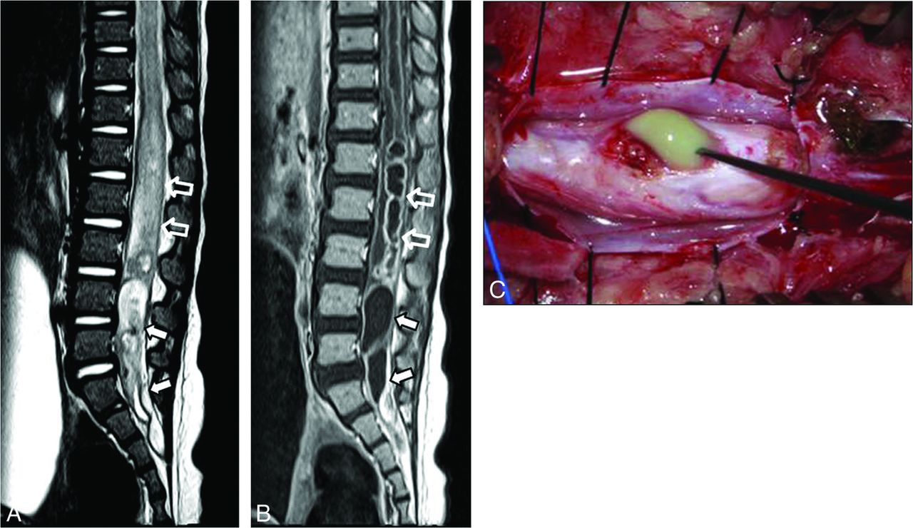
  • Fig 3.
    • Download figure
    • Open in new tab
    • Download powerpoint
    Fig 3.

    CDS in a 12-month-old girl who presented with fever and quadriplegia. Sagittal T2-weighted (A) and contrast-enhanced T1-weighted (B) MR images show ring-enhancing mass lesions nearly filling the entire intrathecal space (arrows), accompanied by spinal cord expansion and intramedullary thick enhancement (open arrows). There is no discernable tract in the intrathecal region on MR imaging (classified into “poorly visible”). These enhancing mass lesions (arrows) and intramedullary enhancement (open arrows) were infected epidermoid tumor and intramedullary abscess at the operation, respectively. C, Photograph obtained during the operation shows a yellowish material, representing infected epidermoid tumor.

Tables

  • Figures
    • View popup
    Table 1:

    Comparison of LDM and CDS: clinical data

    LDM (n = 12)CDS (n = 10)P Value
    Male/female2:106:4.074
    Age (mo) at admissiona11 ± 14.2 (0.5–47)12.9 ± 9 (0.25–34).207
    Clinical presentation
        Skin abnormalityb<.0001
            Skin-covered crater10 (83%)0
            Pit2 (17%)10 (100%)
        Infectionb06 (60%).003
        Neurologic deficitb2 (17%)4 (40%).348
    • ↵a Data are mean value, with ranges in parentheses.

    • ↵b Data are number of patients, with percentages in parentheses.

    • View popup
    Table 2:

    Visibility of tract: MRI and operative findingsa

    TractVisibilityLDM (n = 12)CDS (n = 10)P Value
    Subcutaneous12 (100%)10 (100%)–
    IntrathecalbEntirely visible10 (83%)1 (10%).003
    Partially visible2 (17%)4 (40%)
    Poorly visible0 (0%)4 (40%)
    • ↵a Data are number of patients, with percentages in parentheses.

    • ↵b Because the tract of 1 patient with CDS was revealed to end in the dura mater at the operation and there was no tract in the intrathecal area, only 9 CDS tracts were assessed with regard to visibility and attachment site in its intrathecal portion.

    • View popup
    Table 3:

    Attachment site of tract: MRI and operative findingsa

    TractAttachment SiteLDM (n = 12)CDS (n = 10)P Value
    MRIOPMRIOP
    IntrathecalbConus medullaris12 (100%)12 (100%)2 (20%)3 (30%).004
    Filum terminale/nerve root––2 (20%)2 (20%)
    Dermoid/epidermoid tumor–––4 (40%)
    Not availablec5 (50%)
    No extension into the spinal canal––1 (10%)1 (10%)
    • Note:—Op indicates operative findings.

    • ↵a Data are number of patients, with percentages in parentheses.

    • ↵b Because the tract of 1 patient with CDS was revealed to end in the dura mater at the operation and there was no tract in the intrathecal area, only 9 CDS tracts were assessed with regard to visibility and attachment site in its intrathecal portion.

    • ↵c The attachment sites of the tracts could not be evaluated on MRI.

    • View popup
    Table 4:

    Configuration of spinal cord: MRI and operative findingsa

    Spinal CordLDM (n = 12)CDS (n = 10)P Value
    Level of the conus medullarisNormal3 (25%)4 (40%).652
    Low-lying9 (75%)6 (60%)
    Shape of the spinal cordDorsal tenting10 (83%)1 (10%).001
    • ↵a Data are number of patients, with percentages in parentheses.

    • View popup
    Table 5:

    Intradural lesions: MRI and operative findingsa

    Associated Intradural LesionsMRIOPMRIOPP Value
    Dermoid-epidermoid0 (0%)0 (0%)5 (50%)6 (60%).003
    Lipoma2 (17%)2 (17%)2 (20%)2 (20%)1.000
    • Note:—OP indicates operative findings.

    • ↵a Data are number of patients, with percentages in parentheses.

PreviousNext
Back to top

In this issue

American Journal of Neuroradiology: 38 (1)
American Journal of Neuroradiology
Vol. 38, Issue 1
1 Jan 2017
  • Table of Contents
  • Index by author
  • Complete Issue (PDF)
Advertisement
Print
Download PDF
Email Article
Cite this article
0 Responses
Respond to this article
Share
Bookmark this article
Limited Dorsal Myeloschisis and Congenital Dermal Sinus: Comparison of Clinical and MR Imaging Features
S.M. Lee, J.-E. Cheon, Y.H. Choi, I.-O. Kim, W.S. Kim, H.-H. Cho, J.Y. Lee, K.-C. Wang
American Journal of Neuroradiology Jan 2017, 38 (1) 176-182; DOI: 10.3174/ajnr.A4958
del.icio.us logo Twitter logo Facebook logo Mendeley logo
  • Tweet Widget
Purchase

Jump to section

  • Article
    • Abstract
    • ABBREVIATIONS:
    • Materials and Methods
    • Results
    • Discussion
    • Conclusions
    • References
  • Figures & Data
  • Info & Metrics
  • Responses
  • References
  • PDF

Related Articles

  • No related articles found.
  • PubMed
  • Google Scholar

Cited By...

  • No citing articles found.
  • Crossref
  • Google Scholar

This article has not yet been cited by articles in journals that are participating in Crossref Cited-by Linking.

More in this TOC Section

Pediatric Neuroimaging

  • Clinical SVR of Fetal Brain MRI
  • Comparing MRI Perfusion in Pediatric Brain Tumors
  • Sodium MRI in Pediatric Brain Tumors
Show more Pediatric Neuroimaging

Spine Imaging and Spine Image-Guided Interventions

  • Dynamic CT Myelography: Patient Positioning
  • Evaluation of SIH MR Scoring Systems in Normals
  • Management Outcomes For VO Spine Biopsy
Show more Spine Imaging and Spine Image-Guided Interventions

Similar Articles

Advertisement

Indexed Content

  • Current Issue
  • Accepted Manuscripts
  • Article Preview
  • Past Issues
  • Editorials
  • Editor's Choice
  • Fellows' Journal Club
  • Letters to the Editor
  • Video Articles

Cases

  • Case Collection
  • Archive - Case of the Week
  • Archive - Case of the Month
  • Archive - Classic Case

More from AJNR

  • Trainee Corner
  • Imaging Protocols
  • MRI Safety Corner
  • Book Reviews

Multimedia

  • AJNR Podcasts
  • AJNR Scantastics

Resources

  • Turnaround Time
  • Submit a Manuscript
  • Submit a Video Article
  • Submit an eLetter to the Editor/Response
  • Manuscript Submission Guidelines
  • Statistical Tips
  • Fast Publishing of Accepted Manuscripts
  • Graphical Abstract Preparation
  • Imaging Protocol Submission
  • Evidence-Based Medicine Level Guide
  • Publishing Checklists
  • Author Policies
  • Become a Reviewer/Academy of Reviewers
  • News and Updates

About Us

  • About AJNR
  • Editorial Board
  • Editorial Board Alumni
  • Alerts
  • Permissions
  • Not an AJNR Subscriber? Join Now
  • Advertise with Us
  • Librarian Resources
  • Feedback
  • Terms and Conditions
  • AJNR Editorial Board Alumni

American Society of Neuroradiology

  • Not an ASNR Member? Join Now

© 2025 by the American Society of Neuroradiology All rights, including for text and data mining, AI training, and similar technologies, are reserved.
Print ISSN: 0195-6108 Online ISSN: 1936-959X

Powered by HighWire
Email this Article

Thank you for your interest in spreading the word on American Journal of Neuroradiology.

NOTE: We only request your email address so that the person you are recommending the page to knows that you wanted them to see it, and that it is not junk mail. We do not capture any email address.

Enter multiple addresses on separate lines or separate them with commas.
Limited Dorsal Myeloschisis and Congenital Dermal Sinus: Comparison of Clinical and MR Imaging Features
(Your Name) has sent you a message from American Journal of Neuroradiology
(Your Name) thought you would like to see the American Journal of Neuroradiology web site.
CAPTCHA
This question is for testing whether or not you are a human visitor and to prevent automated spam submissions.
Cite this article
S.M. Lee, J.-E. Cheon, Y.H. Choi, I.-O. Kim, W.S. Kim, H.-H. Cho, J.Y. Lee, K.-C. Wang
Limited Dorsal Myeloschisis and Congenital Dermal Sinus: Comparison of Clinical and MR Imaging Features
American Journal of Neuroradiology Jan 2017, 38 (1) 176-182; DOI: 10.3174/ajnr.A4958

Citation Manager Formats

  • BibTeX
  • Bookends
  • EasyBib
  • EndNote (tagged)
  • EndNote 8 (xml)
  • Medlars
  • Mendeley
  • Papers
  • RefWorks Tagged
  • Ref Manager
  • RIS
  • Zotero

We use cookies on this site to enhance your user experience. By clicking any link on this page you are giving your consent for us to set cookies.