
Article Figures & Data
Figures
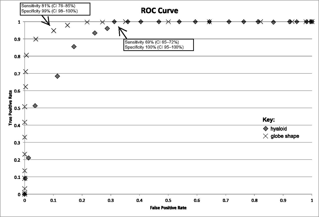
- Fig 1.
Receiver operating characteristic curves demonstrate sensitivity (true-positive) versus 1-specificity (false-positive) of MR imaging of the fetal brain for elliptic globe shape (X) and hyaloid vasculature visibility (diamond) as a function of age.
- Fig 2.
Age-related changes in globe morphology and hyaloid vasculature visibility from 17 to 39 weeks' gestation.
- Fig 3.
Sagittal (A) and axial (B) “fetography” T2WI (TR/TE, 5000/163 ms) of a normal fetal brain at 20 weeks' gestational age. The globe morphology is conical, with angulation of its posterior (arrows, A) and posterolateral (arrows, B) margins.
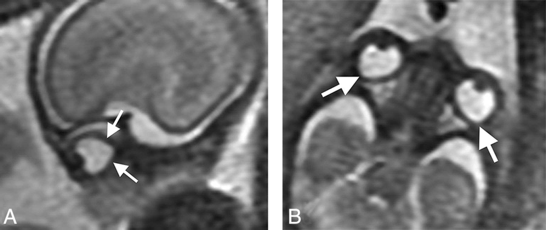
- Fig 4.
Sagittal (A) and axial (B) T2 HASTE (TR/TE, 1270/80 ms) of a normal fetal brain at 18 weeks' gestational age. The globe morphology is conical with angulation of its posterior (arrows, A) and posterolateral (arrows, B) margins. Follow-up sagittal (C) and axial (D) T2 HASTE (TR/TE, 1270/81 ms) of a normal fetal brain from imaging performed on the same patient at 30 weeks' gestational age. Ocular globe morphology is round/elliptic in the axial and sagittal planes. Fetal vasculature is not visible.
- Fig 5.
Sagittal single-shot fast spin-echo T2WI (TR/TE, 1087/56 ms) of a normal fetal brain at 19 weeks' gestation. Linear hypointense signal extending from the posterior margin of the lens through the vitreous toward the apex represents the hyaloid vasculature (arrow).


Tables
Frequency of hyaloid visibility and elliptic, transitional, nonelliptic, and combined transitional and noneliptic globe morphology at various gestational ages (16–39 weeks)
GA F H (F) H (%) E (F) E (%) T (F) T (%) NE (F) NE (%) T + NE (F) T + NE (%) 17 7 6 86 0 0 0 0 7 100 7 100 18 16 9 89 0 0 0 0 16 100 16 100 19 37 23 62 0 0 0 0 37 100 37 100 20 60 13 22 0 0 0 0 60 100 60 100 21 48 13 27 0 0 0 0 48 100 48 100 22 50 6 12 0 0 3 6 47 94 50 100 23 28 2 7 0 0 3 11 25 89 28 100 24 18 3 17 0 0 5 27 13 73 18 100 25 29 0 0 6 21 18 62 5 17 23 79 26 27 0 0 11 46 16 59 0 0 16 59 27 38 0 0 17 45 21 75 0 0 21 75 28 43 0 0 32 74 11 6 0 0 11 6 29 34 0 0 33 97 1 3 0 0 0 0 30 30 0 0 30 100 0 0 0 0 0 0 31 41 0 0 40 98 1 2 0 0 1 2 32 31 0 0 31 100 0 0 0 0 0 0 33 30 0 0 30 100 0 0 0 0 0 0 34 34 0 0 34 100 0 0 0 0 0 0 35 33 0 0 33 100 0 0 0 0 0 0 36 15 0 0 15 100 0 0 0 0 0 0 37 21 0 0 21 100 0 0 0 0 0 0 38 8 0 0 8 100 0 0 0 0 0 0 39 3 0 0 3 100 0 0 0 0 0 0 Note:—GA indicates gestational age in weeks; F, frequency; H, hyaloid vasculature; E, elliptic; T, transitional globe morphology/mild nonellipsoid shape; NE, nonelliptic.


