Advertisement

Main menu

  • Home
  • Content
    • Current Issue
    • Accepted Manuscripts
    • Article Preview
    • Past Issue Archive
    • Video Articles
    • AJNR Case Collection
    • Case of the Week Archive
    • Case of the Month Archive
    • Classic Case Archive
  • Special Collections
    • AJNR Awards
    • Low-Field MRI
    • Alzheimer Disease
    • ASNR Foundation Special Collection
    • Photon-Counting CT
    • View All
  • Multimedia
    • AJNR Podcasts
    • AJNR SCANtastic
    • Trainee Corner
    • MRI Safety Corner
    • Imaging Protocols
  • For Authors
    • Submit a Manuscript
    • Submit a Video Article
    • Submit an eLetter to the Editor/Response
    • Manuscript Submission Guidelines
    • Statistical Tips
    • Fast Publishing of Accepted Manuscripts
    • Graphical Abstract Preparation
    • Imaging Protocol Submission
    • Author Policies
  • About Us
    • About AJNR
    • Editorial Board
    • Editorial Board Alumni
  • More
    • Become a Reviewer/Academy of Reviewers
    • Subscribers
    • Permissions
    • Alerts
    • Feedback
    • Advertisers
    • ASNR Home

User menu

  • Alerts
  • Log in

Search

  • Advanced search
American Journal of Neuroradiology

American Journal of Neuroradiology

ASHNR American Society of Functional Neuroradiology ASHNR American Society of Pediatric Neuroradiology ASSR
  • Alerts
  • Log in

Advanced Search

  • Home
  • Content
    • Current Issue
    • Accepted Manuscripts
    • Article Preview
    • Past Issue Archive
    • Video Articles
    • AJNR Case Collection
    • Case of the Week Archive
    • Case of the Month Archive
    • Classic Case Archive
  • Special Collections
    • AJNR Awards
    • Low-Field MRI
    • Alzheimer Disease
    • ASNR Foundation Special Collection
    • Photon-Counting CT
    • View All
  • Multimedia
    • AJNR Podcasts
    • AJNR SCANtastic
    • Trainee Corner
    • MRI Safety Corner
    • Imaging Protocols
  • For Authors
    • Submit a Manuscript
    • Submit a Video Article
    • Submit an eLetter to the Editor/Response
    • Manuscript Submission Guidelines
    • Statistical Tips
    • Fast Publishing of Accepted Manuscripts
    • Graphical Abstract Preparation
    • Imaging Protocol Submission
    • Author Policies
  • About Us
    • About AJNR
    • Editorial Board
    • Editorial Board Alumni
  • More
    • Become a Reviewer/Academy of Reviewers
    • Subscribers
    • Permissions
    • Alerts
    • Feedback
    • Advertisers
    • ASNR Home
  • Follow AJNR on Twitter
  • Visit AJNR on Facebook
  • Follow AJNR on Instagram
  • Join AJNR on LinkedIn
  • RSS Feeds

AJNR Awards, New Junior Editors, and more. Read the latest AJNR updates

Research ArticlePediatric Neuroimaging

Development of the Fetal Vermis: New Biometry Reference Data and Comparison of 3 Diagnostic Modalities–3D Ultrasound, 2D Ultrasound, and MR Imaging

E. Katorza, E. Bertucci, S. Perlman, S. Taschini, R. Ber, Y. Gilboa, V. Mazza and R. Achiron
American Journal of Neuroradiology July 2016, 37 (7) 1359-1366; DOI: https://doi.org/10.3174/ajnr.A4725
E. Katorza
aFrom the Antenatal Diagnostic Unit (E.K., S.P., R.B., Y.G., R.A.), Department of Obstetrics and Gynecology, Haim Sheba Medical Center, Sackler School of Medicine, Tel Aviv University, Israel
  • Find this author on Google Scholar
  • Find this author on PubMed
  • Search for this author on this site
  • ORCID record for E. Katorza
E. Bertucci
bPrenatal Medicine Unit (E.B., S.T., V.M.), Department of Obstetrics and Gynecology, Modena Hospital, Modena, Italy.
  • Find this author on Google Scholar
  • Find this author on PubMed
  • Search for this author on this site
  • ORCID record for E. Bertucci
S. Perlman
aFrom the Antenatal Diagnostic Unit (E.K., S.P., R.B., Y.G., R.A.), Department of Obstetrics and Gynecology, Haim Sheba Medical Center, Sackler School of Medicine, Tel Aviv University, Israel
  • Find this author on Google Scholar
  • Find this author on PubMed
  • Search for this author on this site
  • ORCID record for S. Perlman
S. Taschini
bPrenatal Medicine Unit (E.B., S.T., V.M.), Department of Obstetrics and Gynecology, Modena Hospital, Modena, Italy.
  • Find this author on Google Scholar
  • Find this author on PubMed
  • Search for this author on this site
  • ORCID record for S. Taschini
R. Ber
aFrom the Antenatal Diagnostic Unit (E.K., S.P., R.B., Y.G., R.A.), Department of Obstetrics and Gynecology, Haim Sheba Medical Center, Sackler School of Medicine, Tel Aviv University, Israel
  • Find this author on Google Scholar
  • Find this author on PubMed
  • Search for this author on this site
  • ORCID record for R. Ber
Y. Gilboa
aFrom the Antenatal Diagnostic Unit (E.K., S.P., R.B., Y.G., R.A.), Department of Obstetrics and Gynecology, Haim Sheba Medical Center, Sackler School of Medicine, Tel Aviv University, Israel
  • Find this author on Google Scholar
  • Find this author on PubMed
  • Search for this author on this site
  • ORCID record for Y. Gilboa
V. Mazza
bPrenatal Medicine Unit (E.B., S.T., V.M.), Department of Obstetrics and Gynecology, Modena Hospital, Modena, Italy.
  • Find this author on Google Scholar
  • Find this author on PubMed
  • Search for this author on this site
  • ORCID record for V. Mazza
R. Achiron
aFrom the Antenatal Diagnostic Unit (E.K., S.P., R.B., Y.G., R.A.), Department of Obstetrics and Gynecology, Haim Sheba Medical Center, Sackler School of Medicine, Tel Aviv University, Israel
  • Find this author on Google Scholar
  • Find this author on PubMed
  • Search for this author on this site
  • ORCID record for R. Achiron
  • Article
  • Figures & Data
  • Supplemental
  • Info & Metrics
  • Responses
  • References
  • PDF
Loading

Article Figures & Data

Figures

  • Tables
  • Fig 1.
    • Download figure
    • Open in new tab
    • Download powerpoint
    Fig 1.

    Magnification of the fetal posterior fossa and vermis and demonstration of biometric parameters. 1) Maximum superoinferior diameter. 2) Anteroposterior diameter. 3) Perimeter and surface area.

  • Fig 2.
    • Download figure
    • Open in new tab
    • Download powerpoint
    Fig 2.

    Midsagittal view of the fetal brain demonstrating the vermis. A, 2D US. B, 3D US. C, MR imaging.
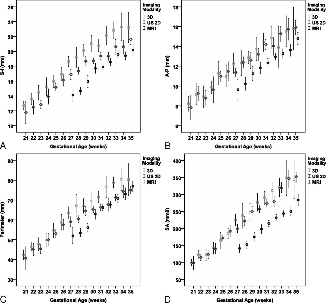
  • Fig 3.
    • Download figure
    • Open in new tab
    • Download powerpoint
    Fig 3.

    Comparison of measurements (mean ± SD, 95% CI) obtained by 2D US, 3D US, and MR imaging. A, SI. B, AP. C, Perimeter. D, SA.

Tables

  • Figures
    • View popup
    Table 1:

    Median, 5th, and 95th percentiles for SI measurements

    Gestational Age (wk)Imaging Modality
    US 2DUS 3DMRI
    %5Median%95%5Median%95%5Median%95
    2110.6911.9512.5711.1012.3014.60
    2210.4212.7214.4011.5013.6014.90
    2311.7112.7913.8413.0014.4016.50
    2413.3313.7914.8213.4015.3517.50
    2513.8515.1716.6614.5016.0017.80
    2613.7516.0918.4515.6016.8019.50
    2714.0517.0718.7816.7018.7020.2013.3014.1315.05
    2816.0817.0919.7216.3018.8023.8013.6614.5815.73
    2917.2918.7220.3318.1020.0022.2013.2516.0217.54
    3017.8318.5619.6218.4020.5023.6015.7717.3820.76
    3118.0319.3420.7518.1021.3023.8016.1618.0219.34
    3217.6819.3320.5919.0021.4026.6016.7518.6720.12
    3316.7820.9322.9420.7022.7024.5018.3719.4821.21
    3419.1120.3823.3420.4022.3028.5017.9019.5621.30
    3519.6221.7524.2920.9022.0027.0018.2719.8323.29
    • Note:—%5 indicates 5th percentile; %95, 95th percentile.

    • View popup
    Table 2:

    Median, 5th, and 95th percentiles for AP measurements

    Gestational Age (wk)Imaging Modality
    US 2DUS 3DMRI
    %5Median%95%5Median%95%5Median%95
    217.087.798.715.708.1010.30
    227.519.5110.627.409.1512.40
    237.278.7210.256.308.8011.00
    248.969.4910.648.609.8511.00
    259.5810.7313.179.9010.5014.60
    268.7611.2814.178.1011.3012.80
    2710.0611.4412.699.3012.5014.809.119.3311.10
    2810.7512.3214.949.2012.3015.908.909.9712.04
    2910.0912.6115.0111.2012.8014.408.9311.0513.28
    3011.0613.3614.9410.7013.7016.8010.3911.9215.01
    3112.6914.0115.8111.8014.6018.0011.0712.3013.96
    3211.9313.8717.3810.7015.2018.4011.4013.0413.90
    3312.6315.1117.8311.8013.0519.7011.9113.2615.22
    3412.3615.9417.8212.0015.6021.4011.1813.7715.10
    3513.8416.4017.4012.6015.7518.9012.7014.8317.17
    • Note:—%5 indicates 5th percentile; %95, 95th percentile.

    • View popup
    Table 3:

    Median, 5th, and 95th percentiles for perimeter measurements

    Gestational Age (wk)Imaging Modality
    US 2DUS 3DMRI
    %5Median%95%5Median%95%5Median%95
    2136.2040.9444.8037.2041.7054.60
    2237.0446.8048.6440.6047.3049.40
    2341.2045.0749.3941.5046.9055.20
    2448.1049.8951.8045.0049.2056.70
    2546.7452.4162.8648.8053.3063.30
    2650.0057.8765.9850.8058.3068.60
    2748.9759.0867.1051.4064.0069.3050.0550.6757.75
    2853.2660.4269.4057.6066.2082.6048.2253.3058.16
    2958.5865.4269.3758.8066.4077.2050.5856.8561.09
    3060.0864.9870.3657.8070.2082.8055.0761.1072.13
    3161.1865.6771.9764.1071.1082.4059.3866.2171.93
    3262.1367.3177.8865.3074.6096.1061.2068.1173.79
    3368.1371.3876.8373.7077.9582.5064.9870.9078.33
    3467.0573.9780.4366.5080.1092.8067.2171.0683.00
    3570.2075.1979.1668.5079.70100.2069.4475.9386.11
    • Note:—%5 indicates 5th percentile; %95, 95th percentile.

    • View popup
    Table 4:

    Median, 5th, and 95th percentiles for SA measurements

    Gestational Age (wk)Imaging Modality
    US 2DUS 3DMRI
    %5Median%95%5Median%95%5Median%95
    2182.4199.27112.7670.0098.00126.00
    2292.43114.88131.1095.00125.00141.00
    23100.38125.18146.1292.00119.00164.00
    24130.89138.14154.26118.00140.50175.00
    25135.91170.76198.12127.00155.00233.00
    26139.84189.49234.09150.00193.00215.00
    27158.35198.22234.51176.00223.00255.00127.08143.50152.34
    28182.56222.26293.91177.00218.00376.00132.66157.32176.64
    29215.82246.64293.68199.00235.00319.00140.89175.90204.92
    30228.12255.40282.77223.00286.00330.00170.35196.48251.86
    31240.25268.21299.06219.00298.00355.00181.44214.24239.03
    32241.17282.90330.94263.00297.00412.00202.60229.60272.65
    33274.90325.12370.45295.00308.00383.00207.49246.18266.11
    34283.93340.38405.17256.00327.00540.00224.95246.21322.73
    35302.87350.00392.47280.00323.00478.00242.75279.97363.32
    • Note:—%5 indicates 5th percentile; %95, 95th percentile.

    • View popup
    Table 5:

    Linear association between gestational age and biometric measurements

    Linear Regression CoefficientModel Summary
    Unstandardized βaStandardized βAdjusted R2
    SI
        US 2D0.6930.9160.839
        US 3D0.8330.8990.806
        MRI0.7420.8110.656
    AP
        US 2D0.5590.8710.757
        US 3D0.5740.8000.637
        MRI0.6060.7910.624
    Perimeter
        US 2D2.3400.9120.831
        US 3D2.9890.8890.790
        MRI3.2430.8430.708
    SA
        US 2D18.4870.9480.897
        US 3D19.6700.8970.804
        MRI17.5870.8440.711
    • ↵a P < .001 for all coefficients.

    • View popup
    Table 6:

    Comparison of imaging modalities for each biometric measurement: estimated marginal means 95% CI

    SIAPPerimeterSA
    Mean95% CIMean95% CIMean95% CIMean95% CI
    At gestational age 29.6 wka
        US 2D18.1017.91–18.3012.9912.80–13.1963.3462.60–64.08247.45243.13–251.77
        US 3D19.8019.58–20.0113.1812.97–13.3967.7766.97–68.5258.43253.77–263.10
        MRI16.4716.23–16.7011.4011.17–11.6460.6859.80–61.55181.04175.90–186.1
    For examinations preformed at ≥27 wk of gestation: mean gestational age, 31.3 wk
        US 2D19.2819.05–19.5113.8813.64–14.1167.2066.31–68.10276.99271.53–282.45
        US 3D17.8017.57–18.0312.3612.13–12.5965.3464.48–66.20212.94207.68–218.20
        MRI21.3421.08–21.6114.2013.93–14.4673.2872.28–74.29294.12288.00–300.23
    • ↵a Mean gestational age for all populations (516 fetuses).

    • View popup
    Table 7:

    Intraobserver reproducibility of measurements

    Vermian MeasurementDifferences in Measurements of Vermian Biometry
    Mean (95% CI of the Difference)ICC (95% CI)
    US 2D, intraobserver variability
        SI−0.34 ± 0.73 (−0.54 to −0.13)0.94 (0.91 to 0.97)
        AP0.71 ± 1.02 (0.42 to 1.00)0.85 (0.75 to 0.91)
        Perimeter0.93 ± 2.7 (0.16 to 1.7)0.93 (0.88 to 0.96)
        SA2.2 ± 14.4 (2.04 to −1.81)0.97 (0.95 to 0.98)
    US 3D, interobserver variability
        SI−0.47 ± 1.5 (−0.89 to −0.43)0.95 (0.92 to 0.97)
        AP−0.26 ± 0.55 (−0.41 to −0.10)0.98 (0.98 to 0.99)
        Perimeter1.05 ± 10.66 (−1.97 to 4.08)0.81 (0.7 to 0.89)
        SA−1.2 ± 2.6 (−1.95 to −0.44)0.999 (0.99 to 1.0)
    MRI, intraobserver variability
        SI0.60 ± 0.66 (0.42 to 0.79)0.94 (0.91 to 0.97)
        AP0.55 ± 1.05 (0.25 to 0.85)0.79 (0.66 to 0.88)
        Perimeter1.2 ± 4.05 (0.05 to 2.3)0.88 (0.81 to 0.93)
        SA−0.36 ± 10.1 (−3.2 to 2.5)0.97 (0.96 to 0.98)
    • View popup
    Table 8:

    Interobserver reproducibility of measurements

    Vermian MeasurementDifferences in Measurements of Vermian Biometry
    Mean (95% CI of the Difference)ICC (95% CI)
    US 2D, interobserver variability
        SI−0.07 ± 1.15 (−0.40 to 0.25)0.86 (0.77 to 0.92)
        AP0.92 ± 1.32 (0.54 to 1.2)0.71 (0.54 to 0.82)
        Perimeter2.4 ± 4.6 (1.15 to 3.81)0.81 (0.69 to 0.88)
        SA8.3 ± 31.18 (−0.47 to 17.25)0.86 (0.76 to 0.91)
    US 3D, interobserver variability
        SI−0.07 ± 1.15 (−0.40 to 0.25)0.86 (0.77 to 0.92)
        AP0.92 ± 1.32 (0.54 to 1.2)0.71 (0.54 to 0.82)
        Perimeter2.4 ± 4.6 (1.15 to 3.81)0.81 (0.69 to 0.88)
        SA8.3 ± 31.18 (−0.47 to 17.25)0.86 (0.76 to 0.91)
    MRI, interobserver variability
        SI−0.47 ± 1.5 (−0.89 to −0.43)0.95 (0.92 to 0.97)
        AP−0.26 ± 0.55 (−0.41 to −0.10)0.98 (0.98 to 0.99)
        Perimeter1.05 ± 10.66 (−1.97 to 4.08)0.81 (0.7 to 0.89)
        SA−1.2 ± 2.6 (−1.95 to −0.44)0.99 (0.99 to 1.0)
PreviousNext
Back to top

In this issue

American Journal of Neuroradiology: 37 (7)
American Journal of Neuroradiology
Vol. 37, Issue 7
1 Jul 2016
  • Table of Contents
  • Index by author
  • Complete Issue (PDF)
Advertisement
Print
Download PDF
Email Article

Thank you for your interest in spreading the word on American Journal of Neuroradiology.

NOTE: We only request your email address so that the person you are recommending the page to knows that you wanted them to see it, and that it is not junk mail. We do not capture any email address.

Enter multiple addresses on separate lines or separate them with commas.
Development of the Fetal Vermis: New Biometry Reference Data and Comparison of 3 Diagnostic Modalities–3D Ultrasound, 2D Ultrasound, and MR Imaging
(Your Name) has sent you a message from American Journal of Neuroradiology
(Your Name) thought you would like to see the American Journal of Neuroradiology web site.
CAPTCHA
This question is for testing whether or not you are a human visitor and to prevent automated spam submissions.
Cite this article
E. Katorza, E. Bertucci, S. Perlman, S. Taschini, R. Ber, Y. Gilboa, V. Mazza, R. Achiron
Development of the Fetal Vermis: New Biometry Reference Data and Comparison of 3 Diagnostic Modalities–3D Ultrasound, 2D Ultrasound, and MR Imaging
American Journal of Neuroradiology Jul 2016, 37 (7) 1359-1366; DOI: 10.3174/ajnr.A4725

Citation Manager Formats

  • BibTeX
  • Bookends
  • EasyBib
  • EndNote (tagged)
  • EndNote 8 (xml)
  • Medlars
  • Mendeley
  • Papers
  • RefWorks Tagged
  • Ref Manager
  • RIS
  • Zotero
0 Responses
Respond to this article
Share
Bookmark this article
Development of the Fetal Vermis: New Biometry Reference Data and Comparison of 3 Diagnostic Modalities–3D Ultrasound, 2D Ultrasound, and MR Imaging
E. Katorza, E. Bertucci, S. Perlman, S. Taschini, R. Ber, Y. Gilboa, V. Mazza, R. Achiron
American Journal of Neuroradiology Jul 2016, 37 (7) 1359-1366; DOI: 10.3174/ajnr.A4725
del.icio.us logo Twitter logo Facebook logo Mendeley logo
  • Tweet Widget
  • Facebook Like
  • Google Plus One
Purchase

Jump to section

  • Article
    • Abstract
    • ABBREVIATIONS:
    • Materials and Methods
    • Results
    • Discussion
    • Conclusions
    • Acknowledgments
    • Footnotes
    • REFERENCES
  • Figures & Data
  • Supplemental
  • Info & Metrics
  • Responses
  • References
  • PDF

Related Articles

  • PubMed
  • Google Scholar

Cited By...

  • Towards automated fetal brain biometry reporting for 3-dimensional T2-weighted 0.55-3T magnetic resonance imaging at 20-40 weeks gestational age range
  • Does 3T Fetal MRI Improve Image Resolution of Normal Brain Structures between 20 and 24 Weeks' Gestational Age?
  • Crossref
  • Google Scholar

This article has not yet been cited by articles in journals that are participating in Crossref Cited-by Linking.

More in this TOC Section

  • Clinical SVR of Fetal Brain MRI
  • Sodium MRI in Pediatric Brain Tumors
  • FRACTURE MR in Congenital Vertebral Anomalies
Show more Pediatric Neuroimaging

Similar Articles

Advertisement

Indexed Content

  • Current Issue
  • Accepted Manuscripts
  • Article Preview
  • Past Issues
  • Editorials
  • Editor's Choice
  • Fellows' Journal Club
  • Letters to the Editor
  • Video Articles

Cases

  • Case Collection
  • Archive - Case of the Week
  • Archive - Case of the Month
  • Archive - Classic Case

More from AJNR

  • Trainee Corner
  • Imaging Protocols
  • MRI Safety Corner
  • Book Reviews

Multimedia

  • AJNR Podcasts
  • AJNR Scantastics

Resources

  • Turnaround Time
  • Submit a Manuscript
  • Submit a Video Article
  • Submit an eLetter to the Editor/Response
  • Manuscript Submission Guidelines
  • Statistical Tips
  • Fast Publishing of Accepted Manuscripts
  • Graphical Abstract Preparation
  • Imaging Protocol Submission
  • Evidence-Based Medicine Level Guide
  • Publishing Checklists
  • Author Policies
  • Become a Reviewer/Academy of Reviewers
  • News and Updates

About Us

  • About AJNR
  • Editorial Board
  • Editorial Board Alumni
  • Alerts
  • Permissions
  • Not an AJNR Subscriber? Join Now
  • Advertise with Us
  • Librarian Resources
  • Feedback
  • Terms and Conditions
  • AJNR Editorial Board Alumni

American Society of Neuroradiology

  • Not an ASNR Member? Join Now

© 2025 by the American Society of Neuroradiology All rights, including for text and data mining, AI training, and similar technologies, are reserved.
Print ISSN: 0195-6108 Online ISSN: 1936-959X

Powered by HighWire