Advertisement

Main menu

  • Home
  • Content
    • Current Issue
    • Accepted Manuscripts
    • Article Preview
    • Past Issue Archive
    • Video Articles
    • AJNR Case Collection
    • Case of the Week Archive
    • Case of the Month Archive
    • Classic Case Archive
  • Special Collections
    • AJNR Awards
    • Low-Field MRI
    • Alzheimer Disease
    • ASNR Foundation Special Collection
    • Photon-Counting CT
    • View All
  • Multimedia
    • AJNR Podcasts
    • AJNR SCANtastic
    • Trainee Corner
    • MRI Safety Corner
    • Imaging Protocols
  • For Authors
    • Submit a Manuscript
    • Submit a Video Article
    • Submit an eLetter to the Editor/Response
    • Manuscript Submission Guidelines
    • Statistical Tips
    • Fast Publishing of Accepted Manuscripts
    • Graphical Abstract Preparation
    • Imaging Protocol Submission
    • Author Policies
  • About Us
    • About AJNR
    • Editorial Board
    • Editorial Board Alumni
  • More
    • Become a Reviewer/Academy of Reviewers
    • Subscribers
    • Permissions
    • Alerts
    • Feedback
    • Advertisers
    • ASNR Home

User menu

  • Alerts
  • Log in

Search

  • Advanced search
American Journal of Neuroradiology

American Journal of Neuroradiology

ASHNR American Society of Functional Neuroradiology ASHNR American Society of Pediatric Neuroradiology ASSR
  • Alerts
  • Log in

Advanced Search

  • Home
  • Content
    • Current Issue
    • Accepted Manuscripts
    • Article Preview
    • Past Issue Archive
    • Video Articles
    • AJNR Case Collection
    • Case of the Week Archive
    • Case of the Month Archive
    • Classic Case Archive
  • Special Collections
    • AJNR Awards
    • Low-Field MRI
    • Alzheimer Disease
    • ASNR Foundation Special Collection
    • Photon-Counting CT
    • View All
  • Multimedia
    • AJNR Podcasts
    • AJNR SCANtastic
    • Trainee Corner
    • MRI Safety Corner
    • Imaging Protocols
  • For Authors
    • Submit a Manuscript
    • Submit a Video Article
    • Submit an eLetter to the Editor/Response
    • Manuscript Submission Guidelines
    • Statistical Tips
    • Fast Publishing of Accepted Manuscripts
    • Graphical Abstract Preparation
    • Imaging Protocol Submission
    • Author Policies
  • About Us
    • About AJNR
    • Editorial Board
    • Editorial Board Alumni
  • More
    • Become a Reviewer/Academy of Reviewers
    • Subscribers
    • Permissions
    • Alerts
    • Feedback
    • Advertisers
    • ASNR Home
  • Follow AJNR on Twitter
  • Visit AJNR on Facebook
  • Follow AJNR on Instagram
  • Join AJNR on LinkedIn
  • RSS Feeds

AJNR Awards, New Junior Editors, and more. Read the latest AJNR updates

Research ArticleEXTRACRANIAL VASCULAR
Open Access

Optimal MR Plaque Imaging for Cervical Carotid Artery Stenosis in Predicting the Development of Microembolic Signals during Exposure of Carotid Arteries in Endarterectomy: Comparison of 4 T1-Weighted Imaging Techniques

Y. Sato, K. Ogasawara, S. Narumi, M. Sasaki, A. Saito, E. Tsushima, T. Namba, M. Kobayashi, K. Yoshida, Y. Terayama and A. Ogawa
American Journal of Neuroradiology June 2016, 37 (6) 1146-1154; DOI: https://doi.org/10.3174/ajnr.A4674
Y. Sato
aFrom the Departments of Neurosurgery (Y.S., K.O., T.N., M.K., K.Y., A.O.)
  • Find this author on Google Scholar
  • Find this author on PubMed
  • Search for this author on this site
K. Ogasawara
aFrom the Departments of Neurosurgery (Y.S., K.O., T.N., M.K., K.Y., A.O.)
  • Find this author on Google Scholar
  • Find this author on PubMed
  • Search for this author on this site
S. Narumi
bNeurology and Gerontology (S.N., A.S., Y.T.)
  • Find this author on Google Scholar
  • Find this author on PubMed
  • Search for this author on this site
M. Sasaki
cDivision of Ultra-High Field MRI and Department of Radiology (M.S.), Iwate Medical University School of Medicine, Morioka, Japan
  • Find this author on Google Scholar
  • Find this author on PubMed
  • Search for this author on this site
  • ORCID record for M. Sasaki
A. Saito
bNeurology and Gerontology (S.N., A.S., Y.T.)
  • Find this author on Google Scholar
  • Find this author on PubMed
  • Search for this author on this site
E. Tsushima
dGraduate School of Health Sciences (E.T.), Hirosaki University, Hirosaki, Japan.
  • Find this author on Google Scholar
  • Find this author on PubMed
  • Search for this author on this site
T. Namba
aFrom the Departments of Neurosurgery (Y.S., K.O., T.N., M.K., K.Y., A.O.)
  • Find this author on Google Scholar
  • Find this author on PubMed
  • Search for this author on this site
M. Kobayashi
aFrom the Departments of Neurosurgery (Y.S., K.O., T.N., M.K., K.Y., A.O.)
  • Find this author on Google Scholar
  • Find this author on PubMed
  • Search for this author on this site
K. Yoshida
aFrom the Departments of Neurosurgery (Y.S., K.O., T.N., M.K., K.Y., A.O.)
  • Find this author on Google Scholar
  • Find this author on PubMed
  • Search for this author on this site
Y. Terayama
bNeurology and Gerontology (S.N., A.S., Y.T.)
  • Find this author on Google Scholar
  • Find this author on PubMed
  • Search for this author on this site
A. Ogawa
aFrom the Departments of Neurosurgery (Y.S., K.O., T.N., M.K., K.Y., A.O.)
  • Find this author on Google Scholar
  • Find this author on PubMed
  • Search for this author on this site
  • Article
  • Figures & Data
  • Info & Metrics
  • Responses
  • References
  • PDF
Loading

Article Figures & Data

Figures

  • Tables
  • Fig 1.
    • Download figure
    • Open in new tab
    • Download powerpoint
    Fig 1.

    The relationship between contrast ratio from each MR sequence for plaque imaging and the development of microembolic signals during exposure of the carotid arteries. Dashed horizontal lines denote the cutoff points lying closest to the left upper corners of the receiver operating characteristic curves in predicting the development of MES during exposure of the carotid arteries. The horizontal line denotes the cutoff points lying closest to the left upper corners of the ROC curve in predicting the development of ≥6 MES during exposure of the carotid arteries.
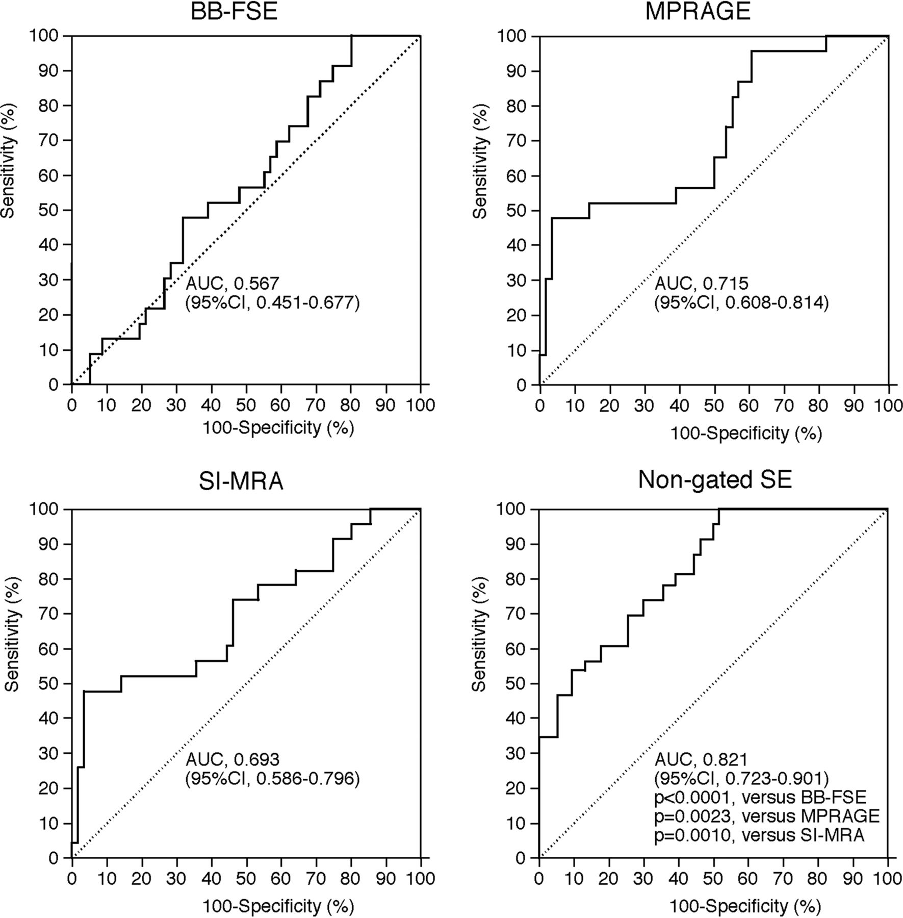
  • Fig 2.
    • Download figure
    • Open in new tab
    • Download powerpoint
    Fig 2.

    ROC curves used to compare accuracy among contrast ratios in each MR image for predicting the development of MES during exposure of the carotid arteries. Pair-wise comparison analysis shows a significantly greater area under the ROC curve for nongated SE compared with AUCs for BB-FSE, MPRAGE, or SI-MRA.

  • Fig 3.
    • Download figure
    • Open in new tab
    • Download powerpoint
    Fig 3.

    Comparisons of contrast ratios in each MR image and the number of MES in patients with MES during exposure of the carotid arteries. In negative binominal regression analysis, CR was associated with the number of MES only in nongated SE. The dashed horizontal line denotes 6 MES as the optimal cutoff point for predicting the development of new postoperative ischemic events.34

  • Fig 4.
    • Download figure
    • Open in new tab
    • Download powerpoint
    Fig 4.

    The relationship between the percentage area of each component in quantitative color-coded MR plaque imaging and the development of MES during exposure of the carotid arteries. The dashed horizontal lines denote the cutoff points lying closest to the left upper corners of the ROC curves in predicting the development of MES during exposure of the carotid arteries.

  • Fig 5.
    • Download figure
    • Open in new tab
    • Download powerpoint
    Fig 5.

    ROC curves used to compare accuracy among percentage areas of each component in quantitative color-coded MR plaque imaging for predicting the development of MES during exposure of the carotid arteries. Pair-wise comparison analysis shows significantly greater AUCs for hemorrhage or fibrous tissue than for lipid/necrosis.

  • Fig 6.
    • Download figure
    • Open in new tab
    • Download powerpoint
    Fig 6.

    Four kinds of MR plaque images (upper) and quantitative color-coded MR plaque image (right lower) in the symptomatically stenosed (95%) right internal carotid artery of a 72-year-old man showing MES during exposure of the carotid arteries in endarterectomy. A larger FOV image (left lower) indicates anatomic relationships among each vessel (arrow, internal carotid artery; yellow circle, lumen of the internal carotid artery filled with plaque; single arrowhead, internal jugular vein; double arrowhead, external carotid artery; and white square, FOV identical to that of other images). Signal intensities of the plaque in the internal carotid artery (arrows) relative to those of the sternocleidomastoid muscle (asterisks) are, in ascending order, BB-FES, MPRAGE, SI-MRA, and nongated SE. The plaque comprises mainly red and partially yellow areas on the color-coded MR plaque image. On the basis of contrast ratios, hemorrhage, lipid/necrosis, and fibrous tissue are displayed as red, yellow, and green, respectively.

Tables

  • Figures
  • Risk factors related to the development of MES during exposure of the carotid arteries

    FactorsDevelopment of MESP Value
    Yes (n = 23)No (n = 57)
    Age (yr) (mean)69.8 ± 5.669.2 ± 7.3.5507
    Male sex22 (95.7%)55 (96.5%)>.9999
    Hypertension18 (78.3%)46 (80.7%)>.9999
    Diabetes mellitus6 (26.1%)20 (35.1%).5989
    Dyslipidemia7 (30.4%)14 (24.6%).5867
    Symptomatic lesions19 (82.6%)31 (54.4%).0223
    Degree of ICA stenosis (%) (mean)86.8 ± 9.388.8 ± 8.1.4913
    Length of stenotic lesion (mm) (mean)52.2 ± 11.354.3 ± 11.9.4003
    Height of distal end of stenotic lesion relative to cervical vertebra (mean)2.8 ± 0.92.9 ± 0.8.6046
    Tortuosity of stenotic lesion (mean)107.0° ± 23.3°111.1° ± 24.0°.4755
    Ulceration of stenotic lesion12 (52.2%)15 (26.3%).0372
PreviousNext
Back to top

In this issue

American Journal of Neuroradiology: 37 (6)
American Journal of Neuroradiology
Vol. 37, Issue 6
1 Jun 2016
  • Table of Contents
  • Index by author
  • Complete Issue (PDF)
Advertisement
Print
Download PDF
Email Article

Thank you for your interest in spreading the word on American Journal of Neuroradiology.

NOTE: We only request your email address so that the person you are recommending the page to knows that you wanted them to see it, and that it is not junk mail. We do not capture any email address.

Enter multiple addresses on separate lines or separate them with commas.
Optimal MR Plaque Imaging for Cervical Carotid Artery Stenosis in Predicting the Development of Microembolic Signals during Exposure of Carotid Arteries in Endarterectomy: Comparison of 4 T1-Weighted Imaging Techniques
(Your Name) has sent you a message from American Journal of Neuroradiology
(Your Name) thought you would like to see the American Journal of Neuroradiology web site.
CAPTCHA
This question is for testing whether or not you are a human visitor and to prevent automated spam submissions.
Cite this article
Y. Sato, K. Ogasawara, S. Narumi, M. Sasaki, A. Saito, E. Tsushima, T. Namba, M. Kobayashi, K. Yoshida, Y. Terayama, A. Ogawa
Optimal MR Plaque Imaging for Cervical Carotid Artery Stenosis in Predicting the Development of Microembolic Signals during Exposure of Carotid Arteries in Endarterectomy: Comparison of 4 T1-Weighted Imaging Techniques
American Journal of Neuroradiology Jun 2016, 37 (6) 1146-1154; DOI: 10.3174/ajnr.A4674

Citation Manager Formats

  • BibTeX
  • Bookends
  • EasyBib
  • EndNote (tagged)
  • EndNote 8 (xml)
  • Medlars
  • Mendeley
  • Papers
  • RefWorks Tagged
  • Ref Manager
  • RIS
  • Zotero
0 Responses
Respond to this article
Share
Bookmark this article
Optimal MR Plaque Imaging for Cervical Carotid Artery Stenosis in Predicting the Development of Microembolic Signals during Exposure of Carotid Arteries in Endarterectomy: Comparison of 4 T1-Weighted Imaging Techniques
Y. Sato, K. Ogasawara, S. Narumi, M. Sasaki, A. Saito, E. Tsushima, T. Namba, M. Kobayashi, K. Yoshida, Y. Terayama, A. Ogawa
American Journal of Neuroradiology Jun 2016, 37 (6) 1146-1154; DOI: 10.3174/ajnr.A4674
del.icio.us logo Twitter logo Facebook logo Mendeley logo
  • Tweet Widget
  • Facebook Like
  • Google Plus One
Purchase

Jump to section

  • Article
    • Abstract
    • ABBREVIATIONS:
    • Materials and Methods
    • Results
    • Discussion
    • Conclusions
    • Footnotes
    • References
  • Figures & Data
  • Info & Metrics
  • Responses
  • References
  • PDF

Related Articles

  • No related articles found.
  • PubMed
  • Google Scholar

Cited By...

  • Diagnostic Accuracy of Preoperative Quantitative Susceptibility Mapping for Detecting Histologic Intraplaque Hemorrhage in Cervical ICA Stenosis in Patients Undergoing Carotid Endarterectomy
  • Crossref (17)
  • Google Scholar

This article has been cited by the following articles in journals that are participating in Crossref Cited-by Linking.

  • Characteristics of Wall Shear Stress and Pressure of Intracranial Atherosclerosis Analyzed by a Computational Fluid Dynamics Model: A Pilot Study
    Zimo Chen, Haiqiang Qin, Jia Liu, Bokai Wu, Zaiheng Cheng, Yong Jiang, Liping Liu, Lina Jing, Xinyi Leng, Jing Jing, Yilong Wang, Yongjun Wang
    Frontiers in Neurology 2020 10
  • Efficacy of Carotid Endarterectomy for Mild (<50%) Symptomatic Carotid Stenosis with Unstable Plaque
    Daina Kashiwazaki, Keitaro Shiraishi, Shusuke Yamamoto, Tetsuhiro Kamo, Haruto Uchino, Hisayasu Saito, Naoki Akioka, Naoya Kuwayama, Kyo Noguchi, Satoshi Kuroda
    World Neurosurgery 2019 121
  • Preoperative cervical carotid artery contrast-enhanced ultrasound findings are associated with development of microembolic signals on transcranial Doppler during carotid exposure in endarterectomy
    Kohki Oikawa, Tadayoshi Kato, Kazumasa Oura, Shinsuke Narumi, Makoto Sasaki, Shunrou Fujiwara, Masakazu Kobayashi, Yoshiyasu Matsumoto, Jun-ichi Nomura, Kenji Yoshida, Yasuo Terayama, Kuniaki Ogasawara
    Atherosclerosis 2017 260
  • Near-infrared spectroscopy carotid plaque characteristics and cerebral embolism in carotid artery stenting
    Ichiro Nakagawa, Masashi Kotsugi, Hun Soo Park, Takanori Furuta, Fumiya Sato, Kaoru Myochin, Fumihiko Nishimura, Syuichi Yamada, Yasushi Motoyama, Hiroyuki Nakase
    EuroIntervention 2021 17 7
  • Wall Shear Stress and T1 Contrast Ratio Are Associated With Embolic Signals During Carotid Exposure in Endarterectomy
    Sotaro Oshida, Futoshi Mori, Makoto Sasaki, Yuiko Sato, Masakazu Kobayshi, Kenji Yoshida, Shunrou Fujiwara, Kuniaki Ogasawara
    Stroke 2018 49 9
  • Comparison of Three-Dimensional T1-Weighted Magnetic Resonance and Contrast-Enhanced Ultrasound Plaque Images for Severe Stenosis of the Cervical Carotid Artery
    Yasuyoshi Shimada, Kohki Oikawa, Shunrou Fujiwara, Yasushi Ogasawara, Yuiko Sato, Shinsuke Narumi, Tadayoshi Kato, Kazumasa Oura, Yasuo Terayama, Makoto Sasaki, Kentaro Fujimoto, Jun Yoshida, Kuniaki Ogasawara
    Journal of Stroke and Cerebrovascular Diseases 2017 26 9
  • Hemorrhagic Plaques in Mild Carotid Stenosis: The Risk of Stroke
    Max Scheffler, Alain Pellaton, José Boto, Isabelle Barnaure, Bénédicte M. Delattre, Jorge Remuinan, Roman Sztajzel, Karl-Olof Lövblad, Maria Isabel Vargas
    Canadian Journal of Neurological Sciences / Journal Canadien des Sciences Neurologiques 2021 48 2
  • Quantitative T1 and T2* carotid atherosclerotic plaque imaging using a three-dimensional multi-echo phase-sensitive inversion recovery sequence: a feasibility study
    Yasuhiro Fujiwara, Hirotoshi Maruyama, Kanako Toyomaru, Yuri Nishizaka, Masahiro Fukamatsu
    Radiological Physics and Technology 2018 11 2
  • Usefulness of rapid MR angiography using two-point Dixon for evaluating carotid and aortic plaques
    Keisuke Morihara, Tatsu Nakano, Kentaro Mori, Issei Fukui, Motohiro Nomura, Keiichiro Suzuki, Kenichi Hirano, Mitsuyuki Takahashi, Hideyuki Takeuchi, Hiroshi Doi, Yoshihisa Kitamura, Fumiaki Tanaka
    Neuroradiology 2022 64 4
  • Preoperative 3D FSE T1-Weighted MR Plaque Imaging for Severely Stenotic Cervical ICA: Accuracy of Predicting Emboli during Carotid Endarterectomy
    Yasushi Ogasawara, Yuiko Sato, Shinsuke Narumi, Makoto Sasaki, Shunrou Fujiwara, Masakazu Kobayashi, Kenji Yoshida, Yasuo Terayama, Kuniaki Ogasawara
    International Journal of Molecular Sciences 2016 17 11

More in this TOC Section

  • Proximal Vertebral Artery Variants and Embryology
  • High-Risk Plaque Features in Carotid MRI
  • Nonstenotic Carotid Plaques and Stroke Review
Show more EXTRACRANIAL VASCULAR

Similar Articles

Advertisement

Indexed Content

  • Current Issue
  • Accepted Manuscripts
  • Article Preview
  • Past Issues
  • Editorials
  • Editor's Choice
  • Fellows' Journal Club
  • Letters to the Editor
  • Video Articles

Cases

  • Case Collection
  • Archive - Case of the Week
  • Archive - Case of the Month
  • Archive - Classic Case

More from AJNR

  • Trainee Corner
  • Imaging Protocols
  • MRI Safety Corner
  • Book Reviews

Multimedia

  • AJNR Podcasts
  • AJNR Scantastics

Resources

  • Turnaround Time
  • Submit a Manuscript
  • Submit a Video Article
  • Submit an eLetter to the Editor/Response
  • Manuscript Submission Guidelines
  • Statistical Tips
  • Fast Publishing of Accepted Manuscripts
  • Graphical Abstract Preparation
  • Imaging Protocol Submission
  • Evidence-Based Medicine Level Guide
  • Publishing Checklists
  • Author Policies
  • Become a Reviewer/Academy of Reviewers
  • News and Updates

About Us

  • About AJNR
  • Editorial Board
  • Editorial Board Alumni
  • Alerts
  • Permissions
  • Not an AJNR Subscriber? Join Now
  • Advertise with Us
  • Librarian Resources
  • Feedback
  • Terms and Conditions
  • AJNR Editorial Board Alumni

American Society of Neuroradiology

  • Not an ASNR Member? Join Now

© 2025 by the American Society of Neuroradiology All rights, including for text and data mining, AI training, and similar technologies, are reserved.
Print ISSN: 0195-6108 Online ISSN: 1936-959X

Powered by HighWire