
Article Figures & Data
Figures
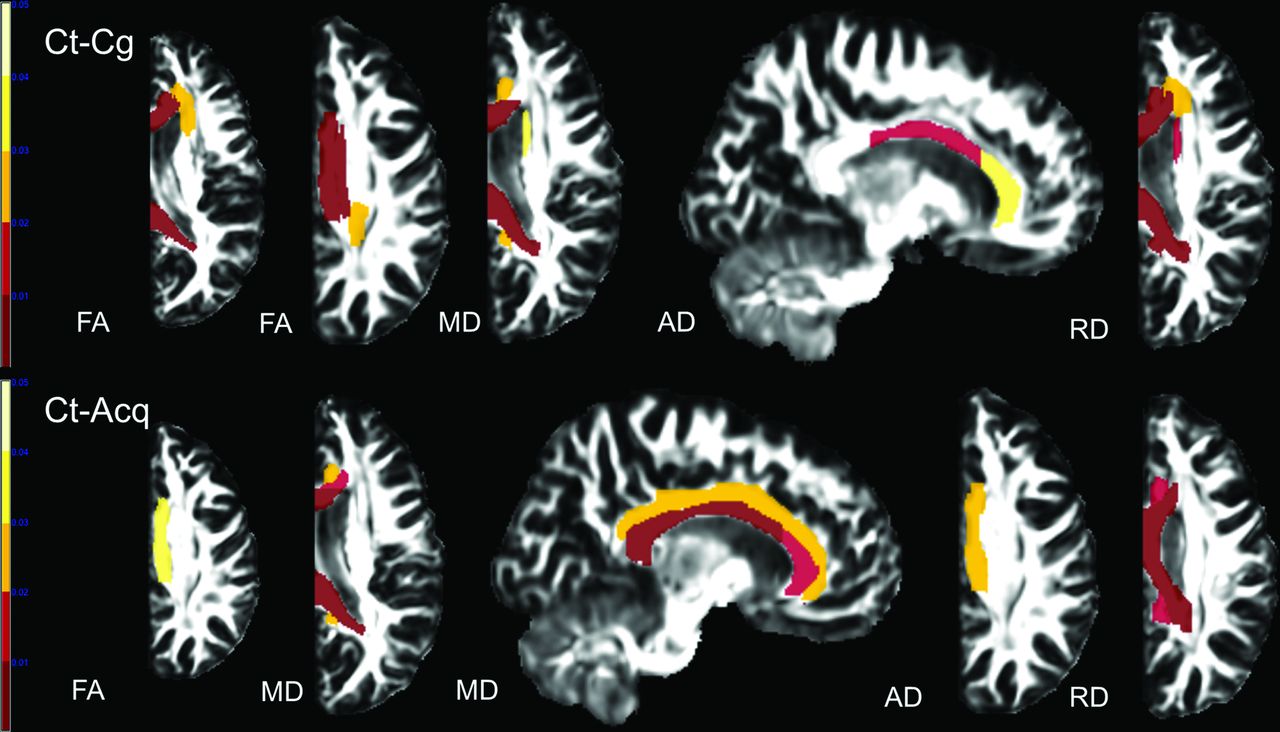
- Fig 1.
Results of ABA. Compared with controls (Ct), patients with anatomic hemispherectomy because of a congenital (Cg) and acquired (Acq) underlying etiology showed a decrease in FA and AD and an increase in MD and RD in multiple WM tracts. Only results that survived the Tukey significant difference test are depicted with P < .05. Color bars represent P = .001–.05 with a color gradient from red to light yellow. Ct-Cg indicates differences between controls and patients with congenital etiology leading to hemispherectomy; Ct-Acq indicates differences between controls and patients with acquired etiology leading to hemispherectomy.
- Fig 2.
Results of the VBA. Compared with controls, patients showed a decrease in FA values and an increase in MD and RD values in all examined WM tracts. Only results that survived false discovery rate correction for multiple comparisons are depicted, with P < .05. Color bars represent the t statistics. PT indicates patients; CT indicates controls.
- Fig 3.
Results of atlas-based analysis of GM structures. Compared with controls, patients in both etiology groups showed decreased FA in all the examined cortical areas with the exception of the putamen, which showed increased FA in the congenital group. Only results that survived the Tukey significant difference test are depicted, with P < .05. The Color bar represents the P value: blue–light blue represents PT < CT, and red–light yellow represents CT < PT. PT indicates patients including Cg (congenital) and Acq (acquired) pathologies; CT, controls..


Tables
Patients Sex Age at Study (yr) Age at Operation (yr) Time since Operation (yr) Diagnosis Etiology Group 1 M 8.1 5.9 2.2 HME left Congenital 2 M 3.3 1.1 2.2 HME left Congenital 3 M 14.8 3.7 11.1 Cortical dysplasia righta Congenital 4 M 2.2 0.9 1.3 HME left Congenital 5 F 2.7 2.7 0.02 Cortical dysplasia righta Congenital 6 M 12.9 5.7 7.2 Cortical dysplasia lefta Congenital 7 F 11.5 6.9 4.6 Cortical dysplasia righta Congenital 8 F 0.9 0.8 0.1 HME left Congenital 9 M 14.0 2.3 11.7 Prenatal stroke right Congenital 10 F 8.9 4.1 4.8 Prenatal stroke left Congenital 11 F 20.5 2.5 18.0 Prenatal stroke right Congenital 12 F 8.7 2.2 6.5 Rasmussen right Acquired 13 F 3.9 3.1 0.8 Rasmussen left Acquired 14 F 18.1 9.8 8.3 Rasmussen right Acquired 15 F 15.2 3.8 11.4 Rasmussen right Acquired 16 F 12.2 4.1 8.1 Rasmussen right Acquired 17 F 25.0 12.9 12.1 Rasmussen right Acquired 18 F 20.7 4.4 16.3 Postnatal stroke right Acquired 19 F 14.5 5.7 8.8 Postnatal stroke right Acquired Note:—HME indicates hemimegalencephaly.
↵a Includes an extensive unilateral malformation of cortical development affecting >1 cerebral lobe requiring full anatomic hemispherectomy rather than a more conservative neurosurgical approach.
- Table 2:
Significant correlations between DTI scalars in WM structures and clinical parameters in patients after hemispherectomy classified on the basis of the underlying etiologya
Clinical Parameter/Etiology Group DTI Scalars WM Tracts Pearson Correlation Coefficient P Value R2 Adjusted R2 Age at the operation Congenital FA CGC 0.840 .001 0.71 0.67 Acquired FA PLIC −0.737 .037 0.54 0.47 RD SCR 0.919 .001 0.84 0.82 MD PCR 0.884 .004 0.78 0.75 RD PCR 0.744 .034 0.55 0.48 Time since the operation Congenital FA CGC −0.632 .037 0.40 0.33 Acquired FA PLIC 0.805 .016 0.65 0.59 RD PLIC −0.792 .019 0.63 0.56 RD ACR −0.802 .017 0.64 0.58 RD SCR −0.906 .002 0.82 0.79 MD PCR −0.788 .020 0.62 0.56 MD CGC −0.728 .041 0.53 0.45 FA GCC 0.722 .043 0.52 0.44 Note:—ACR indicates anterior corona radiata; GCC, genu of the corpus callosum; PCR, posterior corona radiata; SCR, superior corona radiata; PLIC, posterior limb of internal capsule.
↵a Pearson correlation coefficient, r2, adjusted r2, and P value from multivariate linear regression analysis are shown.


Jump to section
Related Articles
Cited By...
- No citing articles found.