
Article Figures & Data
Figures
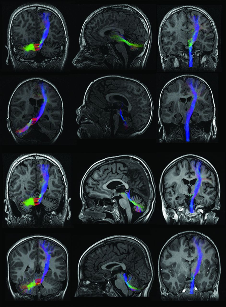
- Fig 1.
Reconstructed bundles for corticopontocerebellar (left), cerebellar-thalamic (middle), and corticospinal (right) tracts in subjects with (from top to bottom) CH, PCH, CA, and controls. Bundles are overlaid on T1-weighted images. Figures are representative of the global shape of the reconstructed bundles, irrespective of the cropping of the anatomic section.
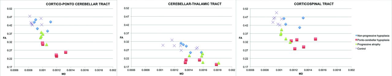
- Fig 2.
Plots of fractional anisotropy and mean diffusivity within the corticopontocerebellar, cerebellar-thalamic, and corticospinal tracts for CH, PCH, CA, and controls.


Tables
Tract/Group FA Mean (SD) MD Mean (SD) (10−3 mm2/s) AD Mean (SD) (10−3 mm2/s) RD Mean (SD) (10−3 mm2/s) CPCT CH 0.42 (0.03) 0.98 (0.11) 1.42 (0.12) 0.76 (0.09) PCH 0.27 (0.04) 1.14 (0.14) 1.41 (0.08) 0.99 (0.14) CA 0.35 (0.03) 0.99 (0.03) 1.36 (0.03) 0.81 (0.04) Control 0.44 (0.02) 0.88 (0.07) 1.37 (0.09) 0.66 (0.06) CTT CH 0.28 (0.03) 1.39 (0.11) 1.79 (0.09) 1.26 (0.17) PCH 0.19 (0.03) 1.49 (0.18) 1.79 (0.23) 1.39 (0.14) CA 0.22 (0.02) 1.59 (0.14) 1.68 (0.21) 1.41 (0.15) Control 0.29 (0.02) 1.29 (0.17) 1.81 (0.19) 1.27 (0.21) CST CH 0.44 (0.02) 1.07 (0.29) 1.69 (0.16) 0.91 (0.11) PCH 0.31 (0.03) 1.29 (0.13) 1.71 (0.19) 1.08 (0.11) CA 0.36 (0.03) 1.06 (0.06) 1.46 (0.05) 0.86 (0.06) Control 0.45 (0.02) 0.97 (0.04) 1.43 (0.06) 0.73 (0.04) PCH CA Control CPCT CH FA (pB < .001)a (pB = .242) (pB = .714) MD (pB = 1.000) (pB = 1.000) (pB = .278) AD (pB = 1.000) (pB = 1.000) (pB = .297) RD (pB = .212) (pB = 1.000) (pB = .277) PCH FA (pB < 0.001)a (pB < .001)a MD (pB = 0.149) (pB = .005)a AD (pB = 1.000) (pB = .952) RD (pB = .045)a (pB < .001)a CA FA (pB = .002)a MD (pB = .821) AD (pB = 1.000) RD (pB = .289) CTT CH FA (pB = .001)a (pB = .019)a (pB = 1.000) MD (pB = 1.000) (pB = 1.000) (pB = 1.000) AD (pB = 1.000) (pB = 1.000) (pB = 1.000) RD (pB = 1.000) (pB = 1.000) (pB = .715) PCH FA (pB = 293) (pB < .001)a MD (pB = 1.000) (pB = .504) AD (pB = .914) (pB = 1.000) RD (pB = 1.000) (pB = .041)a CA FA (pB = .001)a MD (pB = .071) AD (pB = .315) RD (pB = .028)a CST CH FA (pB < .001)a (pB = .082) (pB = .492) MD (pB = .621) (pB = 1.000) (pB = 1.000) AD (pB = 1.000) (pB = .289) (pB = .143) RD (pB = .265) (pB = 1.000) (pB = .018) PCH FA (pB = .041)a (pB < .001)a MD (pB = .221) (pB = .005)a AD (pB = .040)a (pB = .157) RD (pB = .045)a (pB < .001)a CA FA (pB < .001)a MD (pB = 1.000) AD (pB = 1.000) RD (pB = .215) Note:—pB indicates Bonferroni-corrected P value.
↵a Significant.


Jump to section
Related Articles
Cited By...
- Structural and connectivity parameters reveal compensation patterns in young patients with non-progressive and slow-progressive cerebellar ataxia
- Biometry of the Cerebellar Vermis and Brain Stem in Children: MR Imaging Reference Data from Measurements in 718 Children
- Structural Connectivity Analysis in Children with Segmental Callosal Agenesis