
Article Figures & Data
Figures
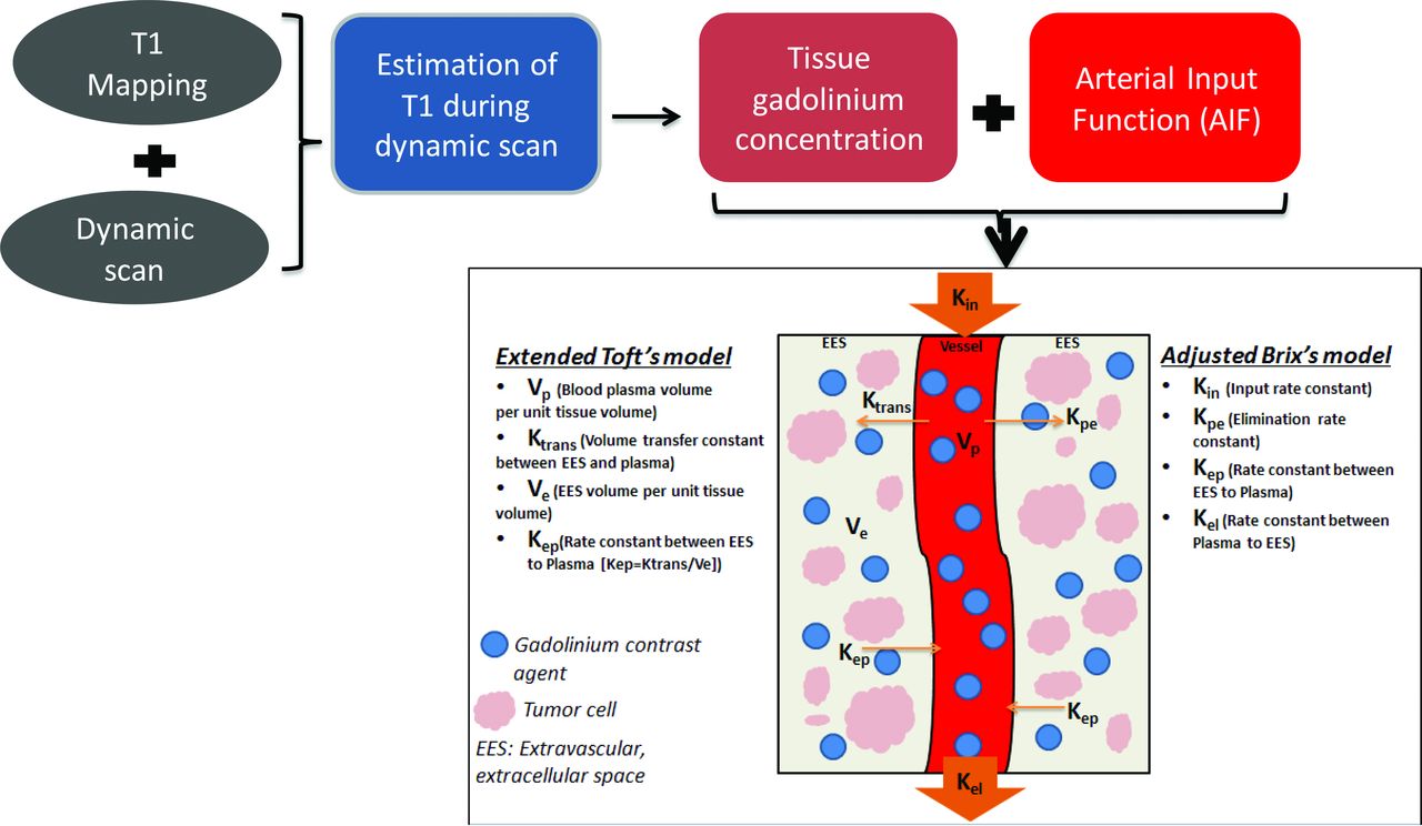
- Fig 1.
Flow chart demonstrating a typical quantitative method of data analysis. The information from the T1 mapping and dynamic data is used to estimate the changes in T1 relaxivity during the dynamic scan, which, in turn, provides the information of tissue gadolinium concentration. By fitting the tissue gadolinium concentration and arterial input function data in to commonly used “2-compartment” models (extended Toft or adjusted Brix model), various parameters can be assessed. The difference between the Toft and extended Toft model is the inclusion of assessment of blood plasma volume per unit tissue volume in the later version.
- Fig 2.
A 57-year-old male patient with T2N3bM0 undifferentiated nasopharyngeal cancer. Pretreatment gadolinium-enhanced axial T1-weighted MR imaging of the neck demonstrates metastatic right level IIb lymph nodes (A). Parametric maps (C, D, and E) show higher volume transfer constant (Ktrans = 0.26/min), Kep, and area under curve, respectively. Axial contrast-enhanced neck CT at 6 months post-chemoradiation treatment demonstrates a favorable response to treatment (B).
- Fig 3.
A 52-year-old male patient with squamous cell carcinoma of the right palatine tonsil. Pretreatment axial contrast-enhanced neck CT demonstrates metastatic right level II lymph nodes (A). Parametric maps (C, D, and E) show a lower volume transfer constant (Ktrans = 0.06/min), Kep, and area under curve, respectively. Gadolinium-enhanced axial T1-weighted MR imaging of the neck at 12 months post-chemoradiation treatment demonstrates an unfavorable response to treatment (B).


Tables
Parameters Philipsa Siemensb GEc Coil 16-Channel neurovascular coil Parallel imaging SENSE iPAT ASSET Sequence 3D-T1WI FFE 3D-T1WI FISP or 3D-T1WI FLASH 3D-T1WI FSPGR TR/TE for T1 mapping 5.2/2.5 ms MFA for T1 mapping 30°, 20°, 15°, 10°, and 2° TR/TE/FA for dynamic imaging 5.2/2.5 ms/5° FOV 212 × 149 mm2 Voxel 0.95/0.95/3.00 mm3 Section thickness 3 mm Signal averaging NSA: 1 ACQ: 1 NEX: 1 Number of sections per dynamic scan/section orientation 20/Axial Temporal resolution 3.6 seconds Total T1 mapping acquisition time 26.5 seconds Total dynamic acquisition time 6.10 minutes Fat saturation No Contrast injection Single dose of 20-mL gadoteridol (ProHanced) injected at a rate of 5 mL/s through a peripheral arm vein, followed by a 20-mL saline flush with a power injector Note:—FFE indicates fast-field echo; FSPGR, fast-spoiled gradient recalled; MFA, multiple flip angles; SENSE, sensitivity encoding; iPAT, integrated parallel acquisition technique; ASSET, array spatial sensitivity encoding technique; NSA, number of signal averages; ACQ, acquisitions; FA, flip angle.
↵a Phillips Healthcare, Best, the Netherlands.
↵b Siemens, Erlangen, Germany.
↵c GE Healthcare, Milwaukee, Wisconsin.
↵d Bracco Diagnostics, Princeton, New Jersey.
Parameter Definition Units Area under curve Area under the signal intensity or gadolinium dynamic curve a.u.min or mmol.min/L Relative signal intensity St/S0 NA Initial slope or enhancement slope/rate Maximum or average slope in the initial enhancement a.u/min Washout slope/rate Maximum or average slope in the washout phase a.u/min Peak enhancement ratio or maximum signal enhancement ratio (Smax − S0)/S0 NA Tmax or time to peak Time from contrast arrival to peak S Maximum intensity–time ratio PER/Tmax S−1 Note:—St indicates MR signal intensity at time t; S0, precontrast signal intensity; Smax, maximum signal intensity; a.u, arbitrary unit; min, minute; PER, peak enhancement ratio; Tmax, time to maximum enhancement; NA, not applicable; S, seconds.
Parameter Definition Units Ktrans Volume transfer constant between EES and blood plasma Min−1 Ve EES volume per unit tissue volume NA Vp Blood plasma volume per unit tissue volume NA Kep or K21 Rate constant from EES to blood plasma Min−1 Kpe or K12 Rate constant from blood plasma to EES Min−1 Kel Elimination rate constant Min−1 Amp or AH Amplitude of the normalized dynamic curve NA Note:—Amp or AH, amplitude of the normalized dynamic curve; NA, not applicable; Min, minute.


Jump to section
Related Articles
Cited By...
- Diagnostic Performance of Dynamic Contrast-Enhanced 3T MR Imaging for Characterization of Orbital Lesions: Validation in a Large Prospective Study
- Differentiation of Skull Base Chondrosarcomas, Chordomas, and Metastases: Utility of DWI and Dynamic Contrast-Enhanced Perfusion MR Imaging
- Prediction of Wound Failure in Patients with Head and Neck Cancer Treated with Free Flap Reconstruction: Utility of CT Perfusion and MR Perfusion in the Early Postoperative Period
- Diagnostic Role of Diffusion-Weighted and Dynamic Contrast-Enhanced Perfusion MR Imaging in Paragangliomas and Schwannomas in the Head and Neck
- Development of a High-Performance Multiparametric MRI Oropharyngeal Primary Tumor Auto-Segmentation Deep Learning Model and Investigation of Input Channel Effects: Results from a Prospective Imaging Registry
- Dynamic Contrast-Enhanced MRI to Differentiate Parotid Neoplasms Using Golden-Angle Radial Sparse Parallel Imaging
- Effect of Tumor Microenvironment on Selective Uptake of Boric Acid in HepG2 Human Hepatoma Cells