
Article Figures & Data
Figures
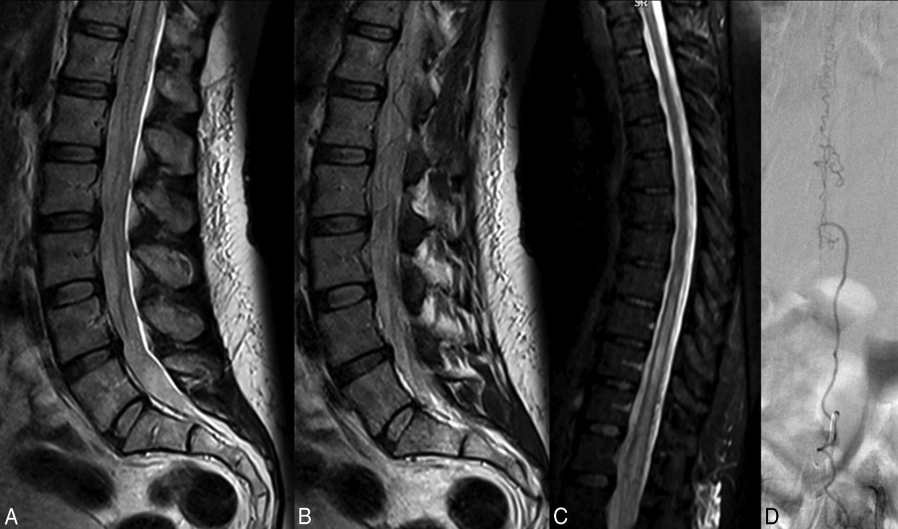
- Fig 1.
A 57-year-old woman with a 3-month history of bilateral lower extremity tingling and progressive lower extremity weakness. A and B, T2-weighted lumbar spine MR images demonstrate high T2 signal in the conus with multiple flow voids in the intradural space. C, T2-weighted MR image of the thoracic spine demonstrates high T2 signal in the lower thoracic cord to the conus. The patient was diagnosed with neuromyelitis optica and received no spinal-vasculature imaging before referral to our institution. Two rounds of IV methylprednisolone (Solu-Medrol) therapy resulted in worsening of symptoms, and rituximab therapy was of no benefit. D, Spinal angiography demonstrates the spinal dural AVF with an arterial feeder from the L3 radiculomeningeal artery.
- Fig 2.
A 68-year-old man with a 3-month history of saddle anesthesia, constipation, difficulty voiding, and numbness in the lower extremities. T2-weighted images of the lumbar and thoracic spine demonstrate high T2 signal in the lower thoracic cord and conus (A and B). Due to clinical suspicion of SDAVF, an angiogram was obtained before referral to our center. C, The angiogram clearly demonstrates the fistula arising from the L2 radiculomeningeal artery; however, it was interpreted as a negative finding. Before the diagnosis was made, the patient underwent an extensive imaging and clinical evaluation, including a panel negative for paraneoplastic syndrome, PET/CT, and lumbar puncture. Two rounds of IV Solu-Medrol therapy resulted in worsening of symptoms. The patient also underwent a T10–T11 laminectomy and 2 spinal cord biopsies. D, Repeat spinal angiography re-demonstrates the fistula.


Tables
No. (%) No. 53 Mean (SD) age 65.0 (10.8) No. (%) male 43 (81.1) Mean delay in diagnosis (mo) 9.2 ± 11.1 Symptoms at presentation Bilateral motor symptoms 48 (90.6) Sensory symptoms 20 (37.7) Bowel or bladder symptoms 13 (24.5) Focal unilateral motor deficit 3 (5.7) Initial working diagnosis Spinal stenosis 13 (24.5) Myelopathy NOS 10 (18.9) Transverse myelitis 9 (17.0) Ischemic myelopathy 4 (7.6) Peripheral neuropathy 3 (5.7) Myopathy 2 (3.8) NMO 2 (3.8) CIDP 2 (3.8) Other 8 (15.1) Additional interventions Systemic steroids 18 (34.0) IVIG 5 (9.4) Surgery 6 (11.3) Biopsy 2 (3.8) Plasma exchange 4 (7.6) Rituximab 2 (3.8) No. of additional spine MRIs or CTs until diagnosis 1 10 (18.9) 2 8 (15.1) 3 12 (22.6) 4 12 (22.6) ≥5 11 (20.8) Note:—NOS indicates not otherwise specified; NMO, neuromyelitis optica; CIDP, chronic inflammatory demyelinating polyneuropathy.
Imaging Findings No. (%) High T2 cord signal (including conus) 46 (95.8) Increased conus signal 44 (91.7) Prominent intradural vessel on CT myelography or MRI 51 (96.2) Cord enhancement 38 (79.2) High T2 signal and flow void 44 (91.7) Presenting (No.) (%) At Diagnosis (No.) (%) 90 Days after Treatment (No.) (%) mRS 0 0 (0.0) 0 (0.0) 2 (3.9) 1 42 (79.2) 2 (3.8) 8 (15.7) 2 8 (15.1) 12 (22.6) 12 (23.5) 3 3 (5.7) 16 (30.2) 16 (31.4) 4 0 (0.0) 21 (39.6) 10 (19.6) 5 0 (0.0) 2 (3.8) 2 (3.9) 6 0 (0.0) 0 (0.0) 1 (2.0) Aminoff motor score 0 (No deficit) 1 (1.9) 1 (1.9) 2 (4.0) 1 (Hyposthenia) 34 (64.2) 9 (17.0) 6 (12.0) 2 (Reduced tolerance) 6 (11.3) 11 (20.8) 5 (10.0) 3 (Need for cane) 12 (22.6) 15 (28.3) 14 (28.0) 4 (Need for crutches or walker) 0 (0) 15 (28.3) 11 (22.0) 5 (Patient in wheelchair) 0 (0) 17 (32.1) 12 (24.0) Bowel or bladder symptoms Yes 13 (24.5) 27 (50.9) 23 (45.1) No 40 (75.5) 26 (49.1) 28 (54.9) Sensory symptoms Yes 20 (37.8) 28 (52.8) 20 (39.2) No 33 (62.2) 25 (47.2) 31 (60.8)


Jump to section
Related Articles
Cited By...
- 'Pressure cooker and 'balloon pressure techniques significantly increase 3-month complete occlusion rate after spinal arteriovenous fistula embolization as compared to glue: single center evaluation on 38 consecutive patients
- Knowledge, attitudes and practices regarding spinal vascular malformations among doctors in China: a cross-sectional study
- Spinal Dorsal Intradural Arteriovenous Fistulas: Natural History, Imaging, and Management
- Spinal Dural Arteriovenous Fistula: The Missing-Piece Sign
- Dilated Vein of the Filum Terminale on MRI: A Marker for Deep Lumbar and Sacral Dural and Epidural Arteriovenous Fistulas
- Clinical outcomes following corticosteroid administration in patients with delayed diagnosis of spinal arteriovenous fistulas
- Comparison of Time-Resolved and First-Pass Contrast-Enhanced MR Angiography in Pretherapeutic Evaluation of Spinal Dural Arteriovenous Fistulas
- First-Pass Contrast-Enhanced MR Angiography in Evaluation of Treated Spinal Arteriovenous Fistulas: Is Catheter Angiography Necessary?