Advertisement

Main menu

  • Home
  • Content
    • Current Issue
    • Accepted Manuscripts
    • Article Preview
    • Past Issue Archive
    • Video Articles
    • AJNR Case Collection
    • Case of the Week Archive
    • Case of the Month Archive
    • Classic Case Archive
  • Special Collections
    • AJNR Awards
    • Low-Field MRI
    • Alzheimer Disease
    • ASNR Foundation Special Collection
    • Photon-Counting CT
    • View All
  • Multimedia
    • AJNR Podcasts
    • AJNR SCANtastic
    • Trainee Corner
    • MRI Safety Corner
    • Imaging Protocols
  • For Authors
    • Submit a Manuscript
    • Submit a Video Article
    • Submit an eLetter to the Editor/Response
    • Manuscript Submission Guidelines
    • Statistical Tips
    • Fast Publishing of Accepted Manuscripts
    • Graphical Abstract Preparation
    • Imaging Protocol Submission
    • Author Policies
  • About Us
    • About AJNR
    • Editorial Board
    • Editorial Board Alumni
  • More
    • Become a Reviewer/Academy of Reviewers
    • Subscribers
    • Permissions
    • Alerts
    • Feedback
    • Advertisers
    • ASNR Home

User menu

  • Alerts
  • Log in

Search

  • Advanced search
American Journal of Neuroradiology

American Journal of Neuroradiology

ASHNR American Society of Functional Neuroradiology ASHNR American Society of Pediatric Neuroradiology ASSR
  • Alerts
  • Log in

Advanced Search

  • Home
  • Content
    • Current Issue
    • Accepted Manuscripts
    • Article Preview
    • Past Issue Archive
    • Video Articles
    • AJNR Case Collection
    • Case of the Week Archive
    • Case of the Month Archive
    • Classic Case Archive
  • Special Collections
    • AJNR Awards
    • Low-Field MRI
    • Alzheimer Disease
    • ASNR Foundation Special Collection
    • Photon-Counting CT
    • View All
  • Multimedia
    • AJNR Podcasts
    • AJNR SCANtastic
    • Trainee Corner
    • MRI Safety Corner
    • Imaging Protocols
  • For Authors
    • Submit a Manuscript
    • Submit a Video Article
    • Submit an eLetter to the Editor/Response
    • Manuscript Submission Guidelines
    • Statistical Tips
    • Fast Publishing of Accepted Manuscripts
    • Graphical Abstract Preparation
    • Imaging Protocol Submission
    • Author Policies
  • About Us
    • About AJNR
    • Editorial Board
    • Editorial Board Alumni
  • More
    • Become a Reviewer/Academy of Reviewers
    • Subscribers
    • Permissions
    • Alerts
    • Feedback
    • Advertisers
    • ASNR Home
  • Follow AJNR on Twitter
  • Visit AJNR on Facebook
  • Follow AJNR on Instagram
  • Join AJNR on LinkedIn
  • RSS Feeds

AJNR Awards, New Junior Editors, and more. Read the latest AJNR updates

Research ArticleNeurointervention
Open Access

Evolution of Flow-Diverter Endothelialization and Thrombus Organization in Giant Fusiform Aneurysms after Flow Diversion: A Histopathologic Study

I. Szikora, E. Turányi and M. Marosfoi
American Journal of Neuroradiology September 2015, 36 (9) 1716-1720; DOI: https://doi.org/10.3174/ajnr.A4336
I. Szikora
aFrom the National Institute of Clinical Neurosciences (I.S., M.M.), Budapest, Hungary
  • Find this author on Google Scholar
  • Find this author on PubMed
  • Search for this author on this site
E. Turányi
b1st Department of Pathology (E.T.), Semmelweis University Medical School, Budapest, Hungary.
  • Find this author on Google Scholar
  • Find this author on PubMed
  • Search for this author on this site
M. Marosfoi
aFrom the National Institute of Clinical Neurosciences (I.S., M.M.), Budapest, Hungary
  • Find this author on Google Scholar
  • Find this author on PubMed
  • Search for this author on this site
  • Article
  • Figures & Data
  • Supplemental
  • Info & Metrics
  • Responses
  • References
  • PDF
Loading

Article Figures & Data

Figures

  • Fig 1.
    • Download figure
    • Open in new tab
    • Download powerpoint
    Fig 1.

    Unruptured, nonthrombosed giant fusiform aneurysm involving the vertebrobasilar junction. A, Volume-rendering 3D reconstruction of a CTA, demonstrating fusiform aneurysm of the vertebrobasilar junction. B, Histologic section of the fusiform aneurysm showing fresh clot inside the aneurysm (star), intact elastic lamina (arrow), and thick aneurysm wall (double arrow) (H&E staining). C, Section of the aneurysm wall showing a thick layer of smooth muscle cells (arrow) (h-Caldesmon staining). D, Section of the aneurysm wall showing a thick subintimal layer of connective tissue (Picrosirius staining) (arrows) and no connective tissue invasion into the thrombus inside the aneurysm, indicating a lack of thrombus organization.
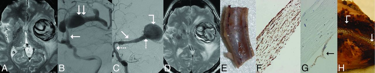
  • Fig 2.
    • Download figure
    • Open in new tab
    • Download powerpoint
    Fig 2.

    Giant, partially thrombosed, fusiform aneurysm of the left MCA treated with a construct of 2 Pipeline embolization devices; the specimen was removed 6 months after PED implantation. A, T2-weighted MR image before treatment, showing large mass of mixed signal intensity, associated with significant mass effect and white matter edema, consistent with a giant aneurysm. B, DSA of the same aneurysm. The arrow points to the proximal, normal portion of the M1 section. and the double arrow points to the fusiform aneurysm expending into the M2 sections. C, Follow-up DSA 6 months later showing “angiographic reconstruction” of the distal M1 section (arrow), significant enlargement of the dilated proximal section of the cranial M2 branch (bent arrow), and lack of filling (occlusion) of this branch distal to the dilation. The PED construct can be seen between the 2 dotted arrows. D, Follow-up T2-weighted MR image from 6 months after treatment showing unchanged mass effect, edema, and mixed signal intensity. E, Longitudinal cut of the proximal landing zone. The luminal surface of the PED is covered by a smooth tissue layer. F, Microscopic section of the layer removed from the luminal surface of the PED showing neointimal growth consisting of smooth muscle cells (h-Caldesmon staining). G, The same layer is covered by a single cell layer of endothelium (arrow) (CD34 staining). H, Macroscopic cross-section of the specimen at the level of the fusiform aneurysm. The implanted PED construct (arrow) is uncovered and surrounded by fresh clot (bent arrow).
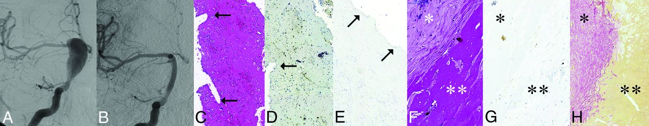
  • Fig 3.
    • Download figure
    • Open in new tab
    • Download powerpoint
    Fig 3.

    Giant, partially thrombosed fusiform aneurysm of the right ICA treated by a construct of 2 PEDs. The specimen was removed 13 months after treatment. A, DSA before treatment showing the circulating portion of the partially thrombosed GFA involving the supraclinoid ICA on the right. B, DSA 1 year after treatment showing angiographic reconstruction of the entire length of the fusiform aneurysm. C. Thin fibrin layer removed from the luminal surface of the PED construct by H&E staining. The arrows in C and D point to the impressions of the flow-diverter struts on the outer surface of the fibrin layer. D, h-Caldesmon staining fails to show smooth muscle cells inside this layer. E, CD34 fails to show endothelial coverage on the luminal surface (arrows) of the layer. F, Histologic section showing a thick aneurysm wall with low cell attenuation (star) and fresh thrombus underneath the wall (double star) by H&E staining. G, h-Caldesmon staining fails to show any smooth muscle cells within the wall (star) or invasion into the clot (double star). H, Picrosirius staining reveals subintimal connective tissue within the thick aneurysm wall (star) but no invasion into the thrombus (double star).

PreviousNext
Back to top

In this issue

American Journal of Neuroradiology: 36 (9)
American Journal of Neuroradiology
Vol. 36, Issue 9
1 Sep 2015
  • Table of Contents
  • Index by author
  • Complete Issue (PDF)
Advertisement
Print
Download PDF
Email Article

Thank you for your interest in spreading the word on American Journal of Neuroradiology.

NOTE: We only request your email address so that the person you are recommending the page to knows that you wanted them to see it, and that it is not junk mail. We do not capture any email address.

Enter multiple addresses on separate lines or separate them with commas.
Evolution of Flow-Diverter Endothelialization and Thrombus Organization in Giant Fusiform Aneurysms after Flow Diversion: A Histopathologic Study
(Your Name) has sent you a message from American Journal of Neuroradiology
(Your Name) thought you would like to see the American Journal of Neuroradiology web site.
CAPTCHA
This question is for testing whether or not you are a human visitor and to prevent automated spam submissions.
Cite this article
I. Szikora, E. Turányi, M. Marosfoi
Evolution of Flow-Diverter Endothelialization and Thrombus Organization in Giant Fusiform Aneurysms after Flow Diversion: A Histopathologic Study
American Journal of Neuroradiology Sep 2015, 36 (9) 1716-1720; DOI: 10.3174/ajnr.A4336

Citation Manager Formats

  • BibTeX
  • Bookends
  • EasyBib
  • EndNote (tagged)
  • EndNote 8 (xml)
  • Medlars
  • Mendeley
  • Papers
  • RefWorks Tagged
  • Ref Manager
  • RIS
  • Zotero
0 Responses
Respond to this article
Share
Bookmark this article
Evolution of Flow-Diverter Endothelialization and Thrombus Organization in Giant Fusiform Aneurysms after Flow Diversion: A Histopathologic Study
I. Szikora, E. Turányi, M. Marosfoi
American Journal of Neuroradiology Sep 2015, 36 (9) 1716-1720; DOI: 10.3174/ajnr.A4336
del.icio.us logo Twitter logo Facebook logo Mendeley logo
  • Tweet Widget
  • Facebook Like
  • Google Plus One
Purchase

Jump to section

  • Article
    • Abstract
    • ABBREVIATIONS:
    • Materials and Methods
    • Results
    • Discussion
    • Conclusions
    • Acknowledgments
    • Footnotes
    • References
  • Figures & Data
  • Supplemental
  • Info & Metrics
  • Responses
  • References
  • PDF

Related Articles

  • PubMed
  • Google Scholar

Cited By...

  • Long-term outcomes and dynamic changes of in-stent stenosis after Pipeline embolization device treatment of intracranial aneurysms
  • Treatment of fusiform aneurysms with a pipeline embolization device: a multicenter cohort study
  • Learning Curve for Flow Diversion of Posterior Circulation Aneurysms: A Long-Term International Multicenter Cohort Study
  • Flow Diversion for ICA Aneurysms with Compressive Neuro-Ophthalmologic Symptoms: Predictors of Morbidity, Mortality, and Incomplete Aneurysm Occlusion
  • Neck Location on the Outer Convexity is a Predictor of Incomplete Occlusion in Treatment with the Pipeline Embolization Device: Clinical and Angiographic Outcomes
  • Implications of the Collar Sign in Incompletely Occluded Aneurysms after Pipeline Embolization Device Implantation: A Follow-Up Study
  • Endothelialization following Flow Diversion for Intracranial Aneurysms: A Systematic Review
  • Risk of Branch Occlusion and Ischemic Complications with the Pipeline Embolization Device in the Treatment of Posterior Circulation Aneurysms
  • Endovascular Treatment of Very Large and Giant Intracranial Aneurysms: Comparison between Reconstructive and Deconstructive Techniques--A Meta-Analysis
  • Predictors of Incomplete Occlusion following Pipeline Embolization of Intracranial Aneurysms: Is It Less Effective in Older Patients?
  • In situ tissue engineering: endothelial growth patterns as a function of flow diverter design
  • Treatment of posterior circulation non-saccular aneurysms with flow diverters: a single-center experience and review of 56 patients
  • Lack of Association between Statin Use and Angiographic and Clinical Outcomes after Pipeline Embolization for Intracranial Aneurysms
  • Republished: Pipeline embolization device induced collateral channels in elective flow diversion treatment
  • Republished: Pipeline embolization device thrombosis induced peri-construct collateral channels
  • Risk Factors for Ischemic Complications following Pipeline Embolization Device Treatment of Intracranial Aneurysms: Results from the IntrePED Study
  • Pipeline embolization device induced collateral channels in elective flow diversion treatment
  • Pipeline embolization device thrombosis induced peri-construct collateral channels
  • Crossref (78)
  • Google Scholar

This article has been cited by the following articles in journals that are participating in Crossref Cited-by Linking.

  • Treatment of posterior circulation non-saccular aneurysms with flow diverters: a single-center experience and review of 56 patients
    P Bhogal, M Aguilar Pérez, O Ganslandt, H Bäzner, H Henkes, S Fischer
    Journal of NeuroInterventional Surgery 2017 9 5
  • Risk Factors for Ischemic Complications following Pipeline Embolization Device Treatment of Intracranial Aneurysms: Results from the IntrePED Study
    W. Brinjikji, G. Lanzino, H.J. Cloft, A.H. Siddiqui, E. Boccardi, S. Cekirge, D. Fiorella, R. Hanel, P. Jabbour, E. Levy, D. Lopes, P. Lylyk, I. Szikora, D.F. Kallmes
    American Journal of Neuroradiology 2016 37 9
  • Mechanism of Action and Biology of Flow Diverters in the Treatment of Intracranial Aneurysms
    Krishnan Ravindran, Amanda M Casabella, Juan Cebral, Waleed Brinjikji, David F Kallmes, Ram Kadirvel
    Neurosurgery 2020 86 Supplement_1
  • Predictors of Incomplete Occlusion following Pipeline Embolization of Intracranial Aneurysms: Is It Less Effective in Older Patients?
    N. Adeeb, J.M. Moore, M. Wirtz, C.J. Griessenauer, P.M. Foreman, H. Shallwani, R. Gupta, A.A. Dmytriw, R. Motiei-Langroudi, A. Alturki, M.R. Harrigan, A.H. Siddiqui, E.I. Levy, A.J. Thomas, C.S. Ogilvy
    American Journal of Neuroradiology 2017 38 12
  • Endothelialization following Flow Diversion for Intracranial Aneurysms: A Systematic Review
    K. Ravindran, M.M. Salem, A.Y. Alturki, A.J. Thomas, C.S. Ogilvy, J.M. Moore
    American Journal of Neuroradiology 2019 40 2
  • Endovascular Treatment of Very Large and Giant Intracranial Aneurysms: Comparison between Reconstructive and Deconstructive Techniques—A Meta-Analysis
    F. Cagnazzo, D. Mantilla, A. Rouchaud, W. Brinjikji, P.-H. Lefevre, C. Dargazanli, G. Gascou, C. Riquelme, P. Perrini, D. di Carlo, A. Bonafe, V. Costalat
    American Journal of Neuroradiology 2018 39 5
  • Thrombosis in Cerebral Aneurysms and the Computational Modeling Thereof: A Review
    Malebogo N. Ngoepe, Alejandro F. Frangi, James V. Byrne, Yiannis Ventikos
    Frontiers in Physiology 2018 9
  • Risk of Branch Occlusion and Ischemic Complications with the Pipeline Embolization Device in the Treatment of Posterior Circulation Aneurysms
    N. Adeeb, C.J. Griessenauer, A.A. Dmytriw, H. Shallwani, R. Gupta, P.M. Foreman, H. Shakir, J. Moore, N. Limbucci, S. Mangiafico, A. Kumar, C. Michelozzi, Y. Zhang, V.M. Pereira, C.C. Matouk, M.R. Harrigan, A.H. Siddiqui, E.I. Levy, L. Renieri, T.R. Marotta, C. Cognard, C.S. Ogilvy, A.J. Thomas
    American Journal of Neuroradiology 2018 39 7
  • Predictive factors of incomplete aneurysm occlusion after endovascular treatment with the Pipeline embolization device
    Georgios A. Maragkos, Luis C. Ascanio, Mohamed M. Salem, Sricharan Gopakumar, Santiago Gomez-Paz, Alejandro Enriquez-Marulanda, Abhi Jain, Clemens M. Schirmer, Paul M. Foreman, Christoph J. Griessenauer, Peter Kan, Christopher S. Ogilvy, Ajith J. Thomas
    Journal of Neurosurgery 2020 132 5
  • Predictors of Complications, Functional Outcome, and Morbidity in a Large Cohort Treated With Flow Diversion
    Ahmad Sweid, Robert M Starke, Nabeel Herial, Nohra Chalouhi, Somnath Das, Michael P Baldassari, Tyler D Alexander, Stavropoula Tjoumakaris, M Reid Gooch, David Hasan, Robert H Rosenwasser, Victor Romo, Pascal Jabbour
    Neurosurgery 2020 87 4

More in this TOC Section

  • Contour Neurovascular System: Five Year Follow Up
  • Effect of SARS-CoV2 on Endovascular Thrombectomy
  • Optimizing Voxel Size in 3D Rotational Angiography
Show more NEUROINTERVENTION

Similar Articles

Advertisement

Indexed Content

  • Current Issue
  • Accepted Manuscripts
  • Article Preview
  • Past Issues
  • Editorials
  • Editor's Choice
  • Fellows' Journal Club
  • Letters to the Editor
  • Video Articles

Cases

  • Case Collection
  • Archive - Case of the Week
  • Archive - Case of the Month
  • Archive - Classic Case

More from AJNR

  • Trainee Corner
  • Imaging Protocols
  • MRI Safety Corner
  • Book Reviews

Multimedia

  • AJNR Podcasts
  • AJNR Scantastics

Resources

  • Turnaround Time
  • Submit a Manuscript
  • Submit a Video Article
  • Submit an eLetter to the Editor/Response
  • Manuscript Submission Guidelines
  • Statistical Tips
  • Fast Publishing of Accepted Manuscripts
  • Graphical Abstract Preparation
  • Imaging Protocol Submission
  • Evidence-Based Medicine Level Guide
  • Publishing Checklists
  • Author Policies
  • Become a Reviewer/Academy of Reviewers
  • News and Updates

About Us

  • About AJNR
  • Editorial Board
  • Editorial Board Alumni
  • Alerts
  • Permissions
  • Not an AJNR Subscriber? Join Now
  • Advertise with Us
  • Librarian Resources
  • Feedback
  • Terms and Conditions
  • AJNR Editorial Board Alumni

American Society of Neuroradiology

  • Not an ASNR Member? Join Now

© 2025 by the American Society of Neuroradiology All rights, including for text and data mining, AI training, and similar technologies, are reserved.
Print ISSN: 0195-6108 Online ISSN: 1936-959X

Powered by HighWire