
Article Figures & Data
Figures
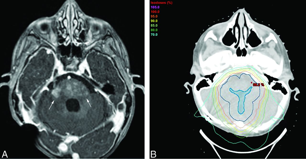
- Fig 1.
A 2-year-old child with a posterior fossa ependymoma status post gross total resection who developed multiple small foci of abnormal enhancement (arrows) in the pons and middle cerebellar peduncles, seen on an axial T1WI+C image (A), located within the radiation field (B) at 6 months following completion of PBT. Shown in the radiation treatment image (B) are the target structures of the gross tumor volume (dark blue filled) and the clinical target volume (darker blue, not filled). The dose lines of the proton beam treatment plan (analogous to the elevation lines of a topographic map) are shown as percentages of the prescription dose (59.4 Gy) in light purple (105%), red (100%), orange (95%), yellow (90%), light green (85%), forest green (80%), and cyan (70%).
- Fig 2.
Examples of radiation necrosis in patients with pediatric brain tumor treated with proton radiation therapy. A, A 4-year-old child with a posterior fossa ependymoma status post subtotal resection who developed multiple small foci of abnormal parenchymal enhancement (arrows) in the pons and cerebellum seen on an axial T1WI+C image at 4 months following completion of PBT. B, A 7-year-old child with a posterior fossa medulloblastoma status post subtotal resection who developed multiple small foci of abnormal parenchymal enhancement (arrows) in the corpus callosum/periventricular white matter and the cerebellum and left superior cerebellar peduncle seen on a coronal T1WI+C image at 7 months following completion of PBT. C, A 2-year-old child with a supratentorial ependymoma status post gross total resection who developed a single small foci of abnormal parenchymal enhancement (arrow) in the right periventricular white matter seen on an axial T1WI+C image at 11 months following completion of PBT.


Tables
- Table 1:
Common Terminology Criteria for Adverse Events, Version 4.0: central nervous system necrosisa
Grade Criteria 1 Asymptomatic; clinical, or diagnostic observations only; intervention not indicated 2 Moderate symptoms; corticosteroids indicated 3 Severe symptoms; medical intervention indicated 4 Life-threatening consequences; urgent intervention indicated 5 Death ↵a Adapted from Department of Health and Human Services.15
Category Characteristics Age Average, 7.2 ± 5.1 yr (range, 0.8–18 yr) 14/52 (27%) 3 years of age or younger Sex Male/female, 2.5:1 Tumor pathology Medulloblastoma and PNET (n = 19) Ependymoma (n = 12) Germinoma (n = 4) Brain stem glioma (n = 3) ATRT (n = 3) Craniopharyngioma (n = 3) Mature teratoma (n = 2) Pilocytic astrocytoma (n = 2) High-grade neuroepithelial tumor (n = 1) Pilomyxoid astrocytoma (n = 1) Pineal parenchymal tumor (n = 1) Chordoid meningioma (n = 1) Total cranial radiation Average, 54.0 Gy (range, 21–59.4 Gy) Note:—ATRT indicates atypical teratoid rhabdoid tumor; PNET, primitive neuroectodermal tumor.
Statistically Significant Not Statistically Significant >3 Chemotherapy agents (P = .03) Age, 2 years or younger (P = .11) ATRT pathology (P = .03) Age, 3 years or younger (P = .34) Sex (P = 1.0) Gross total surgical resection (P = .77) Medulloblastoma tumor pathology (P = .35) Ependymoma tumor pathology (P = .15) Germinomaa (P = .3) Infratentorial tumor location (P = 1.0) Pineal tumor locationa (P = .16) Craniospinal radiation (P = .48) Total radiation dose (P = .66) Note:—ATRT indicates atypical teratoid rhabdoid tumor.
↵a No pineal tumors or germinomas demonstrated radiation necrosis.


Jump to section
Related Articles
Cited By...
- Pretreatment Normal WM Magnetization Transfer Ratio Predicts Risk of Radiation Necrosis in Patients with Medulloblastoma
- New Approaches in Targeted Therapy for Medulloblastoma in Children
- Longitudinal changes in brain diffusion MRI indices during and after proton beam therapy in a child with pilocytic astrocytoma: a case report