Advertisement

Main menu

  • Home
  • Content
    • Current Issue
    • Accepted Manuscripts
    • Article Preview
    • Past Issue Archive
    • Video Articles
    • AJNR Case Collection
    • Case of the Week Archive
    • Case of the Month Archive
    • Classic Case Archive
  • Special Collections
    • AJNR Awards
    • Low-Field MRI
    • Alzheimer Disease
    • ASNR Foundation Special Collection
    • Photon-Counting CT
    • View All
  • Multimedia
    • AJNR Podcasts
    • AJNR SCANtastic
    • Trainee Corner
    • MRI Safety Corner
    • Imaging Protocols
  • For Authors
    • Submit a Manuscript
    • Submit a Video Article
    • Submit an eLetter to the Editor/Response
    • Manuscript Submission Guidelines
    • Statistical Tips
    • Fast Publishing of Accepted Manuscripts
    • Graphical Abstract Preparation
    • Imaging Protocol Submission
    • Author Policies
  • About Us
    • About AJNR
    • Editorial Board
    • Editorial Board Alumni
  • More
    • Become a Reviewer/Academy of Reviewers
    • Subscribers
    • Permissions
    • Alerts
    • Feedback
    • Advertisers
    • ASNR Home

User menu

  • Alerts
  • Log in

Search

  • Advanced search
American Journal of Neuroradiology

American Journal of Neuroradiology

ASHNR American Society of Functional Neuroradiology ASHNR American Society of Pediatric Neuroradiology ASSR
  • Alerts
  • Log in

Advanced Search

  • Home
  • Content
    • Current Issue
    • Accepted Manuscripts
    • Article Preview
    • Past Issue Archive
    • Video Articles
    • AJNR Case Collection
    • Case of the Week Archive
    • Case of the Month Archive
    • Classic Case Archive
  • Special Collections
    • AJNR Awards
    • Low-Field MRI
    • Alzheimer Disease
    • ASNR Foundation Special Collection
    • Photon-Counting CT
    • View All
  • Multimedia
    • AJNR Podcasts
    • AJNR SCANtastic
    • Trainee Corner
    • MRI Safety Corner
    • Imaging Protocols
  • For Authors
    • Submit a Manuscript
    • Submit a Video Article
    • Submit an eLetter to the Editor/Response
    • Manuscript Submission Guidelines
    • Statistical Tips
    • Fast Publishing of Accepted Manuscripts
    • Graphical Abstract Preparation
    • Imaging Protocol Submission
    • Author Policies
  • About Us
    • About AJNR
    • Editorial Board
    • Editorial Board Alumni
  • More
    • Become a Reviewer/Academy of Reviewers
    • Subscribers
    • Permissions
    • Alerts
    • Feedback
    • Advertisers
    • ASNR Home
  • Follow AJNR on Twitter
  • Visit AJNR on Facebook
  • Follow AJNR on Instagram
  • Join AJNR on LinkedIn
  • RSS Feeds

AJNR Awards, New Junior Editors, and more. Read the latest AJNR updates

Research ArticlePediatrics
Open Access

Tract-Based Spatial Statistics in Preterm-Born Neonates Predicts Cognitive and Motor Outcomes at 18 Months

E.G. Duerden, J. Foong, V. Chau, H. Branson, K.J. Poskitt, R.E. Grunau, A. Synnes, J.G. Zwicker and S.P. Miller
American Journal of Neuroradiology August 2015, 36 (8) 1565-1571; DOI: https://doi.org/10.3174/ajnr.A4312
E.G. Duerden
aFrom the Department of Paediatrics (E.G.D., J.F., V.C., H.B., S.P.M.), Hospital for Sick Children and University of Toronto, Toronto, Ontario, Canada
  • Find this author on Google Scholar
  • Find this author on PubMed
  • Search for this author on this site
J. Foong
aFrom the Department of Paediatrics (E.G.D., J.F., V.C., H.B., S.P.M.), Hospital for Sick Children and University of Toronto, Toronto, Ontario, Canada
  • Find this author on Google Scholar
  • Find this author on PubMed
  • Search for this author on this site
V. Chau
bDepartment of Pediatrics (V.C., K.J.P., R.E.G., A.S., J.G.Z., S.P.M.), University of British Columbia, BC Children's and Women's Hospitals, Child and Family Research Institute, Vancouver, British Columbia, Canada
  • Find this author on Google Scholar
  • Find this author on PubMed
  • Search for this author on this site
H. Branson
aFrom the Department of Paediatrics (E.G.D., J.F., V.C., H.B., S.P.M.), Hospital for Sick Children and University of Toronto, Toronto, Ontario, Canada
  • Find this author on Google Scholar
  • Find this author on PubMed
  • Search for this author on this site
K.J. Poskitt
bDepartment of Pediatrics (V.C., K.J.P., R.E.G., A.S., J.G.Z., S.P.M.), University of British Columbia, BC Children's and Women's Hospitals, Child and Family Research Institute, Vancouver, British Columbia, Canada
  • Find this author on Google Scholar
  • Find this author on PubMed
  • Search for this author on this site
R.E. Grunau
bDepartment of Pediatrics (V.C., K.J.P., R.E.G., A.S., J.G.Z., S.P.M.), University of British Columbia, BC Children's and Women's Hospitals, Child and Family Research Institute, Vancouver, British Columbia, Canada
  • Find this author on Google Scholar
  • Find this author on PubMed
  • Search for this author on this site
A. Synnes
bDepartment of Pediatrics (V.C., K.J.P., R.E.G., A.S., J.G.Z., S.P.M.), University of British Columbia, BC Children's and Women's Hospitals, Child and Family Research Institute, Vancouver, British Columbia, Canada
  • Find this author on Google Scholar
  • Find this author on PubMed
  • Search for this author on this site
J.G. Zwicker
bDepartment of Pediatrics (V.C., K.J.P., R.E.G., A.S., J.G.Z., S.P.M.), University of British Columbia, BC Children's and Women's Hospitals, Child and Family Research Institute, Vancouver, British Columbia, Canada
cDepartment of Occupational Science and Occupational Therapy (J.G.Z.), University of British Columbia, Vancouver, British Columbia, Canada.
  • Find this author on Google Scholar
  • Find this author on PubMed
  • Search for this author on this site
S.P. Miller
aFrom the Department of Paediatrics (E.G.D., J.F., V.C., H.B., S.P.M.), Hospital for Sick Children and University of Toronto, Toronto, Ontario, Canada
bDepartment of Pediatrics (V.C., K.J.P., R.E.G., A.S., J.G.Z., S.P.M.), University of British Columbia, BC Children's and Women's Hospitals, Child and Family Research Institute, Vancouver, British Columbia, Canada
  • Find this author on Google Scholar
  • Find this author on PubMed
  • Search for this author on this site
  • Article
  • Figures & Data
  • Supplemental
  • Info & Metrics
  • Responses
  • References
  • PDF
Loading

Article Figures & Data

Figures

  • Tables
  • Fig 1.
    • Download figure
    • Open in new tab
    • Download powerpoint
    Fig 1.

    Participant flow chart. Neonate data (180 very preterm-born infants of <32 weeks' gestation, with 1 or 2 MR imaging scanning sessions including DTI) are separated into 3 groups: group 1, 75 neonates with only 1 early scan near the time of birth (median postmenstrual age at scanning, 32 weeks) (all 75 neonates have neurodevelopmental follow-up data at 18-month corrected age); group 2, 78 neonates with 2 scans, both early (PMA, 32 weeks) and at term-equivalent age (PMA, 39.7 weeks) (75 neonates have follow-up data); group 3, 27 neonates with a late scan (PMA, 39 weeks) (16 neonates have follow-up data).

  • Fig 2.
    • Download figure
    • Open in new tab
    • Download powerpoint
    Fig 2.

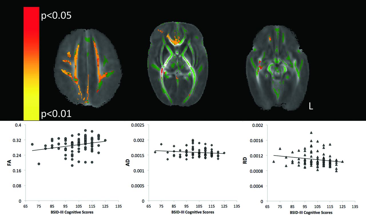
    TBSS analysis of term scans (PMA of 37–41-weeks). Top: Mean FA map (red-yellow) demonstrating the significant positive linear association between cognitive scores on the Bayley-III and FA in the territory of the medial prefrontal cortex (left), the genu of the corpus callosum (middle), and portions of the inferior fronto-occipital fasciculus (right, P < .05, corrected for multiple comparisons). The mean FA skeleton is shown in green. Bottom: FA (R = 0.3, P = .03), AD (R = −0.1, P = .2), and RD (R = −0.2, P = .03) values from the significant clusters in the FA map. Spearman ρ correlation and an α level are set at .05.

Tables

  • Figures
    • View popup
    Table 1:

    Clinical characteristics: separated into groups based on the postmenstrual age at scanninga

    27–29 Weeks (n = 24) (Median) (IQR) or (No.) (%)30–33 Weeks (n = 99) (Median) (IQR) or (No.) (%)34–36 Weeks (n = 34) (Median) (IQR) or (No.) (%)37–41 Weeks (n = 101) (Median) (IQR) or (No.) (%)
    Birth GA (wks)27.3 (26.1–27.8)29.3 (27.5–30.6)26.1 (25–27.7)26.9 (25.9–29.7)
    Age at MRI (wks)29 (28.5–29.4)32 (30.9–32.9)35.1 (34.5–36.3)39.7 (38.6–40.6)
    Sex (male)12 (50%)57 (57%)16 (47%)54 (53%)
    Birth weight (g)1022.5 (911.5–1171.3)1140 (957.5–1377.5)749 (607.5–1002.5)970 (805–1270)
    Days of mechanical ventilation2 (1–5.8)3 (1–10.5)37.5 (20.5–51.3)11.5 (2–51)
    Infectionb11 (46%)31 (31%)19 (56%)49 (49%)
    Patent ductus arteriosus9 (38%)38 (38%)27 (79%)47 (47%)
    Chronic lung disease5 (21%)10 (10%)17 (50%)27 (27%)
    • Note:—GA indicates gestational age.

    • ↵a One hundred eighty neonates participated. Seventy-five neonates had early scans (near birth), 78 neonates were scanned early and late (birth, term-equivalent age), and 27 neonates were scanned late (term-equivalent age), for a total of 258 scans.

    • ↵b Infection, culture-positive infection, confirmed necrotizing enterocolitis.

    • View popup
    Table 2:

    Radiologic findings: separated into groups based on the postmenstrual age at scanninga

    27–29 Weeks (n = 24) (No.) (%)30–33 Weeks (n = 99) (No.) (%)34–36 Weeks (n = 34) (No.) (%)37–41 Weeks (n = 101) (No.) (%)
    WMI (moderate/severe)b6 (25%)15 (15%)4 (12%)12 (12%)
    IVH (grade 1/2)c10 (42%)40 (40%)18 (53%)33 (33%)
    IVH (grade 3/4)c1 (4%)2 (2%)1 (3%)2 (2%)
    Cerebellar hemorrhage2 (8%)11 (11%)5 (15%)12 (12%)
    • Note:—WMI indicates white matter injury.

    • ↵a One hundred eighty neonates participated. Seventy-five neonates had early scans (near birth), 78 neonates were scanned early and late (birth, term-equivalent age), and 27 neonates were scanned late, for a total of 258 scans.

    • ↵b WMI defined as foci exhibiting T1 hyperintensity without T2 hypointensity or by low-intensity T1 foci.

    • ↵c IVH was graded (none = 0, mild = 1–2, and moderate-severe = 3–4) using the Papile system.

    • View popup
    Table 3:

    Neurodevelopmental outcome: separated into groups by postmenstrual age at scanninga

    27–29 Weeks (n = 22) (Median) (IQR)30–33 Weeks (n = 93) (Median) (IQR)34–36 Weeks (n = 32) (Median) (IQR)37–41 Weeks (n = 94) (Median) (IQR)
    Age at follow-up (mo)b18.7 (18.4–19.2)18.7 (18.3–19.7)18.6 (18.3–19.1)18.8 (18.4–19.4)
    Bayley-III cognitivec105 (100–110)110 (100–115)102.5 (92.5–110)105 (95–110)
    Bayley-III languagec101.5 (83.5–111.3)100 (91–109)95.5 (83–109)100 (86.8–108.3)
    Bayley-III motorc98.5 (88.8–105.3)100 (91.8–107)92.5 (84.3–104)97 (88–107)
    PDMS-2 Gross Motorc91 (87–96)94 (87–98)89 (79–96)91 (87–98)
    PDMS-2 Fine Motorc100 (97–106)100 (97–103)97 (89.5–103)100 (94–103)
    PDMS-2 Total Motorc96 (90–101)96 (92–98)92 (84–97)94 (89–98)
    • ↵a One hundred sixty-six neonates returned for neurodevelopmental follow-up. DTI data were acquired in 75 of the neonates early and in 75 of the neonates at early and late time points (150 scans). Sixteen neonates had late scans for a total of 241 scans.

    • ↵b Age corrected for prematurity.

    • ↵c The mean composite score in a normative population is 100 ± 15.

PreviousNext
Back to top

In this issue

American Journal of Neuroradiology: 36 (8)
American Journal of Neuroradiology
Vol. 36, Issue 8
1 Aug 2015
  • Table of Contents
  • Index by author
  • Complete Issue (PDF)
Advertisement
Print
Download PDF
Email Article

Thank you for your interest in spreading the word on American Journal of Neuroradiology.

NOTE: We only request your email address so that the person you are recommending the page to knows that you wanted them to see it, and that it is not junk mail. We do not capture any email address.

Enter multiple addresses on separate lines or separate them with commas.
Tract-Based Spatial Statistics in Preterm-Born Neonates Predicts Cognitive and Motor Outcomes at 18 Months
(Your Name) has sent you a message from American Journal of Neuroradiology
(Your Name) thought you would like to see the American Journal of Neuroradiology web site.
CAPTCHA
This question is for testing whether or not you are a human visitor and to prevent automated spam submissions.
Cite this article
E.G. Duerden, J. Foong, V. Chau, H. Branson, K.J. Poskitt, R.E. Grunau, A. Synnes, J.G. Zwicker, S.P. Miller
Tract-Based Spatial Statistics in Preterm-Born Neonates Predicts Cognitive and Motor Outcomes at 18 Months
American Journal of Neuroradiology Aug 2015, 36 (8) 1565-1571; DOI: 10.3174/ajnr.A4312

Citation Manager Formats

  • BibTeX
  • Bookends
  • EasyBib
  • EndNote (tagged)
  • EndNote 8 (xml)
  • Medlars
  • Mendeley
  • Papers
  • RefWorks Tagged
  • Ref Manager
  • RIS
  • Zotero
0 Responses
Respond to this article
Share
Bookmark this article
Tract-Based Spatial Statistics in Preterm-Born Neonates Predicts Cognitive and Motor Outcomes at 18 Months
E.G. Duerden, J. Foong, V. Chau, H. Branson, K.J. Poskitt, R.E. Grunau, A. Synnes, J.G. Zwicker, S.P. Miller
American Journal of Neuroradiology Aug 2015, 36 (8) 1565-1571; DOI: 10.3174/ajnr.A4312
del.icio.us logo Twitter logo Facebook logo Mendeley logo
  • Tweet Widget
  • Facebook Like
  • Google Plus One
Purchase

Jump to section

  • Article
    • Abstract
    • ABBREVIATIONS:
    • Materials and Methods
    • Results
    • Discussion
    • Conclusions
    • Acknowledgments
    • Footnotes
    • References
  • Figures & Data
  • Supplemental
  • Info & Metrics
  • Responses
  • References
  • PDF

Related Articles

  • PubMed
  • Google Scholar

Cited By...

  • Associations between prenatal adversity and neonatal white matter microstructure on language outcomes at age 2 years
  • Data-driven characterization of Preterm Birth through intramodal Diffusion MRI
  • Early structural connectivity within the sensorimotor network: deviations related to prematurity and association to neurodevelopmental outcome
  • Quantitative Diffusion and Spectroscopic Neuroimaging Combined with a Novel Early-Developmental Assessment Improves Models for 1-Year Developmental Outcomes
  • Neonatal White Matter Microstructure and Emotional Development during the Preschool Years in Children Who Were Born Very Preterm
  • Diffusion MRI Microstructural Abnormalities at Term-Equivalent Age Are Associated with Neurodevelopmental Outcomes at 3 Years of Age in Very Preterm Infants
  • Cyto/myeloarchitectural changes of cortical gray matter and superficial white matter in early neurodevelopment: Multimodal MRI study of preterm neonates
  • Early Procedural Pain Is Associated with Regionally-Specific Alterations in Thalamic Development in Preterm Neonates
  • Severe retinopathy of prematurity predicts delayed white matter maturation and poorer neurodevelopment
  • Quantitative assessment of white matter injury in preterm neonates: Association with outcomes
  • Crossref (59)
  • Google Scholar

This article has been cited by the following articles in journals that are participating in Crossref Cited-by Linking.

  • Early Procedural Pain Is Associated with Regionally-Specific Alterations in Thalamic Development in Preterm Neonates
    Emma G. Duerden, Ruth E. Grunau, Ting Guo, Justin Foong, Alexander Pearson, Stephanie Au-Young, Raphael Lavoie, M. Mallar Chakravarty, Vann Chau, Anne Synnes, Steven P. Miller
    The Journal of Neuroscience 2018 38 4
  • Quantitative assessment of white matter injury in preterm neonates
    Ting Guo, Emma G. Duerden, Elysia Adams, Vann Chau, Helen M. Branson, M. Mallar Chakravarty, Kenneth J. Poskitt, Anne Synnes, Ruth E. Grunau, Steven P. Miller
    Neurology 2017 88 7
  • Exploring the multiple-hit hypothesis of preterm white matter damage using diffusion MRI
    Madeleine L. Barnett, Nora Tusor, Gareth Ball, Andrew Chew, Shona Falconer, Paul Aljabar, Jessica A. Kimpton, Nigel Kennea, Mary Rutherford, A. David Edwards, Serena J. Counsell
    NeuroImage: Clinical 2018 17
  • Early postnatal docosahexaenoic acid levels and improved preterm brain development
    Emily W. Y. Tam, Vann Chau, A. James Barkovich, Donna M. Ferriero, Steven P. Miller, Elizabeth E. Rogers, Ruth E. Grunau, Anne R. Synnes, Duan Xu, Justin Foong, Rollin Brant, Sheila M. Innis
    Pediatric Research 2016 79 5
  • Neonatal Neurology
    Juliane Schneider, Steven P. Miller
    2019 162
  • Advanced neuroimaging and its role in predicting neurodevelopmental outcomes in very preterm infants
    Nehal A. Parikh
    Seminars in Perinatology 2016 40 8
  • Heterogeneity in Brain Microstructural Development Following Preterm Birth
    Ralica Dimitrova, Maximilian Pietsch, Daan Christiaens, Judit Ciarrusta, Thomas Wolfers, Dafnis Batalle, Emer Hughes, Jana Hutter, Lucilio Cordero-Grande, Anthony N Price, Andrew Chew, Shona Falconer, Katy Vecchiato, Johannes K Steinweg, Olivia Carney, Mary A Rutherford, J-Donald Tournier, Serena J Counsell, Andre F Marquand, Daniel Rueckert, Joseph V Hajnal, Grainne McAlonan, A David Edwards, Jonathan O’Muircheartaigh
    Cerebral Cortex 2020 30 9
  • Severe retinopathy of prematurity predicts delayed white matter maturation and poorer neurodevelopment
    Torin J A Glass, Vann Chau, Jane Gardiner, Justin Foong, Jillian Vinall, Jill G Zwicker, Ruth E Grunau, Anne Synnes, Kenneth J Poskitt, Steven P Miller
    Archives of Disease in Childhood - Fetal and Neonatal Edition 2017 102 6
  • Music enhances structural maturation of emotional processing neural pathways in very preterm infants
    Joana Sa de Almeida, Lara Lordier, Benjamin Zollinger, Nicolas Kunz, Matteo Bastiani, Laura Gui, Alexandra Adam-Darque, Cristina Borradori-Tolsa, François Lazeyras, Petra S. Hüppi
    NeuroImage 2020 207
  • Recent advances in diffusion neuroimaging: applications in the developing preterm brain
    Diliana Pecheva, Christopher Kelly, Jessica Kimpton, Alexandra Bonthrone, Dafnis Batalle, Hui Zhang, Serena J. Counsell
    F1000Research 2018 7

More in this TOC Section

  • SyMRI & MR Fingerprinting in Brainstem Myelination
  • Comparison of Image Quality and Radiation Dose in Pediatric Temporal Bone CT Using Photon-Counting Detector CT and Energy-Integrating Detector CT
  • Axenfeld-Rieger Syndrome: Neuroimaging Findings
Show more Pediatrics

Similar Articles

Advertisement

Indexed Content

  • Current Issue
  • Accepted Manuscripts
  • Article Preview
  • Past Issues
  • Editorials
  • Editor's Choice
  • Fellows' Journal Club
  • Letters to the Editor
  • Video Articles

Cases

  • Case Collection
  • Archive - Case of the Week
  • Archive - Case of the Month
  • Archive - Classic Case

More from AJNR

  • Trainee Corner
  • Imaging Protocols
  • MRI Safety Corner
  • Book Reviews

Multimedia

  • AJNR Podcasts
  • AJNR Scantastics

Resources

  • Turnaround Time
  • Submit a Manuscript
  • Submit a Video Article
  • Submit an eLetter to the Editor/Response
  • Manuscript Submission Guidelines
  • Statistical Tips
  • Fast Publishing of Accepted Manuscripts
  • Graphical Abstract Preparation
  • Imaging Protocol Submission
  • Evidence-Based Medicine Level Guide
  • Publishing Checklists
  • Author Policies
  • Become a Reviewer/Academy of Reviewers
  • News and Updates

About Us

  • About AJNR
  • Editorial Board
  • Editorial Board Alumni
  • Alerts
  • Permissions
  • Not an AJNR Subscriber? Join Now
  • Advertise with Us
  • Librarian Resources
  • Feedback
  • Terms and Conditions
  • AJNR Editorial Board Alumni

American Society of Neuroradiology

  • Not an ASNR Member? Join Now

© 2025 by the American Society of Neuroradiology All rights, including for text and data mining, AI training, and similar technologies, are reserved.
Print ISSN: 0195-6108 Online ISSN: 1936-959X

Powered by HighWire