Advertisement

Main menu

  • Home
  • Content
    • Current Issue
    • Accepted Manuscripts
    • Article Preview
    • Past Issue Archive
    • Video Articles
    • AJNR Case Collection
    • Case of the Week Archive
    • Case of the Month Archive
    • Classic Case Archive
  • Special Collections
    • AJNR Awards
    • Low-Field MRI
    • Alzheimer Disease
    • ASNR Foundation Special Collection
    • Photon-Counting CT
    • View All
  • Multimedia
    • AJNR Podcasts
    • AJNR SCANtastic
    • Trainee Corner
    • MRI Safety Corner
    • Imaging Protocols
  • For Authors
    • Submit a Manuscript
    • Submit a Video Article
    • Submit an eLetter to the Editor/Response
    • Manuscript Submission Guidelines
    • Statistical Tips
    • Fast Publishing of Accepted Manuscripts
    • Graphical Abstract Preparation
    • Imaging Protocol Submission
    • Author Policies
  • About Us
    • About AJNR
    • Editorial Board
    • Editorial Board Alumni
  • More
    • Become a Reviewer/Academy of Reviewers
    • Subscribers
    • Permissions
    • Alerts
    • Feedback
    • Advertisers
    • ASNR Home

User menu

  • Alerts
  • Log in

Search

  • Advanced search
American Journal of Neuroradiology

American Journal of Neuroradiology

ASHNR American Society of Functional Neuroradiology ASHNR American Society of Pediatric Neuroradiology ASSR
  • Alerts
  • Log in

Advanced Search

  • Home
  • Content
    • Current Issue
    • Accepted Manuscripts
    • Article Preview
    • Past Issue Archive
    • Video Articles
    • AJNR Case Collection
    • Case of the Week Archive
    • Case of the Month Archive
    • Classic Case Archive
  • Special Collections
    • AJNR Awards
    • Low-Field MRI
    • Alzheimer Disease
    • ASNR Foundation Special Collection
    • Photon-Counting CT
    • View All
  • Multimedia
    • AJNR Podcasts
    • AJNR SCANtastic
    • Trainee Corner
    • MRI Safety Corner
    • Imaging Protocols
  • For Authors
    • Submit a Manuscript
    • Submit a Video Article
    • Submit an eLetter to the Editor/Response
    • Manuscript Submission Guidelines
    • Statistical Tips
    • Fast Publishing of Accepted Manuscripts
    • Graphical Abstract Preparation
    • Imaging Protocol Submission
    • Author Policies
  • About Us
    • About AJNR
    • Editorial Board
    • Editorial Board Alumni
  • More
    • Become a Reviewer/Academy of Reviewers
    • Subscribers
    • Permissions
    • Alerts
    • Feedback
    • Advertisers
    • ASNR Home
  • Follow AJNR on Twitter
  • Visit AJNR on Facebook
  • Follow AJNR on Instagram
  • Join AJNR on LinkedIn
  • RSS Feeds

AJNR Awards, New Junior Editors, and more. Read the latest AJNR updates

Research ArticleHead and Neck Imaging

Parry Romberg Syndrome: 7 Cases and Literature Review

M. Wong, C.D. Phillips, M. Hagiwara and D.R. Shatzkes
American Journal of Neuroradiology July 2015, 36 (7) 1355-1361; DOI: https://doi.org/10.3174/ajnr.A4297
M. Wong
aFrom the Department of Radiology (M.W., D.R.S.), Lenox Hill Hospital, North Shore Long Island Jewish Health System, New York, New York
  • Find this author on Google Scholar
  • Find this author on PubMed
  • Search for this author on this site
C.D. Phillips
bDepartment of Radiology (C.D.P.), Division of Neuroradiology, Weill Medical College of Cornell University, New York Presbyterian Hospital, New York, New York
  • Find this author on Google Scholar
  • Find this author on PubMed
  • Search for this author on this site
M. Hagiwara
cDepartment of Radiology (M.H.), New York University Langone Medical Center, New York, New York.
  • Find this author on Google Scholar
  • Find this author on PubMed
  • Search for this author on this site
D.R. Shatzkes
aFrom the Department of Radiology (M.W., D.R.S.), Lenox Hill Hospital, North Shore Long Island Jewish Health System, New York, New York
  • Find this author on Google Scholar
  • Find this author on PubMed
  • Search for this author on this site
  • Article
  • Figures & Data
  • Info & Metrics
  • Responses
  • References
  • PDF
Loading

Article Figures & Data

Figures

  • Fig 1.
    • Download figure
    • Open in new tab
    • Download powerpoint
    Fig 1.

    Axial CT images of a 44-year-old woman with PRS obtained for cosmetic surgical planning. A, There is prominent hemiatrophy of the skin, subcutaneous fat, and masseter muscle. B, The right maxillary sinus is considerably smaller than the left. C, Marked right enophthalmos is present.
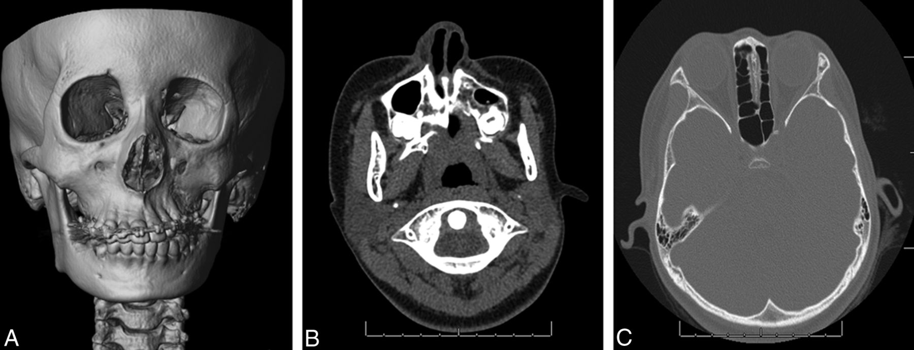
  • Fig 2.
    • Download figure
    • Open in new tab
    • Download powerpoint
    Fig 2.

    A 14-year-old boy who presented with progressive atrophy of the left face. A, T1-weighted image shows striking paucity of fat in the left face. B, 3D reconstruction of a CT series demonstrates left facial hemiatrophy, particularly involving the mandible. C, T1-weighted image at the level of the orbits reveals left enophthalmos.

  • Fig 3.
    • Download figure
    • Open in new tab
    • Download powerpoint
    Fig 3.

    A 24-year-old man with progressive atrophy of the left face. A and B, Axial T1-weighted MR images demonstrate left hemifacial atrophy primarily involving the skin and subcutaneous fat; only subtle asymmetry of the masseter is noted. C, T2-weighted image at the same level demonstrates normal T2 signal in the affected structures.

  • Fig 4.
    • Download figure
    • Open in new tab
    • Download powerpoint
    Fig 4.

    An 11-year-old girl with left-face sensitivity and left-temple pain. A, 3D reconstructions of a CT series show osseous asymmetry, most prominently in the left maxilla. B, Axial CT image at soft-tissue windows demonstrates deviation of the nose toward the affected side. C, Axial bone window demonstrates marked asymmetry in the volume of ethmoidal air cells.
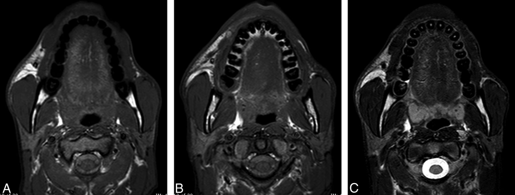
  • Fig 5.
    • Download figure
    • Open in new tab
    • Download powerpoint
    Fig 5.

    A 30-year-old woman with a history of PRS who presented for MR imaging evaluation for weakness and recurrent headaches. A, Coronal enhanced T1-weighted MR image demonstrates mild asymmetry of scalp thickness with relative paucity of subcutaneous fat on the right. B and C, Axial FLAIR images demonstrate abnormal hyperintensity of the white matter in the right corona radiata, internal capsule, and temporal region. D, Axial susceptibility-weighted image demonstrates innumerable punctate foci of susceptibility throughout the right cerebral hemisphere.
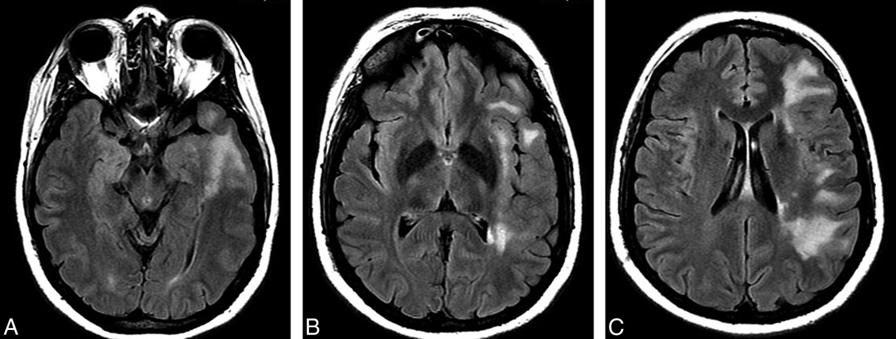
  • Fig 6.
    • Download figure
    • Open in new tab
    • Download powerpoint
    Fig 6.

    A 35-year-old woman with a history of PRS presented for MR imaging evaluation for headaches and weakness. Axial FLAIR images demonstrate hyperinstensity in the left cerebral periventricular to subcortical white matter. The pattern of signal abnormality is geographic, with subtotal involvement of frontal, parietal, and temporal lobes.
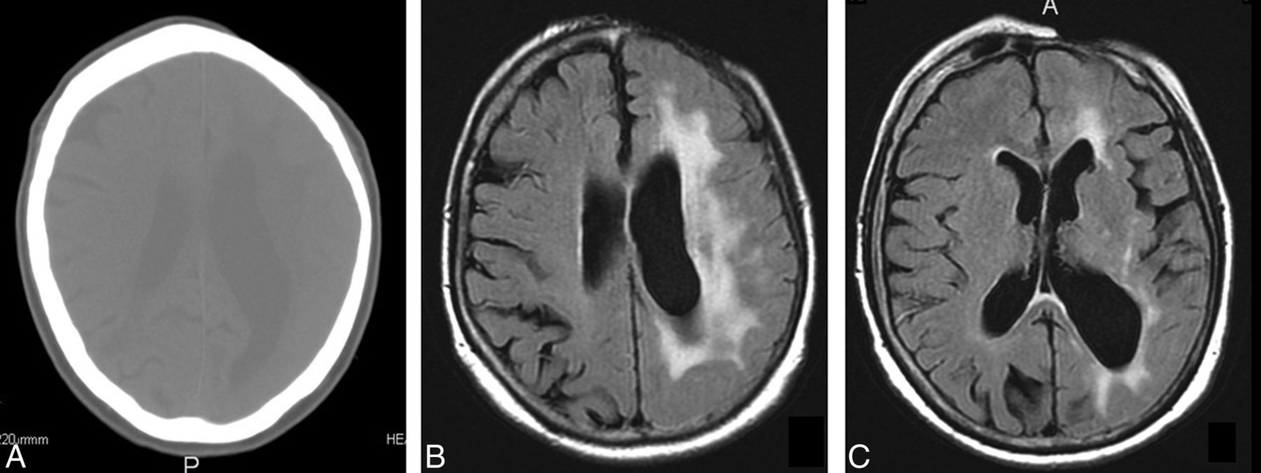
  • Fig 7.
    • Download figure
    • Open in new tab
    • Download powerpoint
    Fig 7.

    A 68-year-old man with PRS who presented with acute right MCA infarct. A, Axial CT image demonstrates striking focal loss of scalp thickness in the left frontal region. B, Axial FLAIR images demonstrate confluent signal abnormality in the high left frontal and parietal white matter. C, Etiology of left ventriculomegaly and sulcal asymmetry was uncertain.

PreviousNext
Back to top

In this issue

American Journal of Neuroradiology: 36 (7)
American Journal of Neuroradiology
Vol. 36, Issue 7
1 Jul 2015
  • Table of Contents
  • Index by author
  • Complete Issue (PDF)
Advertisement
Print
Download PDF
Email Article

Thank you for your interest in spreading the word on American Journal of Neuroradiology.

NOTE: We only request your email address so that the person you are recommending the page to knows that you wanted them to see it, and that it is not junk mail. We do not capture any email address.

Enter multiple addresses on separate lines or separate them with commas.
Parry Romberg Syndrome: 7 Cases and Literature Review
(Your Name) has sent you a message from American Journal of Neuroradiology
(Your Name) thought you would like to see the American Journal of Neuroradiology web site.
CAPTCHA
This question is for testing whether or not you are a human visitor and to prevent automated spam submissions.
Cite this article
M. Wong, C.D. Phillips, M. Hagiwara, D.R. Shatzkes
Parry Romberg Syndrome: 7 Cases and Literature Review
American Journal of Neuroradiology Jul 2015, 36 (7) 1355-1361; DOI: 10.3174/ajnr.A4297

Citation Manager Formats

  • BibTeX
  • Bookends
  • EasyBib
  • EndNote (tagged)
  • EndNote 8 (xml)
  • Medlars
  • Mendeley
  • Papers
  • RefWorks Tagged
  • Ref Manager
  • RIS
  • Zotero
0 Responses
Respond to this article
Share
Bookmark this article
Parry Romberg Syndrome: 7 Cases and Literature Review
M. Wong, C.D. Phillips, M. Hagiwara, D.R. Shatzkes
American Journal of Neuroradiology Jul 2015, 36 (7) 1355-1361; DOI: 10.3174/ajnr.A4297
del.icio.us logo Twitter logo Facebook logo Mendeley logo
  • Tweet Widget
  • Facebook Like
  • Google Plus One
Purchase

Jump to section

  • Article
    • Abstract
    • ABBREVIATION:
    • Discussion
    • REFERENCES
  • Figures & Data
  • Info & Metrics
  • Responses
  • References
  • PDF

Related Articles

  • No related articles found.
  • PubMed
  • Google Scholar

Cited By...

  • Brain Abnormalities and Epilepsy in Patients with Parry-Romberg Syndrome
  • Parry-Romberg syndrome in a patient with scleroderma
  • Visual Diagnosis: Recurrent Seizures and Concomitant Skin Changes in a 16-year-old Boy
  • Late progression of neurological symptoms and MRI T2 hyperintensities in Parry-Romberg syndrome
  • Crossref (66)
  • Google Scholar

This article has been cited by the following articles in journals that are participating in Crossref Cited-by Linking.

  • Ophthalmological manifestations of Parry-Romberg syndrome
    Franziska Bucher, Julia Fricke, Antje Neugebauer, Claus Cursiefen, Ludwig M. Heindl
    Survey of Ophthalmology 2016 61 6
  • Imaging Spectrum of Calvarial Abnormalities
    Iman Khodarahmi, Hamza Alizai, Majid Chalian, Erin F. Alaia, Christopher J. Burke, Shira E. Slasky, Cornelia Wenokor
    RadioGraphics 2021 41 4
  • Acquired Facial, Maxillofacial, and Oral Asymmetries—A Review Highlighting Diagnosis and Management
    Janaki Iyer, Arvind Hariharan, Uyen Minh Nha Cao, Simon D. Tran
    Symmetry 2021 13 9
  • Progressive Hemifacial Atrophy and Linear Scleroderma En Coup de Sabre: A Spectrum of the Same Disease?
    Irina Khamaganova
    Frontiers in Medicine 2018 4
  • Challenging Traditional Thinking: Early Free Tissue Transfer for Active Hemifacial Atrophy in Children
    Jacqueline S. Israel, Jenny T. Chen, Rebecca L. Farmer, John W. Siebert
    Plastic & Reconstructive Surgery 2020 145 2
  • Unilateral Neuroimaging Findings in Pediatric Craniofacial Scleroderma: Parry-Romberg Syndrome and En Coup de Sabre
    Ronald R. Seese, Daniel Glaser, Andre Furtado, Kavita Thakkar, Kathryn S. Torok
    Journal of Child Neurology 2020 35 11
  • Clinico-radiological approach to cerebral hemiatrophy
    Ai Peng Tan, Yen Ling Jocelyn Wong, Bingyuan Jeremy Lin, Hsiang Rong Clement Yong, Kshitij Mankad
    Child's Nervous System 2018 34 12
  • Protean Neurologic Manifestations of Two Rare Dermatologic Disorders: Sweet Disease and Localized Craniofacial Scleroderma
    Asya I. Wallach, Cynthia M. Magro, Andrew G. Franks, Lee Shapiro, Ilya Kister
    Current Neurology and Neuroscience Reports 2019 19 3
  • Color Atlas of Oral and Maxillofacial Diseases
    Brad W. Neville, Douglas D. Damm, Carl M. Allen, Angela C. Chi
    2019
  • Parry–Romberg syndrome in an adolescent: a case report on progressive hemifacial atrophy
    Ranjini Srinivasan, Shasta Theresa Joy
    Oxford Medical Case Reports 2020 2020 1

More in this TOC Section

  • Hydrops Herniation into the Semicircular Canals
  • Post SRS Peritumoral Hyperintense Signal of VSs
  • DIVr of NPC Predicted Outcome
Show more HEAD AND NECK IMAGING

Similar Articles

Advertisement

Indexed Content

  • Current Issue
  • Accepted Manuscripts
  • Article Preview
  • Past Issues
  • Editorials
  • Editor's Choice
  • Fellows' Journal Club
  • Letters to the Editor
  • Video Articles

Cases

  • Case Collection
  • Archive - Case of the Week
  • Archive - Case of the Month
  • Archive - Classic Case

More from AJNR

  • Trainee Corner
  • Imaging Protocols
  • MRI Safety Corner
  • Book Reviews

Multimedia

  • AJNR Podcasts
  • AJNR Scantastics

Resources

  • Turnaround Time
  • Submit a Manuscript
  • Submit a Video Article
  • Submit an eLetter to the Editor/Response
  • Manuscript Submission Guidelines
  • Statistical Tips
  • Fast Publishing of Accepted Manuscripts
  • Graphical Abstract Preparation
  • Imaging Protocol Submission
  • Evidence-Based Medicine Level Guide
  • Publishing Checklists
  • Author Policies
  • Become a Reviewer/Academy of Reviewers
  • News and Updates

About Us

  • About AJNR
  • Editorial Board
  • Editorial Board Alumni
  • Alerts
  • Permissions
  • Not an AJNR Subscriber? Join Now
  • Advertise with Us
  • Librarian Resources
  • Feedback
  • Terms and Conditions
  • AJNR Editorial Board Alumni

American Society of Neuroradiology

  • Not an ASNR Member? Join Now

© 2025 by the American Society of Neuroradiology All rights, including for text and data mining, AI training, and similar technologies, are reserved.
Print ISSN: 0195-6108 Online ISSN: 1936-959X

Powered by HighWire