
Article Figures & Data
Figures
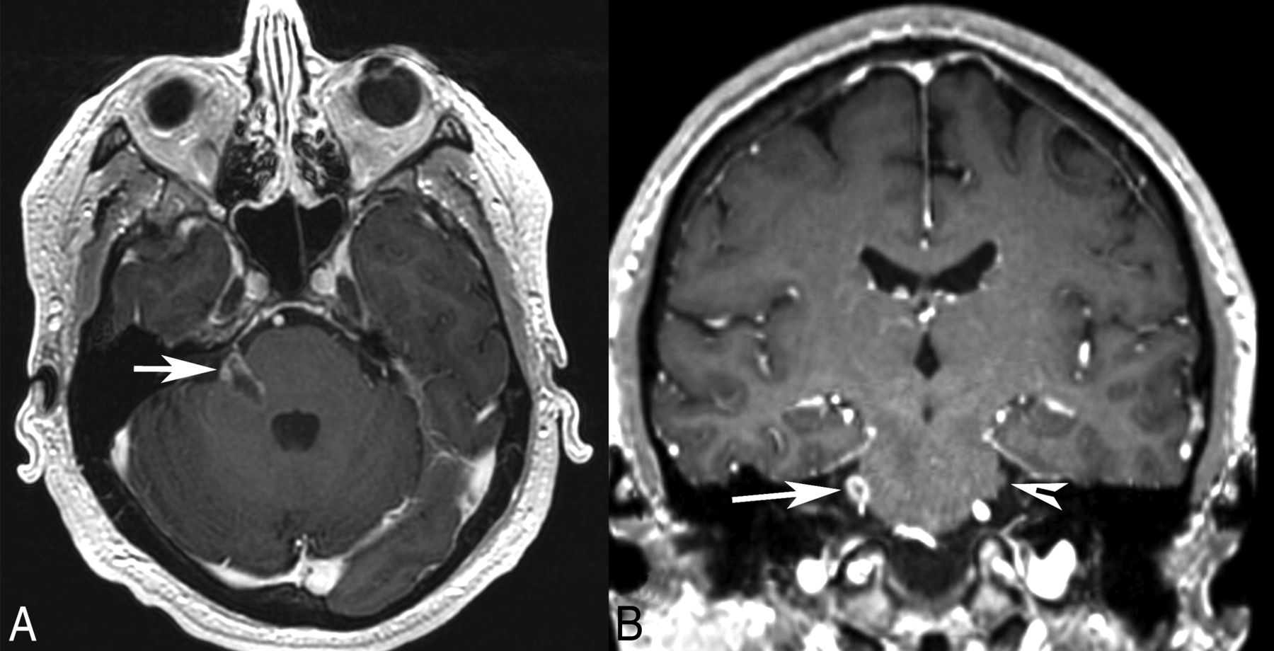
- Fig 1.
Patient 1. Axial (A) and coronal (B) postgadolinium T1-weighted MR images demonstrate an irregular enhancing mass involving the right pons, the expected location of trigeminal nuclei, the root entry zone, and the cisternal segment of the right trigeminal nerve (arrow). The normal left trigeminal nerve is marked on the coronal image (arrowhead). This patient presented with right facial numbness.
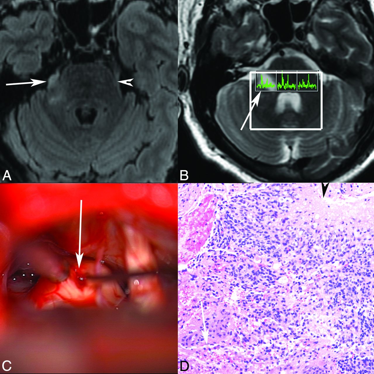
- Fig 2.
Patient 1. Axial T2 FLAIR (A) MR image demonstrates abnormal T2 FLAIR signal in the right lateral pons extending into the right trigeminal nerve (arrow marks the site of biopsy, arrowhead marks the normal left trigeminal nerve). B, Multivoxel MR spectroscopy demonstrates increased choline relative to NAA in the right-sided voxel corresponding to the tumor (arrow). C, Operative photograph shows a right retrosigmoid approach to the expanded right trigeminal nerve (arrow), with biopsy being obtained (courtesy of Jonathan D. Breshears, MD). D, Hematoxylin-eosin stained histology slide (original magnification ×20) demonstrates an infiltrative astrocytic neoplasm with nuclear pleomorphism, brisk mitotic activity, microvascular proliferation (arrow), and pseudopalisading necrosis (arrowhead). Pathologic diagnosis was a glioblastoma, WHO grade IV. No peripheral nerve was identified in the specimen despite the biopsy location, likely reflecting the origin from glial cells in the trigeminal root entry zone.
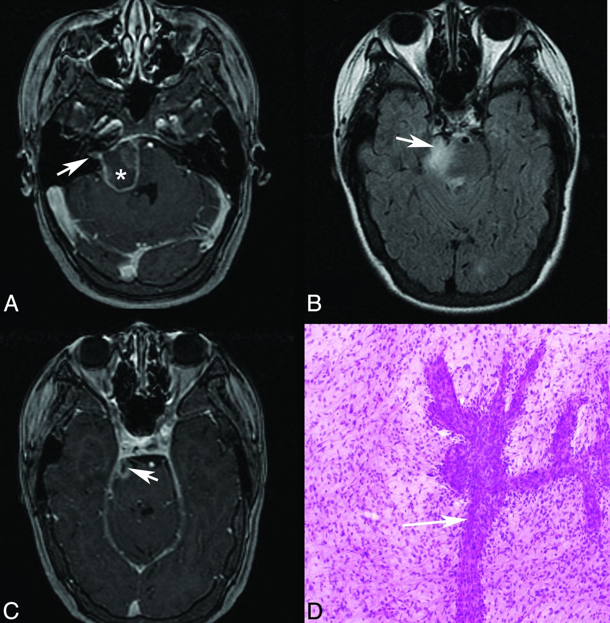
- Fig 3.
Patient 3. Axial T2 FLAIR (A), coronal T2 FLAIR (B), and coronal postgadolinium T1 (C) MR images demonstrate abnormal T2 FLAIR signal in the left lateral pons extending into the expanded left trigeminal nerve (arrow), with enhancement shown at the root entry zone (C). The arrowhead marks the normal right trigeminal nerve, and the asterisk marks the approximate site of biopsy in the pons. D, Hematoxylin-eosin stained histology slide (original magnification ×40) demonstrates an infiltrating population of neoplastic astrocytes with irregular ovoid nuclei (arrow) and coarse chromatin and scant eosinophilic cytoplasm. No mitotic activity, microvascular proliferation, or foci of necrosis are present. The pathologic diagnosis was a diffuse astrocytoma, WHO grade II.
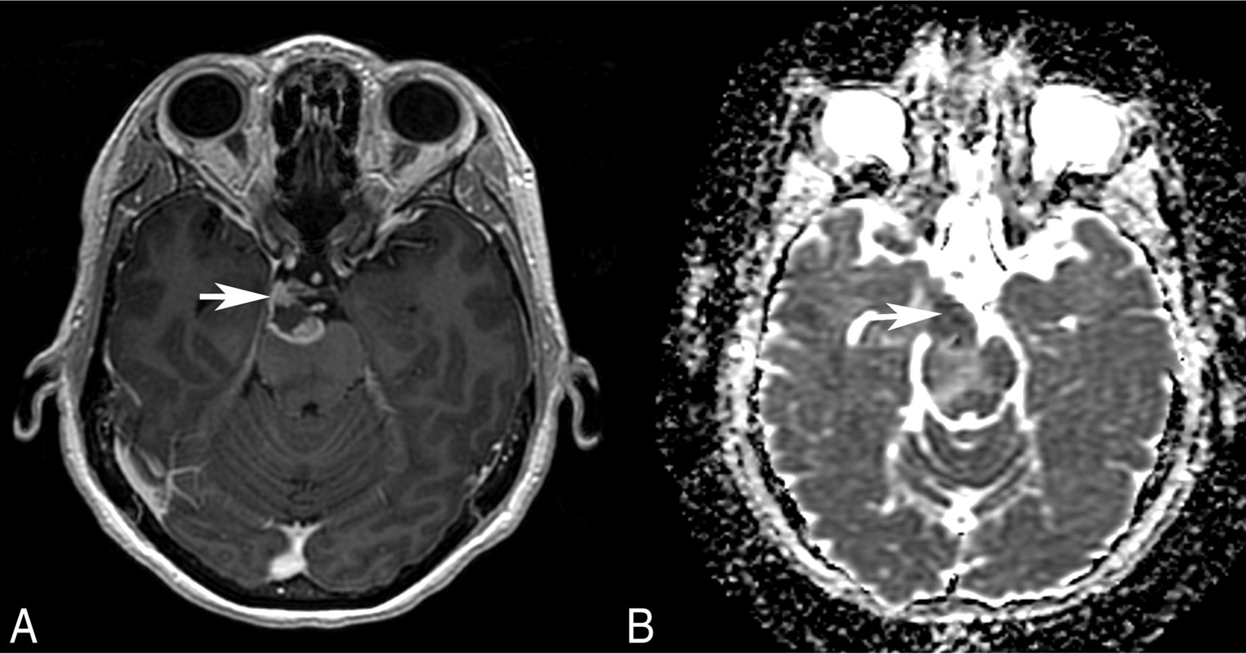
- Fig 4.
Patient 4. Axial postgadolinium T1-weighted MR image (A) and ADC map (B) show an irregularly enhancing, mass with restricted diffusion (arrow) involving the root exit zone and the cisternal course of the right oculomotor nerve in the interpeduncular cistern. This patient presented with right oculomotor palsy.
- Fig 5.
Patient 8. Axial postgadolinium T1 (A and C) and axial T2 FLAIR (B) MR images demonstrate an irregularly enhancing, T2 FLAIR hyperintense mass involving the right lateral pons and expected origins and intracranial course of both the right facial (A) and the trigeminal (B and C, arrow) nerves. Enhancement also extends into the right internal auditory canal along its anterior and superior aspect where the extra-axial facial nerve courses (A, arrow). Asterisk (A) marks the approximate site of biopsy. D, Hematoxylin-eosin-stained intraoperative smear preparation (original magnification ×10) demonstrates a population of neoplastic astrocytes with nuclear pleomorphism and numerous mitotic figures. A vessel with extensive budding and endothelial proliferation is present (arrow). Pathologic diagnosis was a glioblastoma, WHO grade IV. This patient presented with right facial numbness and weakness, but no clinical evidence of vestibulocochlear nerve involvement until later in the disease course.


Tables
Case series of patients with gliomas involving cranial nerves
Case Age (yr) Sex Location Grade (WHO) Neuropathy Maximal Thickness, Involved CN (mm) Maximal Thickness, Contralateral CN (mm) Maximal Length (mm) 1 67 M R pons to trigeminal nerve IV Yes 6 2 8 2 53 F R pons to trigeminal and vestibulocochlear nerves; separate R frontal tumor IV No 3 1 4 3 67 F L pons to trigeminal nerve II Yes 4 1 13 4 49 F R midbrain to R oculomotor nerve; separate R frontal tumor IV Yes 14 1 16 5 22 M R pons to trigeminal nerve; separate R frontal and thalamus tumor IV No 4 2 7 6 9 M R pons to trigeminal nerve; separate R thalamus and midbrain tumor IV No 4 2 4 7 34 M R pons to trigeminal nerve; separate R parietal tumor II No 4 2 8 8 24 F R pons to trigeminal and facial nerves IV Yes 8 1 15 Mean (SD) 41 (20.3) 5.9 (3.6) 1.5 (0.5) 9.4 (4.7) Note:—R indicates right; L, left.


Jump to section
Related Articles
Cited By...
- No citing articles found.