Advertisement

Main menu

  • Home
  • Content
    • Current Issue
    • Accepted Manuscripts
    • Article Preview
    • Past Issue Archive
    • Video Articles
    • AJNR Case Collection
    • Case of the Week Archive
    • Case of the Month Archive
    • Classic Case Archive
  • Special Collections
    • AJNR Awards
    • Low-Field MRI
    • Alzheimer Disease
    • ASNR Foundation Special Collection
    • Photon-Counting CT
    • View All
  • Multimedia
    • AJNR Podcasts
    • AJNR SCANtastic
    • Trainee Corner
    • MRI Safety Corner
    • Imaging Protocols
  • For Authors
    • Submit a Manuscript
    • Submit a Video Article
    • Submit an eLetter to the Editor/Response
    • Manuscript Submission Guidelines
    • Statistical Tips
    • Fast Publishing of Accepted Manuscripts
    • Graphical Abstract Preparation
    • Imaging Protocol Submission
    • Author Policies
  • About Us
    • About AJNR
    • Editorial Board
    • Editorial Board Alumni
  • More
    • Become a Reviewer/Academy of Reviewers
    • Subscribers
    • Permissions
    • Alerts
    • Feedback
    • Advertisers
    • ASNR Home

User menu

  • Alerts
  • Log in

Search

  • Advanced search
American Journal of Neuroradiology

American Journal of Neuroradiology

ASHNR American Society of Functional Neuroradiology ASHNR American Society of Pediatric Neuroradiology ASSR
  • Alerts
  • Log in

Advanced Search

  • Home
  • Content
    • Current Issue
    • Accepted Manuscripts
    • Article Preview
    • Past Issue Archive
    • Video Articles
    • AJNR Case Collection
    • Case of the Week Archive
    • Case of the Month Archive
    • Classic Case Archive
  • Special Collections
    • AJNR Awards
    • Low-Field MRI
    • Alzheimer Disease
    • ASNR Foundation Special Collection
    • Photon-Counting CT
    • View All
  • Multimedia
    • AJNR Podcasts
    • AJNR SCANtastic
    • Trainee Corner
    • MRI Safety Corner
    • Imaging Protocols
  • For Authors
    • Submit a Manuscript
    • Submit a Video Article
    • Submit an eLetter to the Editor/Response
    • Manuscript Submission Guidelines
    • Statistical Tips
    • Fast Publishing of Accepted Manuscripts
    • Graphical Abstract Preparation
    • Imaging Protocol Submission
    • Author Policies
  • About Us
    • About AJNR
    • Editorial Board
    • Editorial Board Alumni
  • More
    • Become a Reviewer/Academy of Reviewers
    • Subscribers
    • Permissions
    • Alerts
    • Feedback
    • Advertisers
    • ASNR Home
  • Follow AJNR on Twitter
  • Visit AJNR on Facebook
  • Follow AJNR on Instagram
  • Join AJNR on LinkedIn
  • RSS Feeds

AJNR Awards, New Junior Editors, and more. Read the latest AJNR updates

Research ArticleHead and Neck Imaging

Visualization of the Peripheral Branches of the Mandibular Division of the Trigeminal Nerve on 3D Double-Echo Steady-State with Water Excitation Sequence

H. Fujii, A. Fujita, A. Yang, H. Kanazawa, K. Buch, O. Sakai and H. Sugimoto
American Journal of Neuroradiology July 2015, 36 (7) 1333-1337; DOI: https://doi.org/10.3174/ajnr.A4288
H. Fujii
aFrom the Department of Radiology (H.F., A.F., H.K., H.S.), Jichi Medical University School of Medicine, Tochigi, Japan
  • Find this author on Google Scholar
  • Find this author on PubMed
  • Search for this author on this site
A. Fujita
aFrom the Department of Radiology (H.F., A.F., H.K., H.S.), Jichi Medical University School of Medicine, Tochigi, Japan
bDepartments of Radiology (A.F., K.B., O.S.)
  • Find this author on Google Scholar
  • Find this author on PubMed
  • Search for this author on this site
A. Yang
eBoston University School of Medicine (A.Y.), Boston, Massachusetts.
  • Find this author on Google Scholar
  • Find this author on PubMed
  • Search for this author on this site
H. Kanazawa
aFrom the Department of Radiology (H.F., A.F., H.K., H.S.), Jichi Medical University School of Medicine, Tochigi, Japan
  • Find this author on Google Scholar
  • Find this author on PubMed
  • Search for this author on this site
K. Buch
bDepartments of Radiology (A.F., K.B., O.S.)
  • Find this author on Google Scholar
  • Find this author on PubMed
  • Search for this author on this site
O. Sakai
bDepartments of Radiology (A.F., K.B., O.S.)
cOtolaryngology–Head and Neck Surgery (O.S.)
dRadiation Oncology (O.S.), Boston Medical Center
  • Find this author on Google Scholar
  • Find this author on PubMed
  • Search for this author on this site
H. Sugimoto
aFrom the Department of Radiology (H.F., A.F., H.K., H.S.), Jichi Medical University School of Medicine, Tochigi, Japan
  • Find this author on Google Scholar
  • Find this author on PubMed
  • Search for this author on this site
  • Article
  • Figures & Data
  • Info & Metrics
  • Responses
  • References
  • PDF
Loading

Article Figures & Data

Figures

  • Tables
  • Fig 1.
    • Download figure
    • Open in new tab
    • Download powerpoint
    Fig 1.

    A and B, Axial source images of the 3D-DESS-WE sequence. C and D, Coronal reformatted images of the 3D-DESS-WE sequence. Axial image shows the masseteric nerve (A, arrow) and buccal nerve (A, solid arrowhead) arising from the V3 trunk. The intermediate point is established, where the masseteric nerve enters the deep surface of the masseter muscle (B, arrow), to divide the nerve into proximal and distal portions. The intermediate point for the buccal nerve is established as the anterolateral edge of the lateral pterygoid muscle (B, solid arrowhead). Reformatted coronal images show the course of the buccal nerve running inferiorly (C, arrows) and the auriculotemporal nerve running inferolaterally (D, arrows). The intermediate point is established where the auriculotemporal nerve enters the pterygoid venous plexus (B and D, open arrowheads).
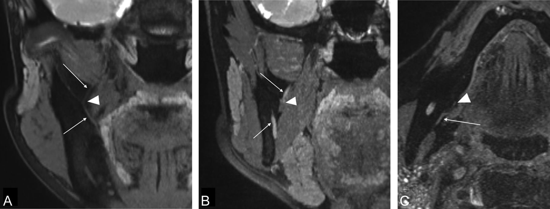
  • Fig 2.
    • Download figure
    • Open in new tab
    • Download powerpoint
    Fig 2.

    A and B, Coronal reformatted image of the 3D-DESS-WE sequence. C, Axial source image of the 3D-DESS-WE sequence. Reformatted coronal images show the lingual nerve (A, arrows) and the inferior alveolar nerve (B, arrows). The intermediate points are established where the lingual nerve running laterally starts to change direction medially (A, arrowhead) and where the inferior alveolar nerve enters the mandibular foramen (B; arrowhead). Axial image shows that the mylohyoid nerve runs at the medial surface of the mandible (C, arrow). The intermediate point is established where the nerve enters the mylohyoid muscle (C, arrowhead).

Tables

  • Figures
    • View popup
    Table 1:

    Average scores of the 3 readers evaluating the V3 nerves

    V3 NervesReader AReader BReader CAverage
    Masseteric3.34 ± 1.0833.36 ± 1.0153.24 ± 1.0163.31 ± 1.038
    Buccal2.64 ± 1.3082.75 ± 1.3322.63 ± 1.2812.67 ± 1.306
    Auriculotemporal3.13 ± 1.3763.07 ± 1.2203.13 ± 1.1853.11 ± 1.261
    Lingual3.77 ± 0.7813.88 ± 0.5443.75 ± 0.6823.80 ± 0.677
    Inferior alveolar4.00 ± 0.0004.00 ± 0.0003.97 ± 0.1573.99 ± 0.091
    Mylohyoid0.46 ± 0.7850.67 ± 0.9170.72 ± 0.7640.62 ± 0.831
    • View popup
    Table 2:

    Interobserver variability of the 3 readers (A, B, and C) evaluating the V3 nerves

    V3 NervesA and BB and CC and AAverage
    Masseteric0.9600.9700.9610.96
    Buccal0.9540.9730.9360.95
    Auriculotemporal0.9420.9610.9550.95
    Lingual0.9740.9940.9770.98
    Inferior alveolar1.0000.9980.9981.00
    Mylohyoid0.9830.9670.9590.97
PreviousNext
Back to top

In this issue

American Journal of Neuroradiology: 36 (7)
American Journal of Neuroradiology
Vol. 36, Issue 7
1 Jul 2015
  • Table of Contents
  • Index by author
  • Complete Issue (PDF)
Advertisement
Print
Download PDF
Email Article

Thank you for your interest in spreading the word on American Journal of Neuroradiology.

NOTE: We only request your email address so that the person you are recommending the page to knows that you wanted them to see it, and that it is not junk mail. We do not capture any email address.

Enter multiple addresses on separate lines or separate them with commas.
Visualization of the Peripheral Branches of the Mandibular Division of the Trigeminal Nerve on 3D Double-Echo Steady-State with Water Excitation Sequence
(Your Name) has sent you a message from American Journal of Neuroradiology
(Your Name) thought you would like to see the American Journal of Neuroradiology web site.
CAPTCHA
This question is for testing whether or not you are a human visitor and to prevent automated spam submissions.
Cite this article
H. Fujii, A. Fujita, A. Yang, H. Kanazawa, K. Buch, O. Sakai, H. Sugimoto
Visualization of the Peripheral Branches of the Mandibular Division of the Trigeminal Nerve on 3D Double-Echo Steady-State with Water Excitation Sequence
American Journal of Neuroradiology Jul 2015, 36 (7) 1333-1337; DOI: 10.3174/ajnr.A4288

Citation Manager Formats

  • BibTeX
  • Bookends
  • EasyBib
  • EndNote (tagged)
  • EndNote 8 (xml)
  • Medlars
  • Mendeley
  • Papers
  • RefWorks Tagged
  • Ref Manager
  • RIS
  • Zotero
0 Responses
Respond to this article
Share
Bookmark this article
Visualization of the Peripheral Branches of the Mandibular Division of the Trigeminal Nerve on 3D Double-Echo Steady-State with Water Excitation Sequence
H. Fujii, A. Fujita, A. Yang, H. Kanazawa, K. Buch, O. Sakai, H. Sugimoto
American Journal of Neuroradiology Jul 2015, 36 (7) 1333-1337; DOI: 10.3174/ajnr.A4288
del.icio.us logo Twitter logo Facebook logo Mendeley logo
  • Tweet Widget
  • Facebook Like
  • Google Plus One
Purchase

Jump to section

  • Article
    • Abstract
    • ABBREVIATIONS:
    • Materials and Methods
    • Results
    • Discussion
    • Conclusions
    • Footnotes
    • REFERENCES
  • Figures & Data
  • Info & Metrics
  • Responses
  • References
  • PDF

Related Articles

  • No related articles found.
  • PubMed
  • Google Scholar

Cited By...

  • Visualization of the Extracranial Branches of the Trigeminal Nerve Using Improved Motion-Sensitized Driven Equilibrium-Prepared 3D Inversion Recovery TSE Sequence
  • 3D Cranial Nerve Imaging, a Novel MR Neurography Technique Using Black-Blood STIR TSE with a Pseudo Steady-State Sweep and Motion-Sensitized Driven Equilibrium Pulse for the Visualization of the Extraforaminal Cranial Nerve Branches
  • Localization of Parotid Gland Tumors in Relation to the Intraparotid Facial Nerve on 3D Double-Echo Steady-State with Water Excitation Sequence
  • Crossref (47)
  • Google Scholar

This article has been cited by the following articles in journals that are participating in Crossref Cited-by Linking.

  • MR Neurographic Evaluation of Facial and Neck Pain: Normal and Abnormal Craniospinal Nerves below the Skull Base
    Avneesh Chhabra, Gitanjali Bajaj, Vibhor Wadhwa, Rehan S. Quadri, Jonathan White, Larry L. Myers, Bardia Amirlak, John R. Zuniga
    RadioGraphics 2018 38 5
  • Localization of Parotid Gland Tumors in Relation to the Intraparotid Facial Nerve on 3D Double-Echo Steady-State with Water Excitation Sequence
    H. Fujii, A. Fujita, H. Kanazawa, E. Sung, O. Sakai, H. Sugimoto
    American Journal of Neuroradiology 2019 40 6
  • Magnetic resonance neurography in the management of peripheral trigeminal neuropathy: experience in a tertiary care centre
    Brian Cox, John R. Zuniga, Neeraj Panchal, Jonathan Cheng, Avneesh Chhabra
    European Radiology 2016 26 10
  • Visualization of the Inferior Alveolar Nerve and Lingual Nerve Using MRI in Oral and Maxillofacial Surgery: A Systematic Review
    Adib Al-Haj Husain, Mark Solomons, Bernd Stadlinger, Rada Pejicic, Sebastian Winklhofer, Marco Piccirelli, Silvio Valdec
    Diagnostics 2021 11 9
  • Neuropathy Score Reporting and Data System (NS-RADS): MRI Reporting Guideline of Peripheral Neuropathy Explained and Reviewed
    Avneesh Chhabra, Swati D. Deshmukh, Amelie M. Lutz, Jan Fritz, Darryl B. Sneag, Bayan Mogharrabi, Mina Guirguis, Gustav Andreisek, Yin Xi, Shivani Ahlawat
    Skeletal Radiology 2022 51 10
  • Visualization of the morphology and pathology of the peripheral branches of the cranial nerves using three-dimensional high-resolution high-contrast magnetic resonance neurography
    Wenjun Wu, Feihong Wu, Dingxi Liu, Chuansheng Zheng, Xiangquan Kong, Shenglei Shu, Dan Li, Xiangchuang Kong, Lixia Wang
    European Journal of Radiology 2020 132
  • Three-dimensional double-echo steady-state with water excitation magnetic resonance imaging to localize the intraparotid facial nerve in patients with deep-seated parotid tumors
    Yikyung Kim, Han-Sin Jeong, Hyung-Jin Kim, Minjung Seong, Yoonha Kim, Sung Tae Kim
    Neuroradiology 2021 63 5
  • 3D Cranial Nerve Imaging, a Novel MR Neurography Technique Using Black-Blood STIR TSE with a Pseudo Steady-State Sweep and Motion-Sensitized Driven Equilibrium Pulse for the Visualization of the Extraforaminal Cranial Nerve Branches
    F. Van der Cruyssen, T.-M. Croonenborghs, R. Hermans, R. Jacobs, J. Casselman
    American Journal of Neuroradiology 2021 42 3
  • Magnetic resonance neurography of the head and neck: state of the art, anatomy, pathology and future perspectives
    Fréderic Van der Cruyssen, Tomas-Marijn Croonenborghs, Tara Renton, Robert Hermans, Constantinus Politis, Reinhilde Jacobs, Jan Casselman
    The British Journal of Radiology 2021 94 1119
  • Preoperative visualization of the lingual nerve by 3D double-echo steady-state MRI in surgical third molar extraction treatment
    Adib Al-Haj Husain, Silvio Valdec, Bernd Stadlinger, Martin Rücker, Marco Piccirelli, Sebastian Winklhofer
    Clinical Oral Investigations 2022 26 2

More in this TOC Section

  • ASL Sensitivity for Head and Neck Paraganglioma
  • Post SRS Peritumoral Hyperintense Signal of VSs
  • DIVr of NPC Predicted Outcome
Show more HEAD AND NECK IMAGING

Similar Articles

Advertisement

Indexed Content

  • Current Issue
  • Accepted Manuscripts
  • Article Preview
  • Past Issues
  • Editorials
  • Editor's Choice
  • Fellows' Journal Club
  • Letters to the Editor
  • Video Articles

Cases

  • Case Collection
  • Archive - Case of the Week
  • Archive - Case of the Month
  • Archive - Classic Case

More from AJNR

  • Trainee Corner
  • Imaging Protocols
  • MRI Safety Corner
  • Book Reviews

Multimedia

  • AJNR Podcasts
  • AJNR Scantastics

Resources

  • Turnaround Time
  • Submit a Manuscript
  • Submit a Video Article
  • Submit an eLetter to the Editor/Response
  • Manuscript Submission Guidelines
  • Statistical Tips
  • Fast Publishing of Accepted Manuscripts
  • Graphical Abstract Preparation
  • Imaging Protocol Submission
  • Evidence-Based Medicine Level Guide
  • Publishing Checklists
  • Author Policies
  • Become a Reviewer/Academy of Reviewers
  • News and Updates

About Us

  • About AJNR
  • Editorial Board
  • Editorial Board Alumni
  • Alerts
  • Permissions
  • Not an AJNR Subscriber? Join Now
  • Advertise with Us
  • Librarian Resources
  • Feedback
  • Terms and Conditions
  • AJNR Editorial Board Alumni

American Society of Neuroradiology

  • Not an ASNR Member? Join Now

© 2025 by the American Society of Neuroradiology All rights, including for text and data mining, AI training, and similar technologies, are reserved.
Print ISSN: 0195-6108 Online ISSN: 1936-959X

Powered by HighWire