Advertisement

Main menu

  • Home
  • Content
    • Current Issue
    • Accepted Manuscripts
    • Article Preview
    • Past Issue Archive
    • Video Articles
    • AJNR Case Collection
    • Case of the Week Archive
    • Case of the Month Archive
    • Classic Case Archive
  • Special Collections
    • AJNR Awards
    • Low-Field MRI
    • Alzheimer Disease
    • ASNR Foundation Special Collection
    • Photon-Counting CT
    • View All
  • Multimedia
    • AJNR Podcasts
    • AJNR SCANtastic
    • Trainee Corner
    • MRI Safety Corner
    • Imaging Protocols
  • For Authors
    • Submit a Manuscript
    • Submit a Video Article
    • Submit an eLetter to the Editor/Response
    • Manuscript Submission Guidelines
    • Statistical Tips
    • Fast Publishing of Accepted Manuscripts
    • Graphical Abstract Preparation
    • Imaging Protocol Submission
    • Author Policies
  • About Us
    • About AJNR
    • Editorial Board
    • Editorial Board Alumni
  • More
    • Become a Reviewer/Academy of Reviewers
    • Subscribers
    • Permissions
    • Alerts
    • Feedback
    • Advertisers
    • ASNR Home

User menu

  • Alerts
  • Log in

Search

  • Advanced search
American Journal of Neuroradiology

American Journal of Neuroradiology

ASHNR American Society of Functional Neuroradiology ASHNR American Society of Pediatric Neuroradiology ASSR
  • Alerts
  • Log in

Advanced Search

  • Home
  • Content
    • Current Issue
    • Accepted Manuscripts
    • Article Preview
    • Past Issue Archive
    • Video Articles
    • AJNR Case Collection
    • Case of the Week Archive
    • Case of the Month Archive
    • Classic Case Archive
  • Special Collections
    • AJNR Awards
    • Low-Field MRI
    • Alzheimer Disease
    • ASNR Foundation Special Collection
    • Photon-Counting CT
    • View All
  • Multimedia
    • AJNR Podcasts
    • AJNR SCANtastic
    • Trainee Corner
    • MRI Safety Corner
    • Imaging Protocols
  • For Authors
    • Submit a Manuscript
    • Submit a Video Article
    • Submit an eLetter to the Editor/Response
    • Manuscript Submission Guidelines
    • Statistical Tips
    • Fast Publishing of Accepted Manuscripts
    • Graphical Abstract Preparation
    • Imaging Protocol Submission
    • Author Policies
  • About Us
    • About AJNR
    • Editorial Board
    • Editorial Board Alumni
  • More
    • Become a Reviewer/Academy of Reviewers
    • Subscribers
    • Permissions
    • Alerts
    • Feedback
    • Advertisers
    • ASNR Home
  • Follow AJNR on Twitter
  • Visit AJNR on Facebook
  • Follow AJNR on Instagram
  • Join AJNR on LinkedIn
  • RSS Feeds

AJNR Awards, New Junior Editors, and more. Read the latest AJNR updates

Research ArticleBrain

Altered Microstructure in Temporal Lobe Epilepsy: A Diffusional Kurtosis Imaging Study

L. Bonilha, C.-Y. Lee, J.H. Jensen, A. Tabesh, M.V. Spampinato, J.C. Edwards, J. Breedlove and J.A. Helpern
American Journal of Neuroradiology April 2015, 36 (4) 719-724; DOI: https://doi.org/10.3174/ajnr.A4185
L. Bonilha
aFrom the Departments of Neurology and Neurosurgery (L.B., J.C.E.)
cComprehensive Epilepsy Center (L.B., J.C.E., J.B.)
dCenter for Biomedical Imaging (L.B., C.-Y.L., J.H.J., A.T., M.V.S.), Medical University of South Carolina, Charleston, South Carolina.
  • Find this author on Google Scholar
  • Find this author on PubMed
  • Search for this author on this site
C.-Y. Lee
bRadiology and Radiological Science (C.-Y.L., J.H.J., A.T., M.V.S., J.A.H.)
dCenter for Biomedical Imaging (L.B., C.-Y.L., J.H.J., A.T., M.V.S.), Medical University of South Carolina, Charleston, South Carolina.
  • Find this author on Google Scholar
  • Find this author on PubMed
  • Search for this author on this site
J.H. Jensen
bRadiology and Radiological Science (C.-Y.L., J.H.J., A.T., M.V.S., J.A.H.)
dCenter for Biomedical Imaging (L.B., C.-Y.L., J.H.J., A.T., M.V.S.), Medical University of South Carolina, Charleston, South Carolina.
  • Find this author on Google Scholar
  • Find this author on PubMed
  • Search for this author on this site
A. Tabesh
bRadiology and Radiological Science (C.-Y.L., J.H.J., A.T., M.V.S., J.A.H.)
dCenter for Biomedical Imaging (L.B., C.-Y.L., J.H.J., A.T., M.V.S.), Medical University of South Carolina, Charleston, South Carolina.
  • Find this author on Google Scholar
  • Find this author on PubMed
  • Search for this author on this site
M.V. Spampinato
bRadiology and Radiological Science (C.-Y.L., J.H.J., A.T., M.V.S., J.A.H.)
dCenter for Biomedical Imaging (L.B., C.-Y.L., J.H.J., A.T., M.V.S.), Medical University of South Carolina, Charleston, South Carolina.
  • Find this author on Google Scholar
  • Find this author on PubMed
  • Search for this author on this site
J.C. Edwards
aFrom the Departments of Neurology and Neurosurgery (L.B., J.C.E.)
cComprehensive Epilepsy Center (L.B., J.C.E., J.B.)
  • Find this author on Google Scholar
  • Find this author on PubMed
  • Search for this author on this site
J. Breedlove
cComprehensive Epilepsy Center (L.B., J.C.E., J.B.)
  • Find this author on Google Scholar
  • Find this author on PubMed
  • Search for this author on this site
J.A. Helpern
bRadiology and Radiological Science (C.-Y.L., J.H.J., A.T., M.V.S., J.A.H.)
  • Find this author on Google Scholar
  • Find this author on PubMed
  • Search for this author on this site
  • Article
  • Figures & Data
  • Supplemental
  • Info & Metrics
  • Responses
  • References
  • PDF
Loading

Abstract

BACKGROUND AND PURPOSE: Temporal lobe epilepsy is associated with regional abnormalities in tissue microstructure, as demonstrated by DTI. However, the full extent of these abnormalities has not yet been defined because DTI conveys only a fraction of the information potentially accessible with diffusion MR imaging. In this study, we assessed the added value of diffusional kurtosis imaging, an extension of DTI, to evaluate microstructural abnormalities in patients with temporal lobe epilepsy.

MATERIALS AND METHODS: Thirty-two patients with left temporal lobe epilepsy and 36 matched healthy subjects underwent diffusion MR imaging. To evaluate abnormalities in patients, we performed voxelwise analyses, assessing DTI-derived mean diffusivity, fractional anisotropy, and diffusional kurtosis imaging–derived mean diffusional kurtosis, as well as diffusional kurtosis imaging and DTI-derived axial and radial components, comparing patients with controls.

RESULTS: We replicated findings from previous studies demonstrating a reduction in fractional anisotropy and an increase in mean diffusivity preferentially affecting, but not restricted to, the temporal lobe ipsilateral to seizure onset. We also noted a pronounced pattern of diffusional kurtosis imaging abnormalities in gray and white matter tissues, often extending into regions that were not detected as abnormal by DTI measures.

CONCLUSIONS: Diffusional kurtosis is a sensitive and complementary measure of microstructural compromise in patients with temporal lobe epilepsy. It provides additional information regarding the anatomic distribution and degree of damage in this condition. Diffusional kurtosis imaging may be used as a biomarker for disease severity, clinical phenotypes, and treatment monitoring in epilepsy.

ABBREVIATIONS:

DKI
diffusional kurtosis imaging
FA
fractional anisotropy
MD
mean diffusivity
TLE
temporal lobe epilepsy

The most common histologic finding in patients with medial temporal lobe epilepsy (TLE) is hippocampal sclerosis, which is defined as neuronal loss and gliosis involving the hippocampus.1⇓–3 Routine clinical MR imaging of patients with TLE can demonstrate signs associated with hippocampal sclerosis.4,5 Recent observations suggest that hippocampal abnormalities are not the only structural injury in TLE.6⇓⇓–9 Imaging studies using whole-brain quantitative morphometry have repeatedly demonstrated that TLE is associated with extrahippocampal abnormalities6,8⇓⇓⇓⇓–13 involving perihippocampal and perilimbic structures.

However, the full extent of structural abnormalities in TLE is still unclear. Therefore, the lack of a sensitive and specific marker of extrahippocampal damage may prevent the assessment of its clinical relevance. Diffusion MR imaging techniques aimed at quantifying tissue microstructure may offer more sensitive tools for determining the extent of brain pathology in TLE. A promising new method called diffusional kurtosis imaging (DKI) can provide information about cerebral microstructural abnormalities beyond that provided by conventional diffusion tensor imaging.14,15 In fact, by providing a more comprehensive characterization of water diffusion properties, DKI may be more suitable for detecting subtle brain damage in TLE.

In this study, we investigated the anatomical pattern of microstructural abnormalities associated with TLE by contrasting the voxel-based analyses of microstructure maps derived from DKI and from well-established DTI.

Materials and Methods

Subjects

Thirty-two consecutive patients with left TLE were recruited from the Comprehensive Epilepsy Center at the Medical University of South Carolina, where they were diagnosed on the basis of a comprehensive history and neurologic evaluation. The TLE diagnosis was defined in accordance with the criteria proposed by the International League Against Epilepsy,16 based on a history compatible with partial seizures with temporal onset, interictal epileptiform discharges on interictal electroencephalography, and hippocampal atrophy on visual inspection of MR imaging. Not all patients included in this study were surgical candidates; thus, ictal recording was not obtained in all patients. All patients exhibited a classic presentation for TLE, based on history, seizure semiology, epileptiform discharges on interictal electroencephalography, as well as routine MRI. Most patients, but not all, exhibited signs of hippocampal sclerosis on MR imaging, and a detailed description of the demographic and clinical information for patients included in this study is provided in the On-line Table.

The mean age of patients was 44.8 ± 16.7 years; 22 patients were women. We also studied a control group of healthy individuals recruited from the local community who had no history of neurologic problems and no risk factors for epilepsy (mean age, 40.4 ± 11.6 years; 24 women). Patients and controls were similar in sex (P = .21) and age (P = .92) distributions.

Image Acquisition

All patients and controls underwent the same imaging protocol performed on a 3T Magnetom Verio MR imaging scanner (Siemens, Erlangen, Germany) equipped with a 12-channel head coil. Diffusion-weighted images were obtained by using a twice-refocused echo-planar sequence with 3 diffusion weightings (b=0, 1000, and 2000 s/mm2) along 30 diffusion-encoding directions with NEX = 1 (NEX = 10 for b=0). Other imaging parameters were TR = 8500 ms, TE = 98 ms, FOV = 222 × 222 mm2, matrix size = 74 × 74, parallel imaging factor = 2, no partial Fourier encoding, section thickness = 3 mm, and 40 axial sections. Acquisition time was 9 minutes and 12 seconds. Structural images were obtained by using a magnetization-prepared rapid acquisition of gradient echo sequence with TR = 2250 ms, TE = 4.18 ms, TI = 900 ms, FOV = 256 × 256 mm2, matrix size = 256 × 256, section thickness = 1 mm, and 176 sagittal sections.

Image Processing

Tissue Volume Maps.

T1-weighted images were submitted to gray and white matter tissue segmentation using the VBM toolbox (http://dbm.neuro.uni-jena.de/vbm/) for SPM8 software (http://www.fil.ion.ucl.ac.uk/spm/software/spm8). Image normalization and segmentation were performed iteratively by using the symmetric tissue probability map of VBM, very light regularization, and affine regularization to the ICBM space template (European Brains; http://bmap.ucla.edu/portfolio/atlases/ICBM_Template/). Spatial normalization was performed with affine and nonlinear transformations yielding modulated normalized gray and white matter probability maps. All maps were transformed into standard space, and their averages (gray and white matter maps separately) were used to provide tissue inclusion masks for statistical analyses.

Microstructure Maps.

DKI postprocessing was performed by using the in-house software Diffusional Kurtosis Estimator (http://www.nitrc.org/projects/dke),17 which performed the following processing steps to generate diffusivity and kurtosis maps: first, motion correction through a 6-parameter rigid-body transformation to spatially align all DWIs. Second, at each voxel, the diffusion and diffusional kurtosis tensors were jointly fitted to the DWIs for b=0, 1000, and 2000 s/mm2 for that voxel, and the mean kurtosis was calculated from the tensors. The diffusion tensor was fitted to the DWIs for b=0 and 1000 s/mm2, and mean diffusivity and (MD) fractional anisotropy (FA) were calculated from the diffusion tensor. Axial and radial diffusion and kurtosis maps were also generated.

FA maps were nonlinearly normalized to a common space by using the fMRI of the Brain Software Library, Version 4.1.7 (http://www.fmrib.ox.ac.uk/fsl) using a symmetric FA template, and the resulting transformation was applied to normalize the other maps. The spatially normalized images were then submitted to spatial smoothing with an 8-mm Gaussian filter.

Statistical Analyses.

We performed whole-brain voxelwise analyses, comparing patients with TLE and controls regarding MD, FA, mean kurtosis, axial diffusion, radial diffusion, axial kurtosis, and radial kurtosis maps. We performed comparisons using voxelwise t tests. All results were corrected for multiple comparisons using a false discovery rate threshold of q < 0.01.18

Results

Gray Matter

Patients with TLE exhibited multiple cortical areas with significant increase in diffusion parameters, along with a concurrent decrease in kurtosis parameters.

Specifically, an increase in MD was observed within the orbitofrontal cortices, frontal poles, medial left temporal lobe region, and midposterior cingulate. No significant changes were observed with axial diffusion, but radial diffusion was increased in patients in the orbitofrontal regions, frontal poles, medial left temporal regions, left lateral temporal regions, and midposterior cingulate cortex.

In contrast, at the same statistical threshold level, a more diffuse pattern of significant reductions in mean kurtosis was observed, encompassing the temporopolar cortices, temporal and frontal opercula, medial temporal lobe regions (more pronounced on the left side), left lateral temporal region, prefrontal cortices and temporal poles, and cingulate and parietal regions. Overall, a higher statistical difference was noted on the left side.

Reductions in axial kurtosis were noted particularly over the frontal polar regions, left temporal pole, left lateral temporal cortex, and left precentral areas. Reductions in radial kurtosis were diffusely distributed across the entire brain but were particularly intense over the left medial temporal cortex, left temporal polar cortex, left insula, left fusiform gyrus, cingulate cortex, and left precentral regions.

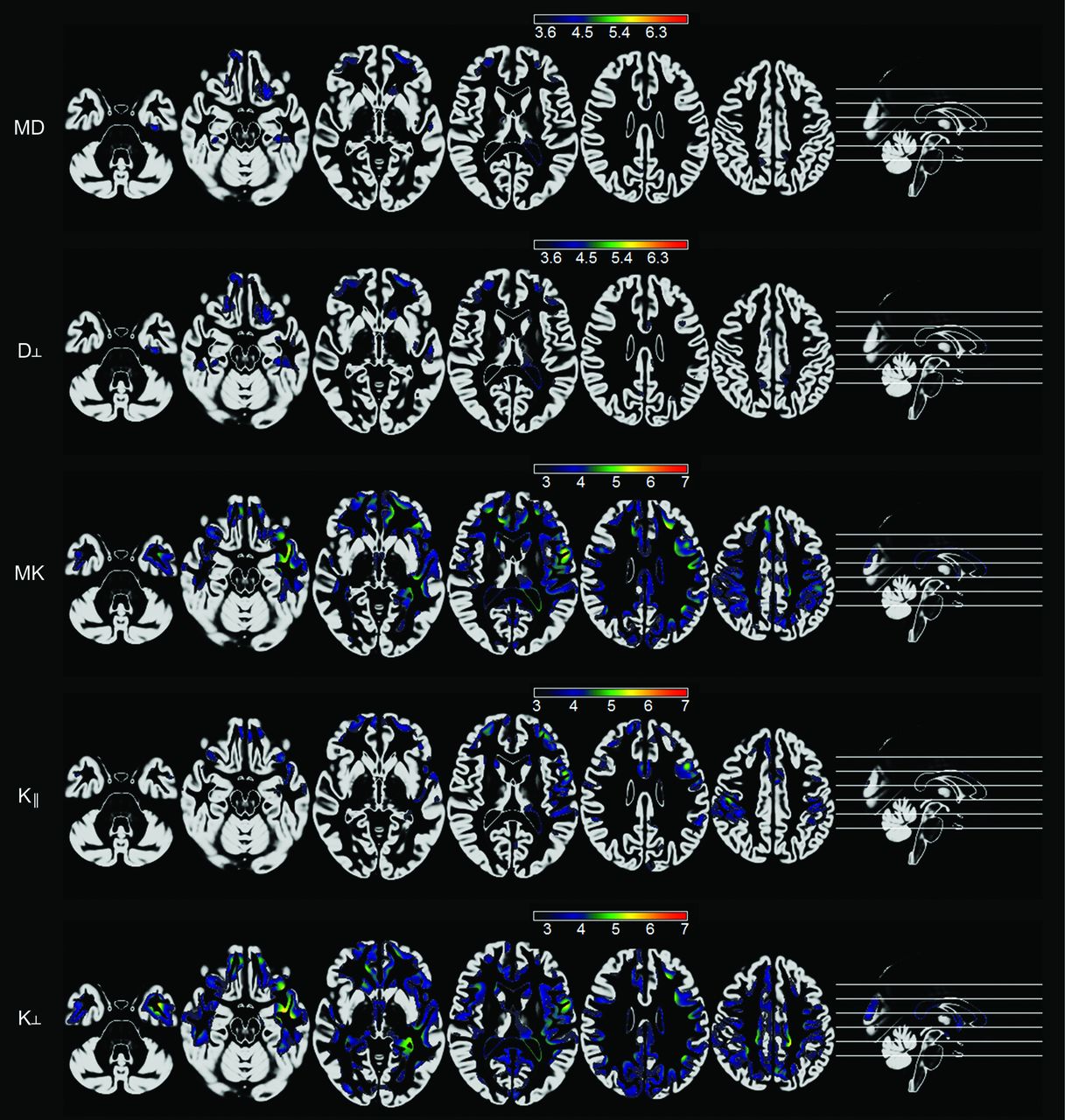
These results are shown in Fig 1.

Fig 1.
  • Download figure
  • Open in new tab
  • Download powerpoint
Fig 1.

Voxelwise maps of cortical abnormalities in patients with TLE compared with controls. Statistical maps are overlaid on an average of probabilistic gray matter maps from all subjects. The scale bars represent absolute values of t scores. The first row demonstrates areas with an increase in MD in patients. The second row demonstrates areas of increased radial diffusion in patients. The third, fourth, and fifth rows demonstrate areas of reduced mean kurtosis and axial and radial kurtosis, respectively, in patients. All results were corrected for multiple comparisons by using a false discovery rate threshold of q < 0.01.

There were no cortical areas of reduced diffusion or increased kurtosis measures in patients.

White Matter

Patients with TLE demonstrated a significant increase in MD within the white matter in the frontal, left medial temporal, perithalamic, left occipitotemporal, and medial frontoparietal regions. There was also an increase in MD within the splenium of the corpus callosum. These abnormalities were also noted in areas of radial diffusion decrement, while axial diffusion did not demonstrate significant abnormalities between groups.

FA decrement was noted in a widespread pattern involving the medial temporal, perithalamic, orbitofrontal, left temporopolar, left occipitotemporal, and medial frontoparietal white matter regions. There was also a notable decrement in FA within the splenium of the corpus callosum, left side of the genu of the corpus callosum, and left uncinate fasciculus.

At the same statistical threshold, there was a remarkable anatomical pattern of mean kurtosis reduction, which involved most of the brain white matter and was most pronounced over the left temporal, bilateral orbitofrontal, and left frontoparietal regions. These abnormalities were similar to the pattern noted with radial kurtosis decrement in patients.

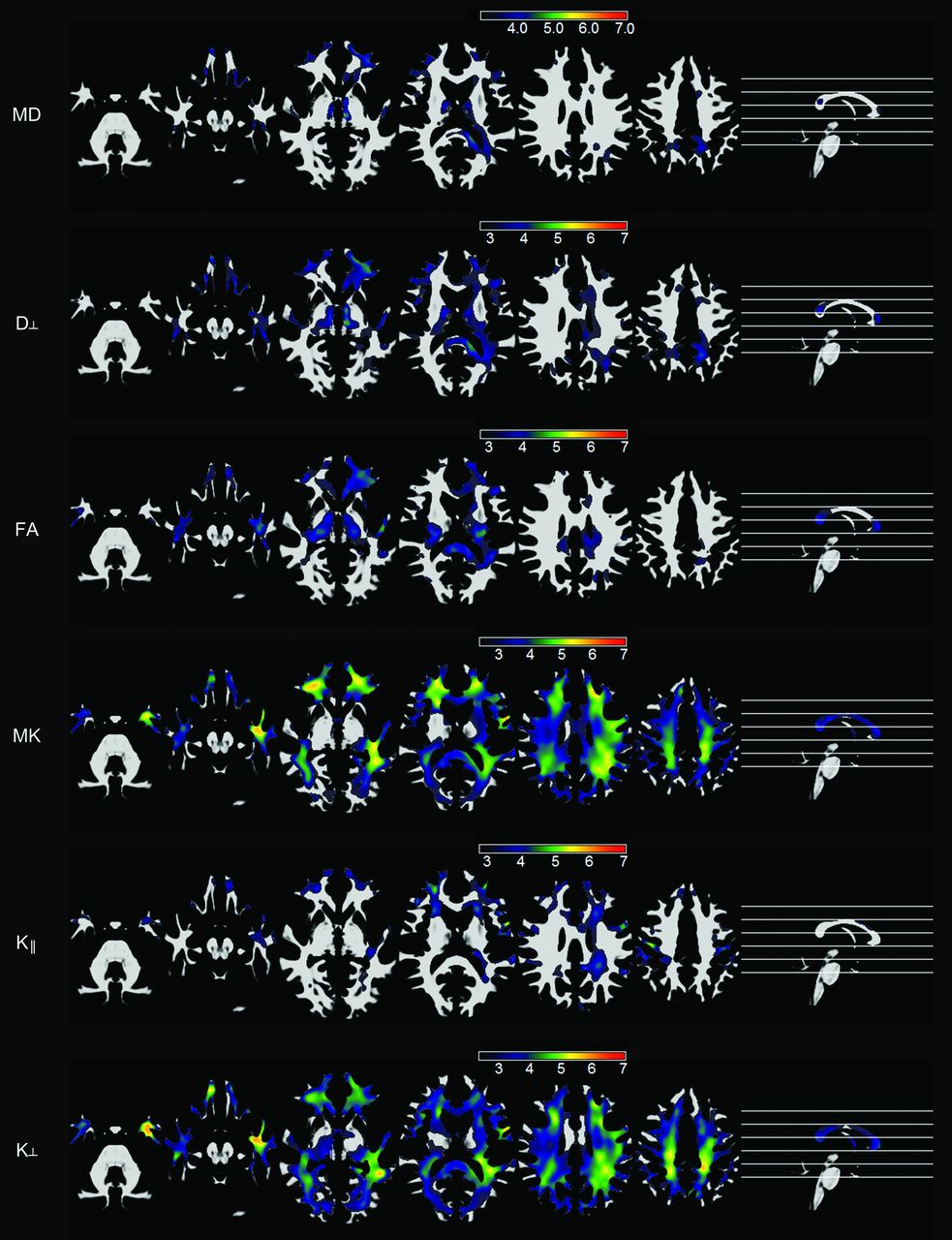
These results are shown in Fig 2.

Fig 2.
  • Download figure
  • Open in new tab
  • Download powerpoint
Fig 2.

Voxelwise maps of white matter abnormalities in patients with TLE compared with controls. Statistical maps are overlaid on an average of probabilistic white matter maps from all subjects. The scale bars represent absolute values of t scores. The first and second rows demonstrate areas with an increase in MD and radial diffusion, respectively, in patients. The third row demonstrates areas with reduced FA in patients, while the fourth, fifth, and sixth rows demonstrate areas of reduced mean kurtosis, axial kurtosis, and radial kurtosis in patients, respectively. All results were corrected for multiple comparisons by using a false discovery rate threshold of q < 0.01.

There were no areas of MD decrement, FA increase, or kurtosis increase in patients.

Discussion

In this study, we investigated microstructural gray and white matter abnormalities in patients with TLE using a novel diffusion MR imaging technique called DKI. DKI is aimed at detecting changes in tissue microstructure, including those arising from non-Gaussian properties of water diffusion, thus providing a potentially more sensitive and specific tool for the evaluation of pathologic changes in TLE.14,15 We contrasted the DKI-derived microstructural measures with the better established DTI-derived measures.

We replicated findings from several previous studies using DTI to assess microstructural abnormalities in TLE. Specifically, we demonstrated areas of increased MD and reduced FA in patients with TLE, which were preferentially located within, but not restricted to, the temporal lobe ipsilateral to the side of seizure onset.19⇓⇓⇓⇓⇓⇓⇓⇓⇓–29

We observed that DKI is capable of detecting a broader anatomic pattern of microstructural abnormalities in patients with TLE, compared with conventional measures. Moreover, these results demonstrate a distribution of network abnormalities that is in direct concordance with the hypothesized network pathology related to TLE.13,30 The presence of abnormalities demonstrated by DKI is more contiguous and anatomically plausible compared with conventional methodologies, and it is consistent with the theory of limbic and perilimbic dysfunction in TLE.31,32

In this study, we also corroborated the findings from Gao et al,33 suggesting that DKI is more sensitive, compared with DTI, to detect microstructural abnormalities in the patients with TLE. Gao et al studied a population of children with TLE; nonetheless, their results are fairly equivalent to our observations from a population of adult subjects with TLE.

DKI and the Pathophysiology of TLE

The hypothesis that TLE is a disease that affects more than just the hippocampus has been corroborated by several imaging studies demonstrating abnormalities in TLE involving limbic structures.6,8⇓⇓⇓⇓–13 However, there is considerable variability across studies regarding the extent and degree of extrahippocampal damage in TLE. These variations may be directly related to the sensitivity of the methods, as further highlighted by the observation that conventional methods are not sensitive to most extrahippocampal histologic changes.34,35 This variability in findings precludes a better understanding of the pathophysiology of TLE.

Our results suggest that TLE is associated with extensive microstructural abnormalities, encompassing a contiguous and extensive network of extrahippocampal and extratemporal regions.

Our results were obtained from a population of consecutive patients with TLE, most of whom demonstrated signs of hippocampal sclerosis on MR imaging (On-line Table). Therefore, it is possible that the temporal and extratemporal microstructural abnormalities demonstrated by DKI are correlated with hippocampal cell loss. Future studies evaluating phenotypical differences in the distribution of DKI abnormalities may provide further insight into this subject.

Moreover, the patient population evaluated in this study was not composed exclusively of surgical candidates (ie, it included patients with relatively well-controlled epilepsy). Most interesting, our strong statistical results suggest that microstructural abnormalities were widespread across all subjects. While these findings support DKI as exquisitely sensitive to abnormalities associated with TLE, it is still unclear whether DKI can provide further information regarding classification into subgroups, particularly as it relates to pharmacologic or surgical treatment outcomes. This study provides initial evidence of DKI as a biomarker of TLE, and we believe that future studies could address the impact of DKI as a clinical biomarker.

DKI as a Biomarker

The results of this study suggest that DKI may provide a sensitive and specific biomarker of TLE. Specifically, it may be a powerful tool for determining the degree of abnormality in cortical regions, which may be related to the clinical or neuropsychological profiles, and for quantifying the burden of overall network abnormalities on clinical progression. Specifically, extrahippocampal microstructural tissue abnormalities may be related to epileptogenesis in some individuals with TLE.31,36,37 These microstructural changes may represent a complex interaction among multiple pathologic mechanisms such as cell loss, inflammation, and axonal and dendritic reorganization, which are known to occur in epilepsy.38

Clinical Feasibility of DKI

DKI provides a convenient platform for the evaluation of microstructural abnormalities related to TLE and is one form of advanced diffusion MR imaging.39 However, DKI may be advantageous to other methods due to its feasibility in routine clinical practice. Other modalities require significantly longer scan times and/or custom hardware. Conversely, DKI can be acquired with a high signal-to-noise ratio in approximately 6–10 minutes, depending on the scanner.

Future Applications

This study provides preliminary evidence in support of the utility of DKI for characterization of microstructural abnormalities in TLE. Our results suggest that DKI–defined microstructural abnormalities are pervasive in the hippocampal, temporal, and extratemporal regions. Future studies should further investigate the histologic correlates of these abnormalities and their relationship with the clinical and neuropsychological profiles in patients with medial temporal lobe epilepsy.

Conclusions

This study demonstrates the added value of kurtosis measures in the assessment of microstructural alterations in adult patients with TLE. The results of this study confirm our hypothesis that DKI may provide complementary information regarding the location and magnitude of structural abnormalities in TLE.

Footnotes

  • Disclosures: Leonardo Bonilha—RELATED: Grant: American Society of Neuroradiology (grant No. 89779-01, Principal Investigator, A. Tabesh).* Jens H. Jensen—OTHER RELATIONSHIPS: Siemens owns a royalty-free nonexclusive license for diffusional kurtosis imaging with the pending patent held by New York University. I am one of the inventors. Maria V. Spampinato—UNRELATED: Grants/Grants Pending: Bracco,* Comments: clinical trial on 2 FDA-approved contrast agents. Ali Tabesh—RELATED: Grant: American Society of Neuroradiology,* Comments: 2013 Research Scientist Award. *Money paid to the institution.

  • This study was supported by the American Society of Neuroradiology (grant No. 89779-01; Principal Investigator, A. Tabesh).

REFERENCES

  1. 1.↵
    1. Sendrowski K,
    2. Sobaniec W
    . Hippocampus, hippocampal sclerosis and epilepsy. Pharmacol Rep 2013;65:555–65
    CrossRefPubMed
  2. 2.↵
    1. Blümcke I,
    2. Coras R,
    3. Miyata H, et al
    . Defining clinico-neuropathological subtypes of mesial temporal lobe epilepsy with hippocampal sclerosis. Brain Pathol 2012;22:402–11
    CrossRefPubMed
  3. 3.↵
    1. Engel JJ
    1. Babb TL,
    2. Brown WJ
    . Pathological findings in epilepsy. In: Engel JJ, ed. Surgical Treatment of the Epilepsies. New York: Raven; 1987:511–40
  4. 4.↵
    1. Cendes F,
    2. Andermann F,
    3. Gloor P, et al
    . MRI volumetric measurement of amygdala and hippocampus in temporal lobe epilepsy. Neurology 1993;43:719–25
    CrossRef
  5. 5.↵
    1. Lencz T,
    2. McCarthy G,
    3. Bronen RA, et al
    . Quantitative magnetic resonance imaging in temporal lobe epilepsy: relationship to neuropathology and neuropsychological function. Ann Neurol 1992;31:629–37
    CrossRefPubMed
  6. 6.↵
    1. Bernasconi N,
    2. Duchesne S,
    3. Janke A, et al
    . Whole-brain voxel-based statistical analysis of gray matter and white matter in temporal lobe epilepsy. Neuroimage 2004;23:717–23
    CrossRefPubMed
  7. 7.↵
    1. Bonilha L,
    2. Kobayashi E,
    3. Rorden C, et al
    . Medial temporal lobe atrophy in patients with refractory temporal lobe epilepsy. J Neurol Neurosurg Psychiatry 2003;74:1627–30
    Abstract/FREE Full Text
  8. 8.↵
    1. Keller SS,
    2. Mackay CE,
    3. Barrick TR, et al
    . Voxel-based morphometric comparison of hippocampal and extrahippocampal abnormalities in patients with left and right hippocampal atrophy. Neuroimage 2002;16:23–31
    CrossRefPubMed
  9. 9.↵
    1. McDonald CR,
    2. Hagler DJ Jr.,
    3. Ahmadi ME, et al
    . Regional neocortical thinning in mesial temporal lobe epilepsy. Epilepsia 2008;49:794–803
    CrossRefPubMed
  10. 10.↵
    1. Bonilha L,
    2. Rorden C,
    3. Castellano G, et al
    . Voxel-based morphometry of the thalamus in patients with refractory medial temporal lobe epilepsy. Neuroimage 2005;25:1016–21
    CrossRefPubMed
  11. 11.↵
    1. Bonilha L,
    2. Rorden C,
    3. Castellano G, et al
    . Voxel-based morphometry reveals gray matter network atrophy in refractory medial temporal lobe epilepsy. Arch Neurol 2004;61:1379–84
    CrossRefPubMed
  12. 12.↵
    1. Bernasconi N,
    2. Bernasconi A,
    3. Caramanos Z, et al
    . Mesial temporal damage in temporal lobe epilepsy: a volumetric MRI study of the hippocampus, amygdala and parahippocampal region. Brain 2003;126:462–69
    Abstract/FREE Full Text
  13. 13.↵
    1. Keller SS,
    2. Roberts N
    . Voxel-based morphometry of temporal lobe epilepsy: an introduction and review of the literature. Epilepsia 2008;49:741–57
    CrossRefPubMed
  14. 14.↵
    1. Jensen JH,
    2. Helpern JA
    . MRI quantification of non-Gaussian water diffusion by kurtosis analysis. NMR Biomed 2010;23:698–710
    CrossRefPubMed
  15. 15.↵
    1. Jensen JH,
    2. Helpern JA,
    3. Ramani A, et al
    . Diffusional kurtosis imaging: the quantification of non-Gaussian water diffusion by means of magnetic resonance imaging. Magn Reson Med 2005;53:1432–40
    CrossRefPubMed
  16. 16.↵
    Proposal for revised classification of epilepsies and epileptic syndromes: Commission on Classification and Terminology of the International League Against Epilepsy. Epilepsia 1989;30:389–99
    CrossRefPubMed
  17. 17.↵
    1. Tabesh A,
    2. Jensen JH,
    3. Ardekani BA, et al
    . Estimation of tensors and tensor-derived measures in diffusional kurtosis imaging. Magn Reson Med 2011;65:823–36
    CrossRefPubMed
  18. 18.↵
    1. Genovese CR,
    2. Lazar NA,
    3. Nichols T
    . Thresholding of statistical maps in functional neuroimaging using the false discovery rate. Neuroimage 2002;15:870–78
    CrossRefPubMed
  19. 19.↵
    1. Keller SS,
    2. Ahrens T,
    3. Mohammadi S, et al
    . Voxel-based statistical analysis of fractional anisotropy and mean diffusivity in patients with unilateral temporal lobe epilepsy of unknown cause. J Neuroimaging 2013;23:352–59
    CrossRefPubMed
  20. 20.↵
    1. Keller SS,
    2. Ahrens T,
    3. Mohammadi S, et al
    . Microstructural and volumetric abnormalities of the putamen in juvenile myoclonic epilepsy. Epilepsia 2011;52:1715–24
    CrossRefPubMed
  21. 21.↵
    1. Concha L,
    2. Beaulieu C,
    3. Collins DL, et al
    . White-matter diffusion abnormalities in temporal-lobe epilepsy with and without mesial temporal sclerosis. J Neurol Neurosurg Psychiatry 2009;80:312–19
    Abstract/FREE Full Text
  22. 22.↵
    1. Gong G,
    2. Concha L,
    3. Beaulieu C, et al
    . Thalamic diffusion and volumetry in temporal lobe epilepsy with and without mesial temporal sclerosis. Epilepsy Res 2008;80:184–93
    CrossRefPubMed
  23. 23.↵
    1. Gross DW,
    2. Concha L,
    3. Beaulieu C
    . Extratemporal white matter abnormalities in mesial temporal lobe epilepsy demonstrated with diffusion tensor imaging. Epilepsia 2006;47:1360–63
    CrossRefPubMed
  24. 24.↵
    1. Concha L,
    2. Beaulieu C,
    3. Gross DW
    . Bilateral limbic diffusion abnormalities in unilateral temporal lobe epilepsy. Ann Neurol 2005;57:188–96
    CrossRefPubMed
  25. 25.↵
    1. Thivard L,
    2. Lehericy S,
    3. Krainik A, et al
    . Diffusion tensor imaging in medial temporal lobe epilepsy with hippocampal sclerosis. Neuroimage 2005;28:682–90
    CrossRefPubMed
  26. 26.↵
    1. Kimiwada T,
    2. Juhasz C,
    3. Makki M, et al
    . Hippocampal and thalamic diffusion abnormalities in children with temporal lobe epilepsy. Epilepsia 2006;47:167–75
    CrossRefPubMed
  27. 27.↵
    1. Focke NK,
    2. Yogarajah M,
    3. Bonelli SB, et al
    . Voxel-based diffusion tensor imaging in patients with mesial temporal lobe epilepsy and hippocampal sclerosis. Neuroimage 2008;40:728–37
    CrossRefPubMed
  28. 28.↵
    1. Nilsson D,
    2. Go C,
    3. Rutka JT, et al
    . Bilateral diffusion tensor abnormalities of temporal lobe and cingulate gyrus white matter in children with temporal lobe epilepsy. Epilepsy Res 2008;81:128–35
    CrossRefPubMed
  29. 29.↵
    1. Bonilha L,
    2. Edwards JC,
    3. Kinsman SL, et al
    . Extrahippocampal gray matter loss and hippocampal deafferentation in patients with temporal lobe epilepsy. Epilepsia 2010;51:519–28
    CrossRefPubMed
  30. 30.↵
    1. Spencer SS
    . Neural networks in human epilepsy: evidence of and implications for treatment. Epilepsia 2002;43:219–27
    CrossRefPubMed
  31. 31.↵
    1. Bonilha L,
    2. Martz GU,
    3. Glazier SS, et al
    . Subtypes of medial temporal lobe epilepsy: influence on temporal lobectomy outcomes? Epilepsia 2012;53:1–6
  32. 32.↵
    1. Richardson MP
    . Large scale brain models of epilepsy: dynamics meets connectomics. J Neurol Neurosurg Psychiatry 2012;83:1238–48
    Abstract/FREE Full Text
  33. 33.↵
    1. Gao Y,
    2. Zhang Y,
    3. Wong CS, et al
    . Diffusion abnormalities in temporal lobes of children with temporal lobe epilepsy: a preliminary diffusional kurtosis imaging study and comparison with diffusion tensor imaging. NMR Biomed 2012;25:1369–77
    CrossRefPubMed
  34. 34.↵
    1. Eriksson SH,
    2. Free SL,
    3. Thom M, et al
    . Quantitative grey matter histological measures do not correlate with grey matter probability values from in vivo MRI in the temporal lobe. J Neurosci Methods 2009;181:111–18
    CrossRefPubMed
  35. 35.↵
    1. Eriksson SH,
    2. Thom M,
    3. Symms MR, et al
    . Cortical neuronal loss and hippocampal sclerosis are not detected by voxel-based morphometry in individual epilepsy surgery patients. Hum Brain Mapp 2009;30:3351–60
    CrossRefPubMed
  36. 36.↵
    1. Rorden C,
    2. Bonilha L,
    3. Nichols TE
    . Rank-order versus mean based statistics for neuroimaging. Neuroimage 2007;35:1531–37
    CrossRefPubMed
  37. 37.↵
    1. Wennberg R,
    2. Arruda F,
    3. Quesney LF, et al
    . Preeminence of extrahippocampal structures in the generation of mesial temporal seizures: evidence from human depth electrode recordings. Epilepsia 2002;43:716–26
    CrossRefPubMed
  38. 38.↵
    1. Thom M,
    2. Eriksson S,
    3. Martinian L, et al
    . Temporal lobe sclerosis associated with hippocampal sclerosis in temporal lobe epilepsy: neuropathological features. J Neuropathol Exp Neurol 2009;68:928–38
    CrossRefPubMed
  39. 39.↵
    1. Wedeen VJ,
    2. Hagmann P,
    3. Tseng WY, et al
    . Mapping complex tissue architecture with diffusion spectrum magnetic resonance imaging. Magn Reson Med 2005;54:1377–86
    CrossRefPubMed
  • Received July 12, 2014.
  • Accepted after revision October 19, 2014.
  • © 2015 by American Journal of Neuroradiology
View Abstract
PreviousNext
Back to top

In this issue

American Journal of Neuroradiology: 36 (4)
American Journal of Neuroradiology
Vol. 36, Issue 4
1 Apr 2015
  • Table of Contents
  • Index by author
  • Complete Issue (PDF)
Advertisement
Print
Download PDF
Email Article

Thank you for your interest in spreading the word on American Journal of Neuroradiology.

NOTE: We only request your email address so that the person you are recommending the page to knows that you wanted them to see it, and that it is not junk mail. We do not capture any email address.

Enter multiple addresses on separate lines or separate them with commas.
Altered Microstructure in Temporal Lobe Epilepsy: A Diffusional Kurtosis Imaging Study
(Your Name) has sent you a message from American Journal of Neuroradiology
(Your Name) thought you would like to see the American Journal of Neuroradiology web site.
CAPTCHA
This question is for testing whether or not you are a human visitor and to prevent automated spam submissions.
Cite this article
L. Bonilha, C.-Y. Lee, J.H. Jensen, A. Tabesh, M.V. Spampinato, J.C. Edwards, J. Breedlove, J.A. Helpern
Altered Microstructure in Temporal Lobe Epilepsy: A Diffusional Kurtosis Imaging Study
American Journal of Neuroradiology Apr 2015, 36 (4) 719-724; DOI: 10.3174/ajnr.A4185

Citation Manager Formats

  • BibTeX
  • Bookends
  • EasyBib
  • EndNote (tagged)
  • EndNote 8 (xml)
  • Medlars
  • Mendeley
  • Papers
  • RefWorks Tagged
  • Ref Manager
  • RIS
  • Zotero
0 Responses
Respond to this article
Share
Bookmark this article
Altered Microstructure in Temporal Lobe Epilepsy: A Diffusional Kurtosis Imaging Study
L. Bonilha, C.-Y. Lee, J.H. Jensen, A. Tabesh, M.V. Spampinato, J.C. Edwards, J. Breedlove, J.A. Helpern
American Journal of Neuroradiology Apr 2015, 36 (4) 719-724; DOI: 10.3174/ajnr.A4185
del.icio.us logo Twitter logo Facebook logo Mendeley logo
  • Tweet Widget
  • Facebook Like
  • Google Plus One
Purchase

Jump to section

  • Article
    • Abstract
    • ABBREVIATIONS:
    • Materials and Methods
    • Results
    • Discussion
    • Conclusions
    • Footnotes
    • REFERENCES
  • Figures & Data
  • Supplemental
  • Info & Metrics
  • Responses
  • References
  • PDF

Related Articles

  • PubMed
  • Google Scholar

Cited By...

  • A correlational study between microstructural, macrostructural and functional age-related changes in the human visual cortex
  • The Role of the Temporal Pole in Temporal Lobe Epilepsy: A Diffusion Kurtosis Imaging Study
  • Evaluating High Spatial Resolution Diffusion Kurtosis Imaging at 3T: Reproducibility and Quality of Fit
  • Epilepsy-related cytoarchitectonic abnormalities along white matter pathways
  • Crossref (45)
  • Google Scholar

This article has been cited by the following articles in journals that are participating in Crossref Cited-by Linking.

  • Getting the best outcomes from epilepsy surgery
    Vejay N. Vakharia, John S. Duncan, Juri‐Alexander Witt, Christian E. Elger, Richard Staba, Jerome Engel
    Annals of Neurology 2018 83 4
  • Diffusion-MRI in neurodegenerative disorders
    Joseph Goveas, Laurence O’Dwyer, Mario Mascalchi, Mirco Cosottini, Stefano Diciotti, Silvia De Santis, Luca Passamonti, Carlo Tessa, Nicola Toschi, Marco Giannelli
    Magnetic Resonance Imaging 2015 33 7
  • Temporal Lobe Epilepsy Surgical Outcomes Can Be Inferred Based on Structural Connectome Hubs: A Machine Learning Study
    Ezequiel Gleichgerrcht, Simon S. Keller, Daniel L. Drane, Brent C. Munsell, Kathryn A. Davis, Erik Kaestner, Bernd Weber, Samantha Krantz, William A. Vandergrift, Jonathan C. Edwards, Carrie R. McDonald, Ruben Kuzniecky, Leonardo Bonilha
    Annals of Neurology 2020 88 5
  • Histological and MRI markers of white matter damage in focal epilepsy
    Francesco Deleo, Maria Thom, Luis Concha, Andrea Bernasconi, Boris C. Bernhardt, Neda Bernasconi
    Epilepsy Research 2018 140
  • White matter abnormalities at a regional and voxel level in focal and generalized epilepsy: A systematic review and meta-analysis
    Geertruida Slinger, Michel R.T. Sinke, Kees P.J. Braun, Willem M. Otte
    NeuroImage: Clinical 2016 12
  • Using machine learning to classify temporal lobe epilepsy based on diffusion MRI
    John Del Gaizo, Neda Mofrad, Jens H. Jensen, David Clark, Russell Glenn, Joseph Helpern, Leonardo Bonilha
    Brain and Behavior 2017 7 10
  • Neuroimaging in Epilepsy
    Erik H. Middlebrooks, Lawrence Ver Hoef, Jerzy P. Szaflarski
    Current Neurology and Neuroscience Reports 2017 17 4
  • Decomposing MRI phenotypic heterogeneity in epilepsy: a step towards personalized classification
    Hyo Min Lee, Fatemeh Fadaie, Ravnoor Gill, Benoit Caldairou, Viviane Sziklas, Joelle Crane, Seok-Jun Hong, Boris C Bernhardt, Andrea Bernasconi, Neda Bernasconi
    Brain 2022 145 3
  • Toward more robust and reproducible diffusion kurtosis imaging
    Rafael N. Henriques, Sune N. Jespersen, Derek K. Jones, Jelle Veraart
    Magnetic Resonance in Medicine 2021 86 3
  • Recent Advances in Neuroimaging of Epilepsy
    Adam M. Goodman, Jerzy P. Szaflarski
    Neurotherapeutics 2021 18 2

More in this TOC Section

  • Multimodal CT Provides Improved Performance for Lacunar Infarct Detection
  • Optimal MRI Sequence for Identifying Occlusion Location in Acute Stroke: Which Value of Time-Resolved Contrast-Enhanced MRA?
  • Evaluating the Effects of White Matter Multiple Sclerosis Lesions on the Volume Estimation of 6 Brain Tissue Segmentation Methods
Show more BRAIN

Similar Articles

Advertisement

Indexed Content

  • Current Issue
  • Accepted Manuscripts
  • Article Preview
  • Past Issues
  • Editorials
  • Editor's Choice
  • Fellows' Journal Club
  • Letters to the Editor
  • Video Articles

Cases

  • Case Collection
  • Archive - Case of the Week
  • Archive - Case of the Month
  • Archive - Classic Case

More from AJNR

  • Trainee Corner
  • Imaging Protocols
  • MRI Safety Corner
  • Book Reviews

Multimedia

  • AJNR Podcasts
  • AJNR Scantastics

Resources

  • Turnaround Time
  • Submit a Manuscript
  • Submit a Video Article
  • Submit an eLetter to the Editor/Response
  • Manuscript Submission Guidelines
  • Statistical Tips
  • Fast Publishing of Accepted Manuscripts
  • Graphical Abstract Preparation
  • Imaging Protocol Submission
  • Evidence-Based Medicine Level Guide
  • Publishing Checklists
  • Author Policies
  • Become a Reviewer/Academy of Reviewers
  • News and Updates

About Us

  • About AJNR
  • Editorial Board
  • Editorial Board Alumni
  • Alerts
  • Permissions
  • Not an AJNR Subscriber? Join Now
  • Advertise with Us
  • Librarian Resources
  • Feedback
  • Terms and Conditions
  • AJNR Editorial Board Alumni

American Society of Neuroradiology

  • Not an ASNR Member? Join Now

© 2025 by the American Society of Neuroradiology All rights, including for text and data mining, AI training, and similar technologies, are reserved.
Print ISSN: 0195-6108 Online ISSN: 1936-959X

Powered by HighWire