
Article Figures & Data
Figures
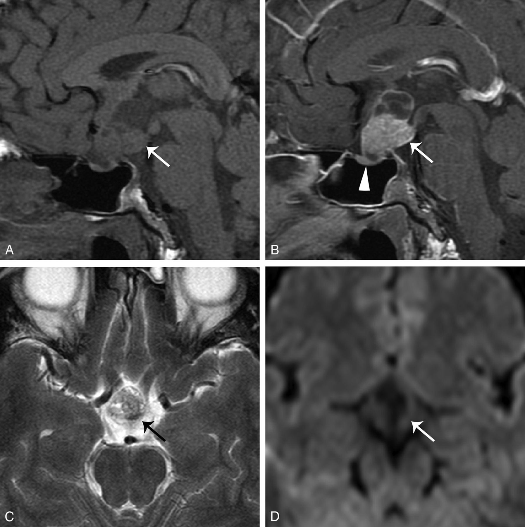
- Fig 1.
A 28-year-old man had bitemporal hemianopsia for several weeks. Surgical resection revealed PCP. A, Sagittal T1WI shows a mixed solid and cystic tumor (arrow) at the sellar and suprasellar regions. B, Contrast-enhanced sagittal T1WI shows strong enhancement at the solid component and its cystic wall (arrow). The lower end of the tumor is spheric. The pituitary gland is compressed but intact (arrowhead). C, Axial T2WI demonstrates that the tumor (arrow) has mixed isointense-to-hyperintense signals compared with the adjacent temporal gray matter. D, DWI (b=800) shows no restricted diffusion in the tumor (arrow).
- Fig 2.
A 38-year-old man had intermittent dizziness for 2 months and right-sided limb weakness for 1 week. Surgical resection revealed PCP. Brain MR imaging. A, Contrast-enhanced sagittal T1WI shows a heterogeneous enhancing tumor (arrow) at the suprasellar region and third ventricular floor. The lower end of the tumor is spheric. The size of the pineal gland is within normal range (arrowhead). B, The 2.5-year follow-up contrast-enhanced sagittal T1WI shows a mixed cystic and solid recurrent tumor (arrow). The size of the pineal gland (arrowhead) remains unchanged. C, DWI (b=800) reveals no restricted diffusion in the tumor (arrow).
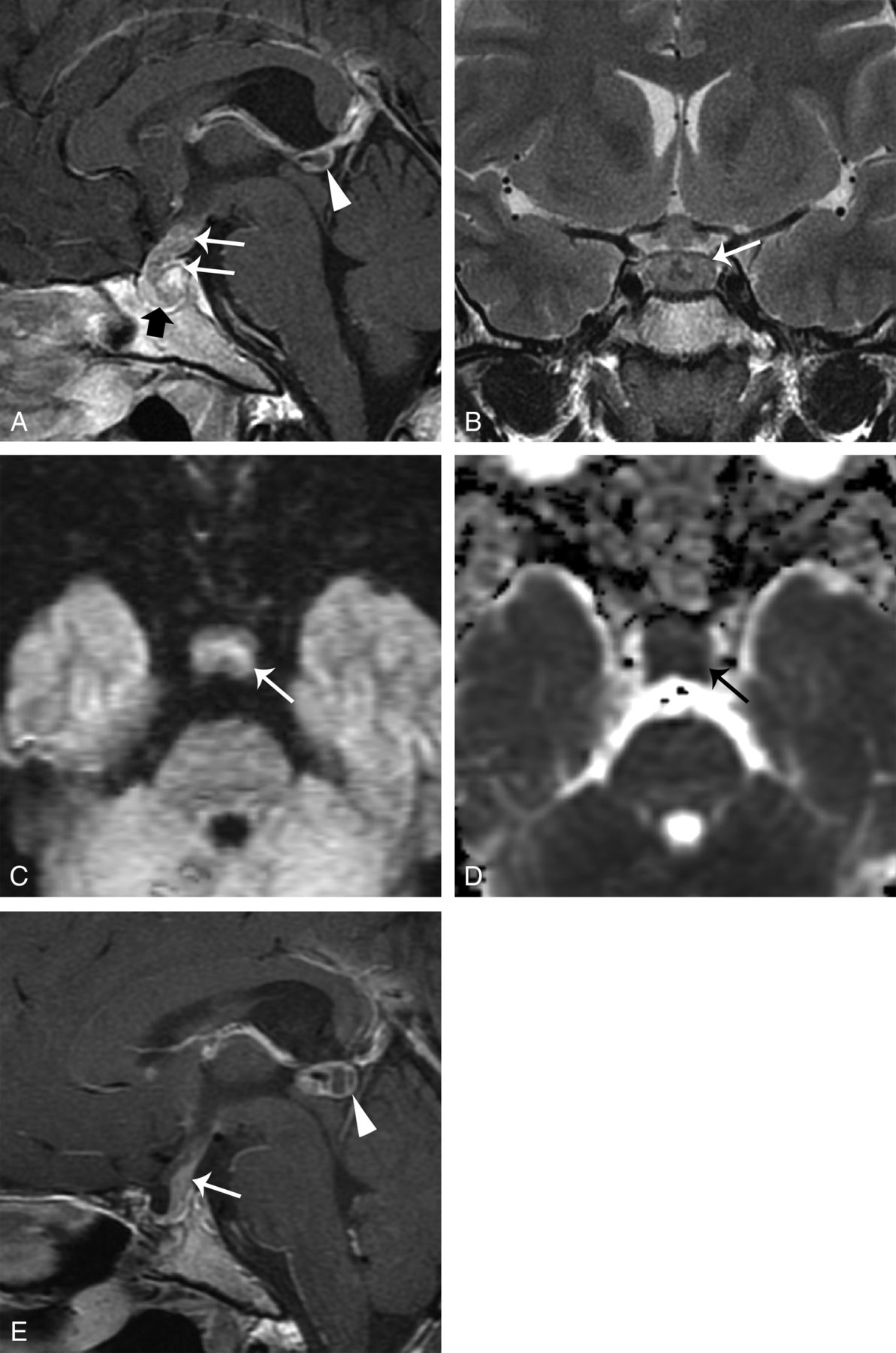
- Fig 3.
A 29-year-old woman who had amenorrhea and diabetes insipidus was diagnosed with germinoma after partial resection of the tumor. A, Sagittal contrast-enhanced T1WI with fat saturation shows that the tumor infiltrates along the infundibular recess down to the sellar region (arrows). The tumor shows transinfundibular growth (thick arrow). Note the 8-mm pineal cystic structure (arrowhead). B, Coronal T2WI demonstrates the isointense tumor (arrow). C and D, DWI (b=800) and an ADC map demonstrate restricted diffusion of the tumor (arrows). E, The 5-year follow-up sagittal contrast-enhanced T1WI shows a recurrent suprasellar GCT (arrow). An enlarged pineal GCT is noted in the original pineal cystic structure (arrowhead).
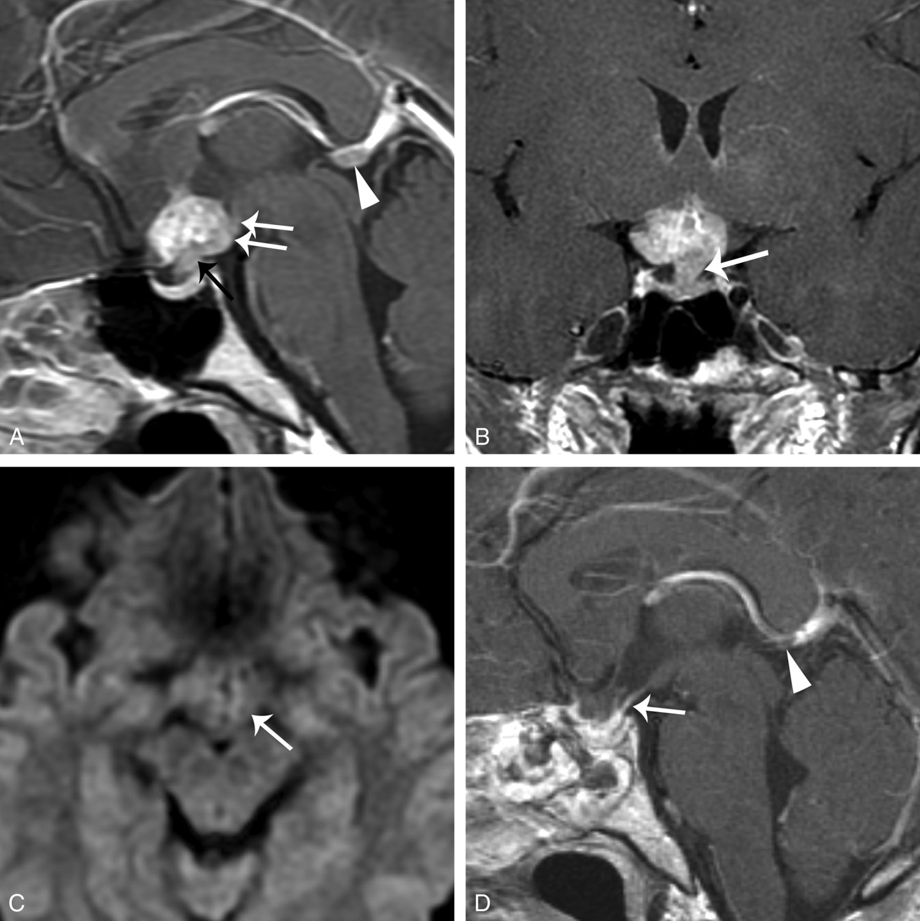
- Fig 4.
A 23-year-old woman had amenorrhea for 2 years and biopsy-proved germinoma. A, Sagittal contrast-enhanced T1WI with fat saturation shows a heterogeneous enhancing suprasellar and third ventricular floor tumor (white arrows) with infundibular stalk thickening (black arrow). The pineal gland is 8 mm in the largest diameter (arrowhead). B, Coronal contrast-enhanced T1WI shows the growth of the tumor along the infundibular recess (arrow) and the indistinct margin between the tumor and pituitary gland. C, DWI (b=800) demonstrates mildly high signal in the tumor (arrow). D, The 3-month follow-up sagittal contrast-enhanced T1WI after surgical and radiation treatment shows shrinkage of both the suprasellar (arrow) and pineal tumors (arrowhead).
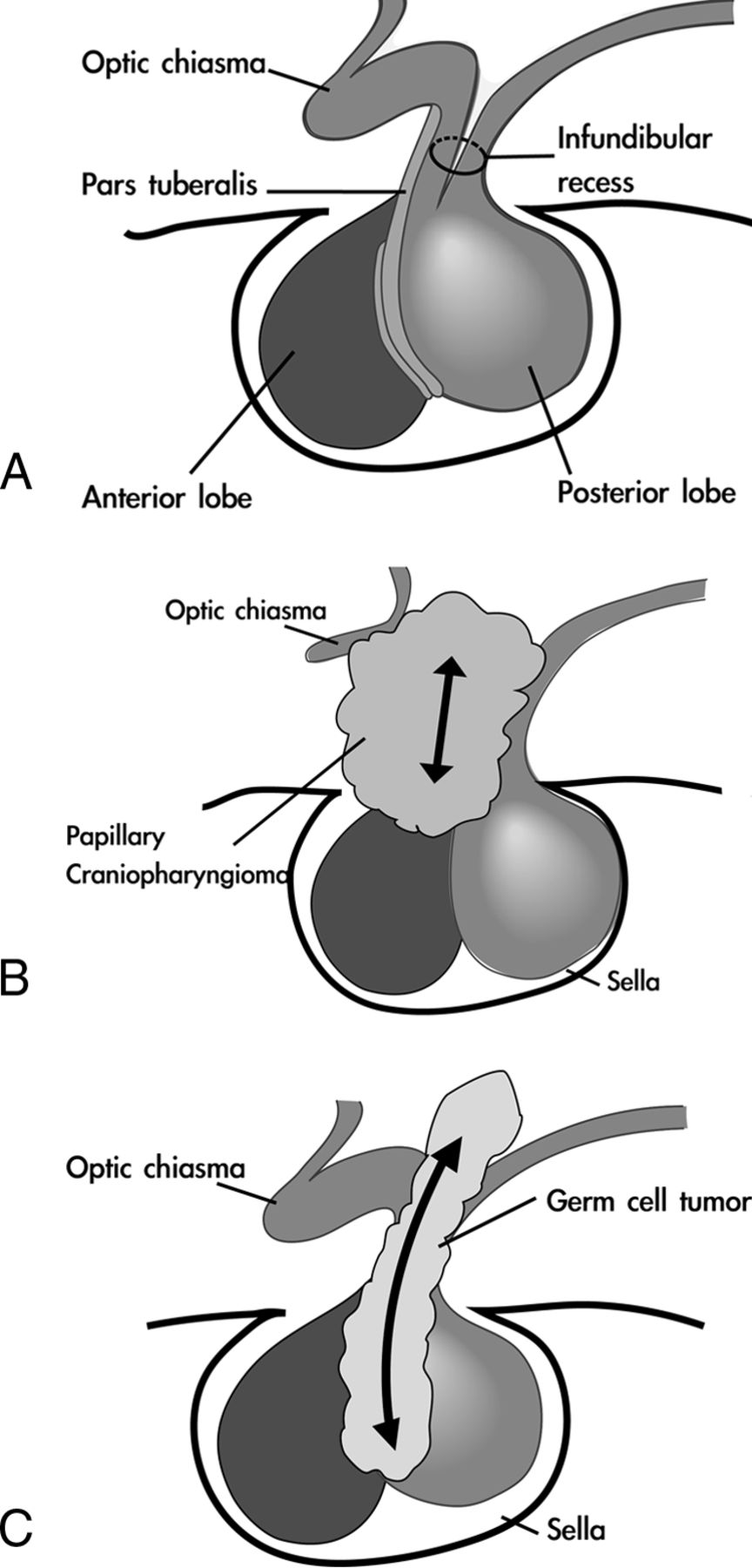
- Fig 5.
Hypothetic pathogenesis of suprasellar GCT and PCP. A, Normal anatomy of the sellar and suprasellar regions. The pars tuberalis, a small part of the pars anterior, extends ventrally and wraps around the pituitary stalk; the neurohypophysis is embryologically and anatomically continuous with the hypothalamus. B, The PCP originates from squamous epithelial cells in the pars tuberalis of the adenohypophysis and is located extraventricularly. C, The GCT originates from the hypothalamic-infundibular axis or pineal region. It is located intraventricularly and infiltrates along the infundibular recess down to the sellar region.


Tables
- Table 1:
Analysis of clinical symptoms and outcomes of suprasellar papillary craniopharyngioma and suprasellar germ cell tumors
Variables PCP (n = 18) GCT (n = 17) OR 95% CI P Valuea Age (yr) 46 ± 13.9 (21–70) 23 ± 7.1 (16–43) <.0001 Sex (male:female) 13:5 13:4 1.3 .27–5.7 .7738 Symptoms Visual field deficits 12 (67%) 6 (35%) .27 .07–1.1 .0634 Hypopituitarismb 12 (71%) 12 (75%) 1.3 .27–5.8 .7761 Hyperprolactinemiac 14 (93%) 11 (67%) .16 .02–1.5 .0834 Hypothalamic dysfunction, including diabetes insipidus 2 (11%) 11 (65%) 15 2.5–87 .0010 Initial management .0303 Total resection 7 (39%) 3 (18%) 1.0 – Subtotal resection 6 (33%) 1 (6%) .39 .03–4.8 Partial resection or biopsy 3 (17%) 6 (35%) 4.7 .67–32 Radiation or radiosurgery 2 (11%) 7 (41%) 8.2 1.03–65 Outcome Recurrenced 13 (81%) 4 (24%) .10 .02–.50 .0031 Survival (mean) 39.6 ± 32.02 (2–118) 52.4 ± 40.32 (13–123) .3815 - Table 2:
Specific clinical findings and MR imaging characteristics favoring papillary craniopharyngiomas or suprasellar germ cell tumors
Papillary Craniopharyngioma Germ Cell Tumor Clinical findings Hypothalamic dysfunction, including diabetes insipidus – Yes MR imaging characteristics Component Cystic predominance Solid predominance DWI signals in the solid part Hypointense Isointense Marginal contrast enhancement Yes – Shape Spheric Transinfundibular Pituitary stalk – Thickening Tumor seedings – Yes Size change of the pineal gland after radiotherapy – Yes Main management strategy Surgery Radiation therapy Outcomes Higher recurrence rate – Note:—indicates the feature was significantly lower or less in the specific tumor group.


