Advertisement

Main menu

  • Home
  • Content
    • Current Issue
    • Accepted Manuscripts
    • Article Preview
    • Past Issue Archive
    • Video Articles
    • AJNR Case Collection
    • Case of the Week Archive
    • Case of the Month Archive
    • Classic Case Archive
  • Special Collections
    • Advancing NeuroMRI with High-Relaxivity Contrast Agents
    • AJNR Awards
    • Low-Field MRI
    • Alzheimer Disease
    • ASNR Foundation Special Collection
    • Photon-Counting CT
    • View All
  • Multimedia
    • AJNR Podcasts
    • AJNR SCANtastic
    • Trainee Corner
    • MRI Safety Corner
    • Imaging Protocols
  • For Authors
    • Submit a Manuscript
    • Submit a Video Article
    • Submit an eLetter to the Editor/Response
    • Manuscript Submission Guidelines
    • Statistical Tips
    • Fast Publishing of Accepted Manuscripts
    • Graphical Abstract Preparation
    • Imaging Protocol Submission
    • Author Policies
  • About Us
    • About AJNR
    • Editorial Board
    • Editorial Board Alumni
  • More
    • Become a Reviewer/Academy of Reviewers
    • Subscribers
    • Permissions
    • Alerts
    • Feedback
    • Advertisers
    • ASNR Home

User menu

  • Alerts
  • Log in

Search

  • Advanced search
American Journal of Neuroradiology

American Journal of Neuroradiology

ASHNR American Society of Functional Neuroradiology ASHNR American Society of Pediatric Neuroradiology ASSR
  • Alerts
  • Log in

Advanced Search

  • Home
  • Content
    • Current Issue
    • Accepted Manuscripts
    • Article Preview
    • Past Issue Archive
    • Video Articles
    • AJNR Case Collection
    • Case of the Week Archive
    • Case of the Month Archive
    • Classic Case Archive
  • Special Collections
    • Advancing NeuroMRI with High-Relaxivity Contrast Agents
    • AJNR Awards
    • Low-Field MRI
    • Alzheimer Disease
    • ASNR Foundation Special Collection
    • Photon-Counting CT
    • View All
  • Multimedia
    • AJNR Podcasts
    • AJNR SCANtastic
    • Trainee Corner
    • MRI Safety Corner
    • Imaging Protocols
  • For Authors
    • Submit a Manuscript
    • Submit a Video Article
    • Submit an eLetter to the Editor/Response
    • Manuscript Submission Guidelines
    • Statistical Tips
    • Fast Publishing of Accepted Manuscripts
    • Graphical Abstract Preparation
    • Imaging Protocol Submission
    • Author Policies
  • About Us
    • About AJNR
    • Editorial Board
    • Editorial Board Alumni
  • More
    • Become a Reviewer/Academy of Reviewers
    • Subscribers
    • Permissions
    • Alerts
    • Feedback
    • Advertisers
    • ASNR Home
  • Follow AJNR on Twitter
  • Visit AJNR on Facebook
  • Follow AJNR on Instagram
  • Join AJNR on LinkedIn
  • RSS Feeds

AJNR Awards, New Junior Editors, and more. Read the latest AJNR updates


Improved Turnaround Times | Median time to first decision: 12 days

Research ArticlePediatric Neuroimaging

Comparison of Spin-Echo and Gradient-Echo T1-Weighted and Spin-Echo T2-Weighted Images at 3T in Evaluating Term-Neonatal Myelination

A.E. Tyan, A.M. McKinney, T.J. Hanson and C.L. Truwit
American Journal of Neuroradiology February 2015, 36 (2) 411-416; DOI: https://doi.org/10.3174/ajnr.A4099
A.E. Tyan
aFrom the Department of Radiology, University of Minnesota-Fairview and Hennepin County Medical Centers, Minneapolis, Minnesota.
  • Find this author on Google Scholar
  • Find this author on PubMed
  • Search for this author on this site
A.M. McKinney
aFrom the Department of Radiology, University of Minnesota-Fairview and Hennepin County Medical Centers, Minneapolis, Minnesota.
  • Find this author on Google Scholar
  • Find this author on PubMed
  • Search for this author on this site
T.J. Hanson
aFrom the Department of Radiology, University of Minnesota-Fairview and Hennepin County Medical Centers, Minneapolis, Minnesota.
  • Find this author on Google Scholar
  • Find this author on PubMed
  • Search for this author on this site
C.L. Truwit
aFrom the Department of Radiology, University of Minnesota-Fairview and Hennepin County Medical Centers, Minneapolis, Minnesota.
  • Find this author on Google Scholar
  • Find this author on PubMed
  • Search for this author on this site
  • Article
  • Figures & Data
  • Info & Metrics
  • Responses
  • References
  • PDF
Loading

Article Figures & Data

Figures

  • Tables
  • Fig 1.
    • Download figure
    • Open in new tab
    • Download powerpoint
    Fig 1.

    A 3-day-old male neonate with respiratory depression. A–C, MR images at the level of the centrum semiovale demonstrate mildly bright signal of the perirolandic cortex (black arrows) on GRE-T1WI (A) and SE-T1WI (B). The perirolandic cortex appears darker on SE-T2WI (C) relative to the remainder of the frontal and parietal cortices in most, but not all, infants in this study. The CST (asterisk) are also quite bright on both T1WI sequences and are mildly dark on SE-T2WI. D–F, On images at a lower level through the basal ganglia, the PLIC (arrows) and the habenular commissure (split arrows) appear visibly myelinated (ie, bright) on GRE-T1WI (D), SE-T1WI (E), and on SE-T2WI (F), though the PLIC appears much smaller on SE-T2WI. Both the habenular commissure and the PLIC appear myelinated on GRE-T1WI in all term neonates in this study.

  • Fig 2.
    • Download figure
    • Open in new tab
    • Download powerpoint
    Fig 2.

    A 3-day-old male neonate with respiratory depression. MR images demonstrate clearly visible bright signal on GRE-T1WI (A and B) and clearly visible dark signal on SE-T2WI (C and D), indicative of normal early myelination within the BIC (wide arrows) and the DSCP (curved arrows). The optic tracts (thin arrows) are visibly myelinated on GRE-T1WI but are questionably myelinated on SE-T2WI. The BIC and the DSCP were both clearly myelinated in at least ≥88% of patients on both GRE-T1WI and SE-T2WI, while the SCP was myelinated in ≥88% of patients on GRE-T1WI only. Note the apparently myelinated lateral geniculate nuclei (arrowheads).
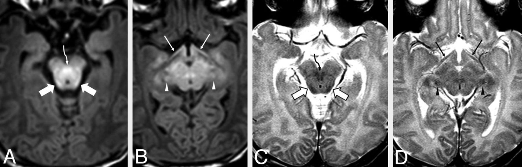
  • Fig 3.
    • Download figure
    • Open in new tab
    • Download powerpoint
    Fig 3.

    A 6-day-old male neonate with respiratory depression. Axial MR images illustrate bright myelination of the medial lemnisci (long arrows), lateral lemnisci (dotted arrows), cranial nerve V fascicle (dashed arrows), and SCP (beveled arrows) on GRE-T1WI (A and B), which were not clearly visible on SE-T1WI (not shown), but all appeared dark (myelinated) on SE-T2WI (C and D). The medial lemnisci were visibly myelinated in ≥94% of neonates on both GRE-T1WI and SE-T2WI. In most patients, the lateral lemnisci and cranial nerve V fascicle were best visualized as being myelinated on GRE-T1WI.

  • Fig 4.
    • Download figure
    • Open in new tab
    • Download powerpoint
    Fig 4.

    A 4-day-old female neonate with respiratory depression and possible seizures. The pyramidal decussation (circle tipped arrows) is visibly myelinated (bright) on GRE-T1WI (A and B) but was rated as “questionably myelinated” (ie, scored ± by both reviewers on SE-T2WI (C and D). In contrast, the spinal tract of V (open arrowheads) was considered to be “definitely myelinated” (ie, scored +) by both reviewers on SE-T2WI, while it was considered to be only “questionably myelinated” on GRE-T1WI. The ICP (double arrows) appeared clearly myelinated on both sequences. On SE-T1WI (not shown), myelination was not visible (ie, scored as −) by either reviewer in the aforementioned structures in this patient. The ICP was the 1 structure that was clearly myelinated in all neonates on SE-T2WI and was better visualized as being “definitely myelinated” (ie, scored +) on SE-T2WI compared with both GRE-T1WI and SE-T1WI.

Tables

  • Figures
  • Percentages and numbers of term infants with myelination

    StructureGRE-T1WISE-T1WISE-T2WI
    Observer 1Observer 2Observer 1Observer 2Observer 1Observer 2
    BIC100%,a 16/1694%,a 15/1675%, 12/1650%, 8/16100%,a 16/1688%,a 14/16
    CCS0%, 0/160%, 0/160%, 0/160%, 0/166%, 1/1638%, 6/16
    CNV94%,a 15/1656%, 9/1650%, 8/166%, 1/1644%, 7/1613%, 2/16
    CST-BS25%, 4/160%, 0/160%, 0/160%, 0/160%, 0/160%, 0/16
    CST-CS75%, 12/1663%, 10/1675%, 12/1631%, 5/166%, 1/166%, 1/16
    DSCP100%,a 16/16100%,a 16/1681%, 13/1631%, 5/16100%,a 16/1694%,a 15/16
    HC100%,a 16/16100%,a 16/16100%,a 16/1694%,a 15/1669%, 11/1663%, 10/16
    ICP88%,a 14/1631%, 5/1656%, 9/160%, 0/16100%,a 16/16100%,a 16/16
    LGN38%, 6/1625%, 4/166%, 1/160%, 0/1650%, 8/1656%, 9/16
    LL94%,a 15/1669%, 11/1638%, 6/1656%, 9/1675%, 12/1644%, 7/16
    ML100%,a 16/1694%,a 15/1688%,a 14/1625%, 4/16100%,a 16/16100%,a 16/16
    MLF25%, 4/160%, 0/160%, 0/160%, 0/1619%, 3/1631%, 5/16
    ON44%, 7/1644%, 7/160%, 0/160%, 0/1619%, 3/1644%, 7/16
    OT88%,a 14/1681%, 13/1669%, 11/1656%, 9/1656%, 9/1656%, 9/16
    PD94%,a 15/1688%,a 14/1631%, 5/166%, 1/1688%,a 14/1638%, 6/16
    PLIC100%,a 16/16100%,a 16/16100%,a 16/1644%, 7/1688%,a 14/1688%,a 14/16
    PRC56%, 9/1619%, 3/1631%, 5/166%, 1/1688%,a 14/1663%, 10/16
    SCP100%,a 16/16100%,a 16/1675%, 12/1644%, 7/1688%,a 14/1675%, 12/16
    STV56%, 9/1625%, 4/1644%, 7/1625%, 4/1681%, 13/1669%, 11/16
    • Note:—CCS indicates callosal splenium; CNV, cranial nerve V fascicle; CST-BS, CST in the brain stem; CST-CS, CST in the centrum semiovale; HC, habenular commissure; LGN, lateral geniculate nucleus; LL, lateral lemniscus; ML, medial lemniscus; MLF, medial longitudinal fasciculus; ON, optic nerve; OT, optic tract; PD, pyramidal decussation; PRC, perirolandic cortex; STV, spinal tract of V.

    • ↵a Structures with ≥88% of patients with myelination.

PreviousNext
Back to top

In this issue

American Journal of Neuroradiology: 36 (2)
American Journal of Neuroradiology
Vol. 36, Issue 2
1 Feb 2015
  • Table of Contents
  • Index by author
  • Complete Issue (PDF)
Advertisement
Print
Download PDF
Email Article

Thank you for your interest in spreading the word on American Journal of Neuroradiology.

NOTE: We only request your email address so that the person you are recommending the page to knows that you wanted them to see it, and that it is not junk mail. We do not capture any email address.

Enter multiple addresses on separate lines or separate them with commas.
Comparison of Spin-Echo and Gradient-Echo T1-Weighted and Spin-Echo T2-Weighted Images at 3T in Evaluating Term-Neonatal Myelination
(Your Name) has sent you a message from American Journal of Neuroradiology
(Your Name) thought you would like to see the American Journal of Neuroradiology web site.
CAPTCHA
This question is for testing whether or not you are a human visitor and to prevent automated spam submissions.
Cite this article
A.E. Tyan, A.M. McKinney, T.J. Hanson, C.L. Truwit
Comparison of Spin-Echo and Gradient-Echo T1-Weighted and Spin-Echo T2-Weighted Images at 3T in Evaluating Term-Neonatal Myelination
American Journal of Neuroradiology Feb 2015, 36 (2) 411-416; DOI: 10.3174/ajnr.A4099

Citation Manager Formats

  • BibTeX
  • Bookends
  • EasyBib
  • EndNote (tagged)
  • EndNote 8 (xml)
  • Medlars
  • Mendeley
  • Papers
  • RefWorks Tagged
  • Ref Manager
  • RIS
  • Zotero
0 Responses
Respond to this article
Share
Bookmark this article
Comparison of Spin-Echo and Gradient-Echo T1-Weighted and Spin-Echo T2-Weighted Images at 3T in Evaluating Term-Neonatal Myelination
A.E. Tyan, A.M. McKinney, T.J. Hanson, C.L. Truwit
American Journal of Neuroradiology Feb 2015, 36 (2) 411-416; DOI: 10.3174/ajnr.A4099
del.icio.us logo Twitter logo Facebook logo Mendeley logo
  • Tweet Widget
  • Facebook Like
  • Google Plus One
Purchase

Jump to section

  • Article
    • Abstract
    • ABBREVIATIONS:
    • Materials and Methods
    • Results
    • Discussion
    • Conclusions
    • Footnotes
    • REFERENCES
  • Figures & Data
  • Info & Metrics
  • Responses
  • References
  • PDF

Related Articles

  • No related articles found.
  • PubMed
  • Google Scholar

Cited By...

  • Imaging Findings and MRI Patterns in a Cohort of 18q Chromosomal Abnormalities
  • Crossref
  • Google Scholar

This article has not yet been cited by articles in journals that are participating in Crossref Cited-by Linking.

More in this TOC Section

  • Pediatric Glioma:Imaging Clusters,Molecular Traits
  • Clinical SVR of Fetal Brain MRI
  • FRACTURE MR in Congenital Vertebral Anomalies
Show more PEDIATRIC NEUROIMAGING

Similar Articles

Advertisement

Indexed Content

  • Current Issue
  • Accepted Manuscripts
  • Article Preview
  • Past Issues
  • Editorials
  • Editor's Choice
  • Fellows' Journal Club
  • Letters to the Editor
  • Video Articles

Cases

  • Case Collection
  • Archive - Case of the Week
  • Archive - Case of the Month
  • Archive - Classic Case

More from AJNR

  • Trainee Corner
  • Imaging Protocols
  • MRI Safety Corner
  • Book Reviews

Multimedia

  • AJNR Podcasts
  • AJNR Scantastics

Resources

  • Turnaround Time
  • Submit a Manuscript
  • Submit a Video Article
  • Submit an eLetter to the Editor/Response
  • Manuscript Submission Guidelines
  • Statistical Tips
  • Fast Publishing of Accepted Manuscripts
  • Graphical Abstract Preparation
  • Imaging Protocol Submission
  • Evidence-Based Medicine Level Guide
  • Publishing Checklists
  • Author Policies
  • Become a Reviewer/Academy of Reviewers
  • News and Updates

About Us

  • About AJNR
  • Editorial Board
  • Editorial Board Alumni
  • Alerts
  • Permissions
  • Not an AJNR Subscriber? Join Now
  • Advertise with Us
  • Librarian Resources
  • Feedback
  • Terms and Conditions
  • AJNR Editorial Board Alumni

American Society of Neuroradiology

  • Not an ASNR Member? Join Now

© 2025 by the American Society of Neuroradiology All rights, including for text and data mining, AI training, and similar technologies, are reserved.
Print ISSN: 0195-6108 Online ISSN: 1936-959X

Powered by HighWire