Advertisement

Main menu

  • Home
  • Content
    • Current Issue
    • Accepted Manuscripts
    • Article Preview
    • Past Issue Archive
    • Video Articles
    • AJNR Case Collection
    • Case of the Week Archive
    • Case of the Month Archive
    • Classic Case Archive
  • Special Collections
    • AJNR Awards
    • Low-Field MRI
    • Alzheimer Disease
    • ASNR Foundation Special Collection
    • Photon-Counting CT
    • View All
  • Multimedia
    • AJNR Podcasts
    • AJNR SCANtastic
    • Trainee Corner
    • MRI Safety Corner
    • Imaging Protocols
  • For Authors
    • Submit a Manuscript
    • Submit a Video Article
    • Submit an eLetter to the Editor/Response
    • Manuscript Submission Guidelines
    • Statistical Tips
    • Fast Publishing of Accepted Manuscripts
    • Graphical Abstract Preparation
    • Imaging Protocol Submission
    • Author Policies
  • About Us
    • About AJNR
    • Editorial Board
    • Editorial Board Alumni
  • More
    • Become a Reviewer/Academy of Reviewers
    • Subscribers
    • Permissions
    • Alerts
    • Feedback
    • Advertisers
    • ASNR Home

User menu

  • Alerts
  • Log in

Search

  • Advanced search
American Journal of Neuroradiology

American Journal of Neuroradiology

ASHNR American Society of Functional Neuroradiology ASHNR American Society of Pediatric Neuroradiology ASSR
  • Alerts
  • Log in

Advanced Search

  • Home
  • Content
    • Current Issue
    • Accepted Manuscripts
    • Article Preview
    • Past Issue Archive
    • Video Articles
    • AJNR Case Collection
    • Case of the Week Archive
    • Case of the Month Archive
    • Classic Case Archive
  • Special Collections
    • AJNR Awards
    • Low-Field MRI
    • Alzheimer Disease
    • ASNR Foundation Special Collection
    • Photon-Counting CT
    • View All
  • Multimedia
    • AJNR Podcasts
    • AJNR SCANtastic
    • Trainee Corner
    • MRI Safety Corner
    • Imaging Protocols
  • For Authors
    • Submit a Manuscript
    • Submit a Video Article
    • Submit an eLetter to the Editor/Response
    • Manuscript Submission Guidelines
    • Statistical Tips
    • Fast Publishing of Accepted Manuscripts
    • Graphical Abstract Preparation
    • Imaging Protocol Submission
    • Author Policies
  • About Us
    • About AJNR
    • Editorial Board
    • Editorial Board Alumni
  • More
    • Become a Reviewer/Academy of Reviewers
    • Subscribers
    • Permissions
    • Alerts
    • Feedback
    • Advertisers
    • ASNR Home
  • Follow AJNR on Twitter
  • Visit AJNR on Facebook
  • Follow AJNR on Instagram
  • Join AJNR on LinkedIn
  • RSS Feeds

AJNR Awards, New Junior Editors, and more. Read the latest AJNR updates

Research ArticleHead & Neck
Open Access

Visualization of Human Inner Ear Anatomy with High-Resolution MR Imaging at 7T: Initial Clinical Assessment

M.A. van der Jagt, W.M. Brink, M.J. Versluis, S.C.A. Steens, J.J. Briaire, A.G. Webb, J.H.M. Frijns and B.M. Verbist
American Journal of Neuroradiology February 2015, 36 (2) 378-383; DOI: https://doi.org/10.3174/ajnr.A4084
M.A. van der Jagt
aFrom the Departments of Otorhinolaryngology (M.A.v.d.J., J.J.B., J.H.M.F.)
  • Find this author on Google Scholar
  • Find this author on PubMed
  • Search for this author on this site
W.M. Brink
bRadiology (W.M.B., M.J.V., A.G.W., B.M.V.)
cC.J. Gorter Center for High-Field MRI (W.M.B., M.J.V., A.G.W.)
  • Find this author on Google Scholar
  • Find this author on PubMed
  • Search for this author on this site
M.J. Versluis
bRadiology (W.M.B., M.J.V., A.G.W., B.M.V.)
cC.J. Gorter Center for High-Field MRI (W.M.B., M.J.V., A.G.W.)
  • Find this author on Google Scholar
  • Find this author on PubMed
  • Search for this author on this site
S.C.A. Steens
eDepartment of Radiology (S.C.A.S., B.M.V.), Radboud University Medical Center, Nijmegen, the Netherlands.
  • Find this author on Google Scholar
  • Find this author on PubMed
  • Search for this author on this site
J.J. Briaire
aFrom the Departments of Otorhinolaryngology (M.A.v.d.J., J.J.B., J.H.M.F.)
  • Find this author on Google Scholar
  • Find this author on PubMed
  • Search for this author on this site
A.G. Webb
bRadiology (W.M.B., M.J.V., A.G.W., B.M.V.)
cC.J. Gorter Center for High-Field MRI (W.M.B., M.J.V., A.G.W.)
  • Find this author on Google Scholar
  • Find this author on PubMed
  • Search for this author on this site
J.H.M. Frijns
aFrom the Departments of Otorhinolaryngology (M.A.v.d.J., J.J.B., J.H.M.F.)
dLeiden Institute for Brain and Cognition (J.H.M.F.), Leiden University Medical Center, Leiden, the Netherlands
  • Find this author on Google Scholar
  • Find this author on PubMed
  • Search for this author on this site
B.M. Verbist
bRadiology (W.M.B., M.J.V., A.G.W., B.M.V.)
eDepartment of Radiology (S.C.A.S., B.M.V.), Radboud University Medical Center, Nijmegen, the Netherlands.
  • Find this author on Google Scholar
  • Find this author on PubMed
  • Search for this author on this site
  • Article
  • Figures & Data
  • Info & Metrics
  • Responses
  • References
  • PDF
Loading

Article Figures & Data

Figures

  • Tables
  • Fig 1.
    • Download figure
    • Open in new tab
    • Download powerpoint
    Fig 1.

    Mean differences in scoring of anatomic structures depicted at 3T and 7T. The bars on the right side of the zero line indicate differences in favor of the 7T images. The bars on the left side indicate differences in favor of the 3T images. The structures showing significant differences are marked with an asterisk on the left, and P values are mentioned if significant.
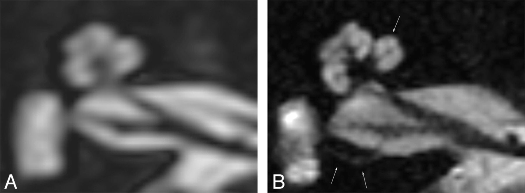
  • Fig 2.
    • Download figure
    • Open in new tab
    • Download powerpoint
    Fig 2.

    Axial cross-section of a right inner ear, rendered at 3T (A) and 7T (B); improved discrimination of the intracochlear structures and compartments is shown. In addition, sharper delineation of the nerves in the internal auditory canal is demonstrated. The single arrow indicates the scala media at the first turn. The double arrows indicate the superior ampullary nerve.
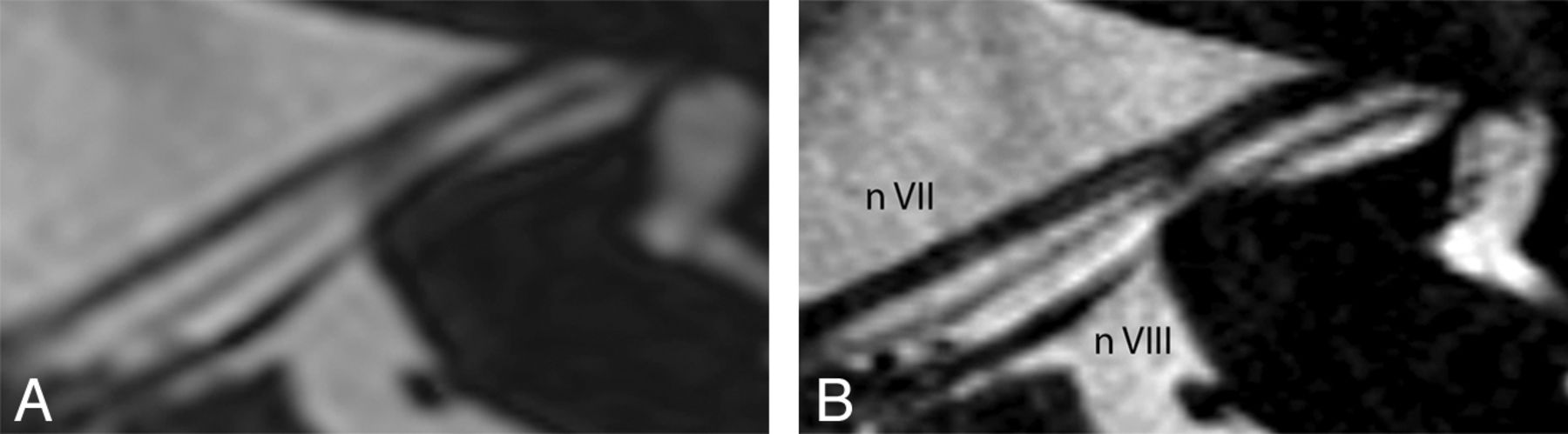
  • Fig 3.
    • Download figure
    • Open in new tab
    • Download powerpoint
    Fig 3.

    Axial cross-section along the course of the facial nerve of a left inner ear, rendered at 3T (A) and 7T (B). A sharp delineation of the neural structures and clear depiction of the intermediate nerve between cranial nerves VII and VIII are demonstrated on the 7T image.
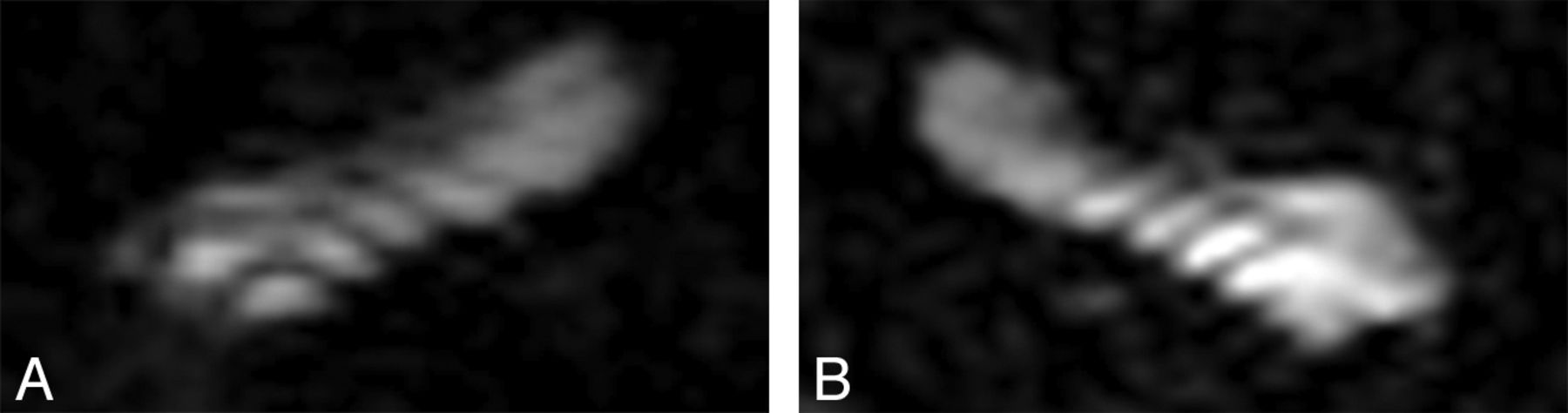
  • Fig 4.
    • Download figure
    • Open in new tab
    • Download powerpoint
    Fig 4.

    In 2 different patients, 7T images showing stripelike-artifacts at the level of the first turn of right (A) and left (B) cochleas, disturbing the quality of the representation and impeding the distinction of the scala vestibuli and tympani.

Tables

  • Figures
  • Demographic details of studied patients (N = 17)

    No.
    Sex
        Male8
        Female9
    Pathologic imaging reporting
        Cochlea malformation1a
        Hypoplasia acoustic nerve1a
        Fenestrel otosclerosis1
        Labyrinthitis ossificans1
        None14
    Etiology
        Congenital
            Pendred syndrome1
            Of unknown origin5
        Acquired
            Sudden deafness2
            MIDD1
            Otosclerosis2
            Rubella infection2
        Unknown4
    Duration of deafness, years (mean)23.2
    • Note:—MIDD indicates maternally inherited diabetes and deafness.

    • ↵a Same patient.

PreviousNext
Back to top

In this issue

American Journal of Neuroradiology: 36 (2)
American Journal of Neuroradiology
Vol. 36, Issue 2
1 Feb 2015
  • Table of Contents
  • Index by author
  • Complete Issue (PDF)
Advertisement
Print
Download PDF
Email Article

Thank you for your interest in spreading the word on American Journal of Neuroradiology.

NOTE: We only request your email address so that the person you are recommending the page to knows that you wanted them to see it, and that it is not junk mail. We do not capture any email address.

Enter multiple addresses on separate lines or separate them with commas.
Visualization of Human Inner Ear Anatomy with High-Resolution MR Imaging at 7T: Initial Clinical Assessment
(Your Name) has sent you a message from American Journal of Neuroradiology
(Your Name) thought you would like to see the American Journal of Neuroradiology web site.
CAPTCHA
This question is for testing whether or not you are a human visitor and to prevent automated spam submissions.
Cite this article
M.A. van der Jagt, W.M. Brink, M.J. Versluis, S.C.A. Steens, J.J. Briaire, A.G. Webb, J.H.M. Frijns, B.M. Verbist
Visualization of Human Inner Ear Anatomy with High-Resolution MR Imaging at 7T: Initial Clinical Assessment
American Journal of Neuroradiology Feb 2015, 36 (2) 378-383; DOI: 10.3174/ajnr.A4084

Citation Manager Formats

  • BibTeX
  • Bookends
  • EasyBib
  • EndNote (tagged)
  • EndNote 8 (xml)
  • Medlars
  • Mendeley
  • Papers
  • RefWorks Tagged
  • Ref Manager
  • RIS
  • Zotero
0 Responses
Respond to this article
Share
Bookmark this article
Visualization of Human Inner Ear Anatomy with High-Resolution MR Imaging at 7T: Initial Clinical Assessment
M.A. van der Jagt, W.M. Brink, M.J. Versluis, S.C.A. Steens, J.J. Briaire, A.G. Webb, J.H.M. Frijns, B.M. Verbist
American Journal of Neuroradiology Feb 2015, 36 (2) 378-383; DOI: 10.3174/ajnr.A4084
del.icio.us logo Twitter logo Facebook logo Mendeley logo
  • Tweet Widget
  • Facebook Like
  • Google Plus One
Purchase

Jump to section

  • Article
    • Abstract
    • ABBREVIATION:
    • Materials and Methods
    • Results
    • Discussion
    • Conclusions
    • Footnotes
    • REFERENCES
  • Figures & Data
  • Info & Metrics
  • Responses
  • References
  • PDF

Related Articles

  • No related articles found.
  • PubMed
  • Google Scholar

Cited By...

  • Imaging the Internal Auditory Canal with an 8 x 2 Transceiver Array Head Coil at 7T
  • IE-Map: A novel in-vivo atlas and template of the human inner ear
  • Crossref (33)
  • Google Scholar

This article has been cited by the following articles in journals that are participating in Crossref Cited-by Linking.

  • Novel materials in magnetic resonance imaging: high permittivity ceramics, metamaterials, metasurfaces and artificial dielectrics
    Andrew Webb, Alena Shchelokova, Alexey Slobozhanyuk, Irena Zivkovic, Rita Schmidt
    Magnetic Resonance Materials in Physics, Biology and Medicine 2022 35 6
  • MRI of the Internal Auditory Canal, Labyrinth, and Middle Ear: How We Do It
    John C. Benson, Matthew L. Carlson, John I. Lane
    Radiology 2020 297 2
  • Hearing Restoration in Cochlear Nerve Deficiency: the Choice Between Cochlear Implant or Auditory Brainstem Implant, a Meta-analysis
    Annemarie Vesseur, Rolien Free, Chantal Snels, Friedo Dekker, Emmanuel Mylanus, Berit Verbist, Johan Frijns
    Otology & Neurotology 2018 39 4
  • Recent advances in MRI of the head and neck, skull base and cranial nerves: new and evolving sequences, analyses and clinical applications
    Philip Touska, Steve E. J. Connor
    The British Journal of Radiology 2019 92 1104
  • Practical improvements in the design of high permittivity pads for dielectric shimming in neuroimaging at 7 T
    T.P.A. O’Reilly, A.G. Webb, W.M. Brink
    Journal of Magnetic Resonance 2016 270
  • Visualization of the Membranous Labyrinth and Nerve Fiber Pathways in Human and Animal Inner Ears Using MicroCT Imaging
    Rudolf Glueckert, Lejo Johnson Chacko, Dominik Schmidbauer, Thomas Potrusil, Elisabeth J. Pechriggl, Romed Hoermann, Erich Brenner, Alen Reka, Anneliese Schrott-Fischer, Stephan Handschuh
    Frontiers in Neuroscience 2018 12
  • Micro-optical coherence tomography of the mammalian cochlea
    Janani S. Iyer, Shelley A. Batts, Kengyeh K. Chu, Mehmet I. Sahin, Hui Min Leung, Guillermo J. Tearney, Konstantina M. Stankovic
    Scientific Reports 2016 6 1
  • Ultra-High-Field Magnetic Resonance Imaging of the Human Inner Ear at 11.7 Tesla
    David S. Thylur, Russell E. Jacobs, John L. Go, Arthur W. Toga, John K. Niparko
    Otology & Neurotology 2017 38 1
  • Contemporary imaging of auditory implants
    S.E.J. Connor
    Clinical Radiology 2018 73 1
  • Improvements in RF Shimming in High Field MRI Using High Permittivity Materials With Low Order Pre-Fractal Geometries
    Rita Schmidt, Andrew Webb
    IEEE Transactions on Medical Imaging 2016 35 8

More in this TOC Section

  • Chondrosarcoma vs Synovial Chondromatosis: Imaging
  • WHO Classification Update: Nasal&Skull Base Tumors
  • Peritumoral Signal in Vestibular Schwannomas
Show more Head & Neck

Similar Articles

Advertisement

Indexed Content

  • Current Issue
  • Accepted Manuscripts
  • Article Preview
  • Past Issues
  • Editorials
  • Editor's Choice
  • Fellows' Journal Club
  • Letters to the Editor
  • Video Articles

Cases

  • Case Collection
  • Archive - Case of the Week
  • Archive - Case of the Month
  • Archive - Classic Case

More from AJNR

  • Trainee Corner
  • Imaging Protocols
  • MRI Safety Corner
  • Book Reviews

Multimedia

  • AJNR Podcasts
  • AJNR Scantastics

Resources

  • Turnaround Time
  • Submit a Manuscript
  • Submit a Video Article
  • Submit an eLetter to the Editor/Response
  • Manuscript Submission Guidelines
  • Statistical Tips
  • Fast Publishing of Accepted Manuscripts
  • Graphical Abstract Preparation
  • Imaging Protocol Submission
  • Evidence-Based Medicine Level Guide
  • Publishing Checklists
  • Author Policies
  • Become a Reviewer/Academy of Reviewers
  • News and Updates

About Us

  • About AJNR
  • Editorial Board
  • Editorial Board Alumni
  • Alerts
  • Permissions
  • Not an AJNR Subscriber? Join Now
  • Advertise with Us
  • Librarian Resources
  • Feedback
  • Terms and Conditions
  • AJNR Editorial Board Alumni

American Society of Neuroradiology

  • Not an ASNR Member? Join Now

© 2025 by the American Society of Neuroradiology All rights, including for text and data mining, AI training, and similar technologies, are reserved.
Print ISSN: 0195-6108 Online ISSN: 1936-959X

Powered by HighWire