
Article Figures & Data
Figures
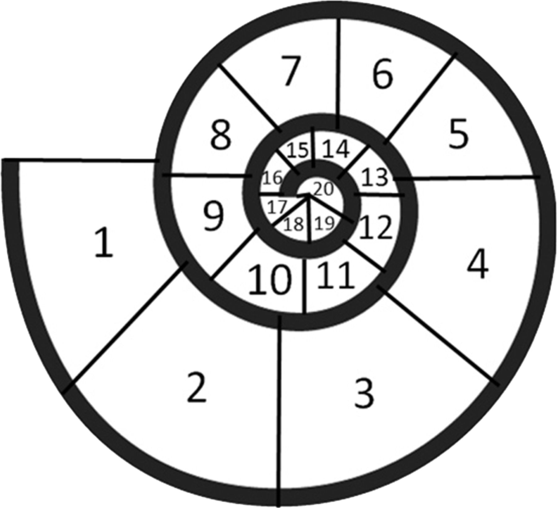
- Fig 1.
Definition of the insertion depth of the CI according to the radial position of the tip: 1 = 0°–45°, 2 = 46°–90°, 3 = 91°–135°, 4 = 136°–180°, 5 = 181°–225°, 6 = 226°–270°, 7 = 271°–315°, 8 = 316°–360°, 9 = 361°–405°, 10 = 406°–450°, 11 = 451°–495°, 12 = 496°–540°, 13 = 541°–585°, 14 = 586°–630°, 15 = 631°–675°, 16 = 676°–720°, 17 = 721°–765°, 18 = 766°–810°, 19 = 811°–855°, 20 = 856°–900°.
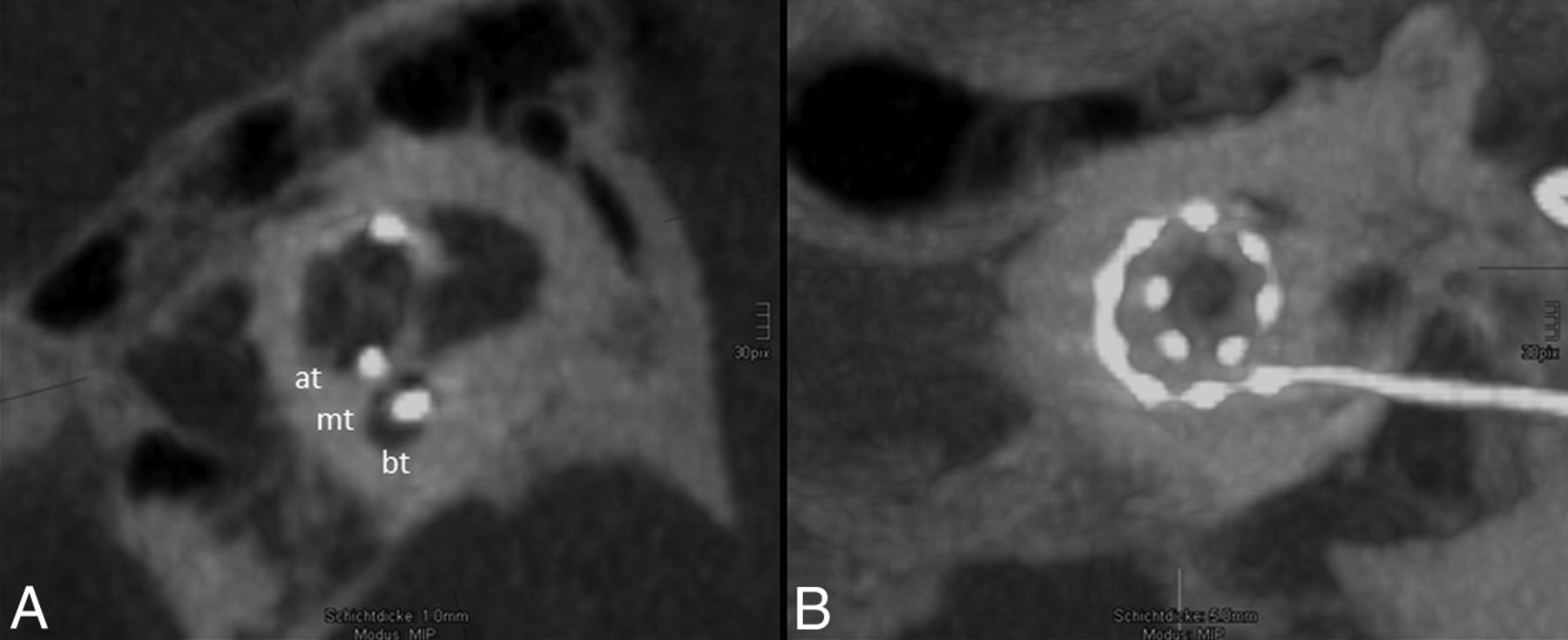
- Fig 2.
Regular position of the electrode array. A, One-millimeter MIP reconstruction of the cochlea shows the basal turn (bt), medial turn (mt), and apical turn (at). The position of the electrode array is clearly identified in the scala tympani (the scala tympani is basal; the scala vestibuli is apical). B, The insertion depth by using 5-mm MIP reconstruction in the horizontal reference plane reaches position 11, which refers to 451°–495° (Fig 1). All 12 electrodes (dots) can be identified on the electrode array.
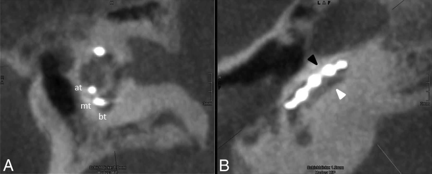
- Fig 3.
Scalar dislocation. A, One-millimeter MIP reconstruction of the cochlea showing the basal turn (bt), medial turn (mt), and apical turn (at). The position of the electrode array is clearly identified in the scala tympani. B, A cut along the basal turn demonstrates the scalar dislocation from the scala tympani (basal, white arrowhead) into the scala vestibuli (apical, black arrowhead) within the first 45° of insertion.
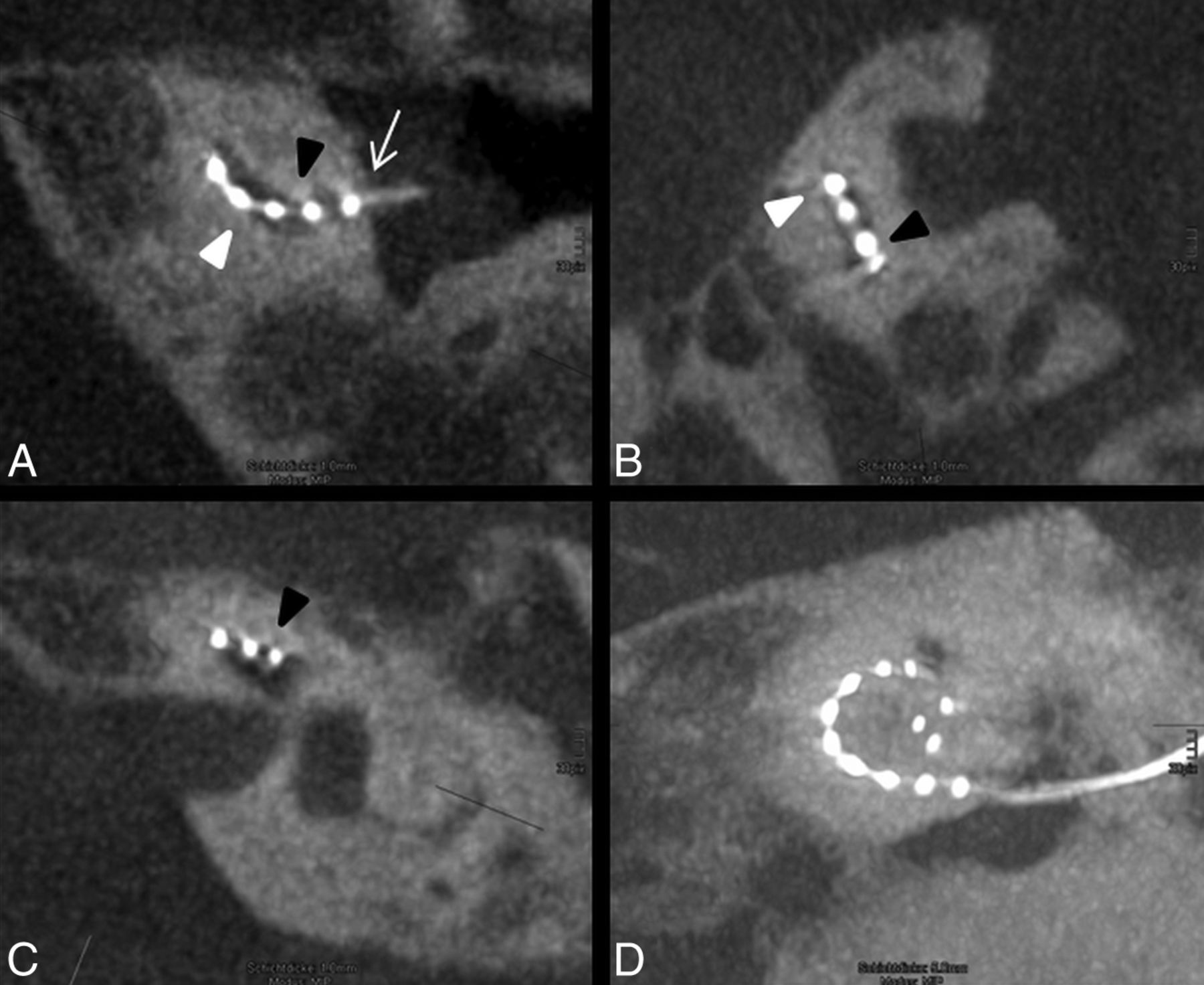
- Fig 4.
Atypical position of the electrode array. A, The electrode is inserted via cochleostomy (white arrow) and enters the basal cochlear turn from the scala vestibuli (apical, black arrowhead) into scala tympani (basal, white arrowhead). B, In the following course of the basal turn, the electrode array dislocates into the scala vestibuli. C, In the middle turn, the electrode array is clearly identified in the scala vestibuli. D, The insertion depth reaches position 8, which refers to 316°–360°, and the tip shows a kinking at the last electrode element.
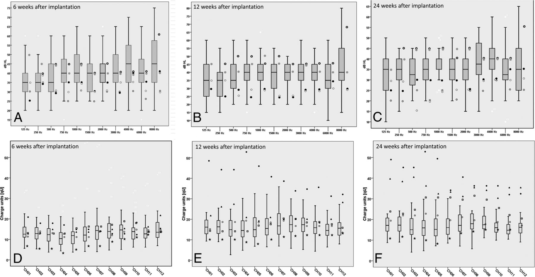
- Fig 5.
A–C, The boxplots represent the hearing thresholds of the control group. The hearing thresholds of the 5 patients with electrode dislocation are added as points beside the boxplots. Frequency-specific hearing thresholds of controls at 6 weeks after implantation (A), at 3 months after implantation (B), and at 6 months after implantation (C). D–F, The boxplots show the charge units of maximum comfortable loudness levels. The symbols beside the boxplots show the 5 patients with electrode dislocation. The charge units after 6 weeks (D), 12 weeks (E), and 24 weeks (F).
- Fig 6.
The speech audiometry of a patient with a dislocated CI on the left side 9 months after surgery and a normally inserted electrode array on the right side 18 months after surgery. No differences in the speech performance could be noted in either ear for the understanding of numbers and monosyllables.


Tables
Electrode Array Name Length (mm) Flexibility FLEXsoft 31 Soft, single contact tip FLEX 28 28 Soft, single contact tip FLEX 24 24 Soft, single contact tip Standard 31 Double contact tip 250 Hz 1000 Hz 4000 Hz 8000 Hz After 6 weeks 38.8 ± 10.3 dB 43.6 ± 11.1 dB 44.6 ± 44.6 dB 47.0 ± 15.0 dB After 12 weeks 35.2 ± 9.9 dB 41.0 ± 10.1 dB 40.0 ± 10.1 dB 44.5 ± 16.6 dB After 24 weeks 33.9 ± 8.8 dB 37.8 ± 8.2 dB 38.1 ± 9.8 dB 38.6 ± 12.5 dB


Jump to section
Related Articles
Cited By...
- No citing articles found.