Advertisement

Main menu

  • Home
  • Content
    • Current Issue
    • Accepted Manuscripts
    • Article Preview
    • Past Issue Archive
    • Video Articles
    • AJNR Case Collection
    • Case of the Week Archive
    • Case of the Month Archive
    • Classic Case Archive
  • Special Collections
    • AJNR Awards
    • Low-Field MRI
    • Alzheimer Disease
    • ASNR Foundation Special Collection
    • Photon-Counting CT
    • View All
  • Multimedia
    • AJNR Podcasts
    • AJNR SCANtastic
    • Trainee Corner
    • MRI Safety Corner
    • Imaging Protocols
  • For Authors
    • Submit a Manuscript
    • Submit a Video Article
    • Submit an eLetter to the Editor/Response
    • Manuscript Submission Guidelines
    • Statistical Tips
    • Fast Publishing of Accepted Manuscripts
    • Graphical Abstract Preparation
    • Imaging Protocol Submission
    • Author Policies
  • About Us
    • About AJNR
    • Editorial Board
    • Editorial Board Alumni
  • More
    • Become a Reviewer/Academy of Reviewers
    • Subscribers
    • Permissions
    • Alerts
    • Feedback
    • Advertisers
    • ASNR Home

User menu

  • Alerts
  • Log in

Search

  • Advanced search
American Journal of Neuroradiology

American Journal of Neuroradiology

ASHNR American Society of Functional Neuroradiology ASHNR American Society of Pediatric Neuroradiology ASSR
  • Alerts
  • Log in

Advanced Search

  • Home
  • Content
    • Current Issue
    • Accepted Manuscripts
    • Article Preview
    • Past Issue Archive
    • Video Articles
    • AJNR Case Collection
    • Case of the Week Archive
    • Case of the Month Archive
    • Classic Case Archive
  • Special Collections
    • AJNR Awards
    • Low-Field MRI
    • Alzheimer Disease
    • ASNR Foundation Special Collection
    • Photon-Counting CT
    • View All
  • Multimedia
    • AJNR Podcasts
    • AJNR SCANtastic
    • Trainee Corner
    • MRI Safety Corner
    • Imaging Protocols
  • For Authors
    • Submit a Manuscript
    • Submit a Video Article
    • Submit an eLetter to the Editor/Response
    • Manuscript Submission Guidelines
    • Statistical Tips
    • Fast Publishing of Accepted Manuscripts
    • Graphical Abstract Preparation
    • Imaging Protocol Submission
    • Author Policies
  • About Us
    • About AJNR
    • Editorial Board
    • Editorial Board Alumni
  • More
    • Become a Reviewer/Academy of Reviewers
    • Subscribers
    • Permissions
    • Alerts
    • Feedback
    • Advertisers
    • ASNR Home
  • Follow AJNR on Twitter
  • Visit AJNR on Facebook
  • Follow AJNR on Instagram
  • Join AJNR on LinkedIn
  • RSS Feeds

AJNR Awards, New Junior Editors, and more. Read the latest AJNR updates

Review ArticleReview Articles
Open Access

The Role of Functional Dopamine-Transporter SPECT Imaging in Parkinsonian Syndromes, Part 1

T.C. Booth, M. Nathan, A.D. Waldman, A.-M. Quigley, A.H. Schapira and J. Buscombe
American Journal of Neuroradiology February 2015, 36 (2) 229-235; DOI: https://doi.org/10.3174/ajnr.A3970
T.C. Booth
aFrom the Department of Neuroradiology (T.C.B.), National Hospital for Neurology and Neurosurgery, London, UK
  • Find this author on Google Scholar
  • Find this author on PubMed
  • Search for this author on this site
M. Nathan
bDepartment of Nuclear Medicine (M.N., A.-M.Q.), Royal Free Hospital National Health Service Trust, London, UK
  • Find this author on Google Scholar
  • Find this author on PubMed
  • Search for this author on this site
A.D. Waldman
cDepartment of Imaging (A.D.W.), Imperial College Healthcare National Health Service Trust, London, UK
  • Find this author on Google Scholar
  • Find this author on PubMed
  • Search for this author on this site
A.-M. Quigley
bDepartment of Nuclear Medicine (M.N., A.-M.Q.), Royal Free Hospital National Health Service Trust, London, UK
  • Find this author on Google Scholar
  • Find this author on PubMed
  • Search for this author on this site
A.H. Schapira
dDepartment of Clinical Neurosciences (A.H.S.), Institute of Neurology, University College London, London, UK
  • Find this author on Google Scholar
  • Find this author on PubMed
  • Search for this author on this site
J. Buscombe
eDepartment of Nuclear Medicine (J.B.), Addenbrooke's Hospital, Cambridge University Hospitals NHS Foundation Trust, Cambridge, UK.
  • Find this author on Google Scholar
  • Find this author on PubMed
  • Search for this author on this site
  • Article
  • Figures & Data
  • Info & Metrics
  • Responses
  • References
  • PDF
Loading

Article Figures & Data

Figures

  • Tables
  • Fig 1.
    • Download figure
    • Open in new tab
    • Download powerpoint
    Fig 1.

    Schematic illustration of the dopaminergic pathway within the striatum. Dopamine is synthesized and stored in vesicles until released into the synaptic cleft in response to an action potential. After interacting with the postsynaptic dopamine receptors, dopamine is released back into the synaptic cleft, where it is actively taken up by dopamine transporters and carried into the presynaptic neuron.

  • Fig 2.
    • Download figure
    • Open in new tab
    • Download powerpoint
    Fig 2.

    Normal study findings. The axial 123I-FP-CIT DaT-SPECT image demonstrates symmetric tracer uptake in the caudate nuclei and putamina, with very low-grade, almost absent, background activity.
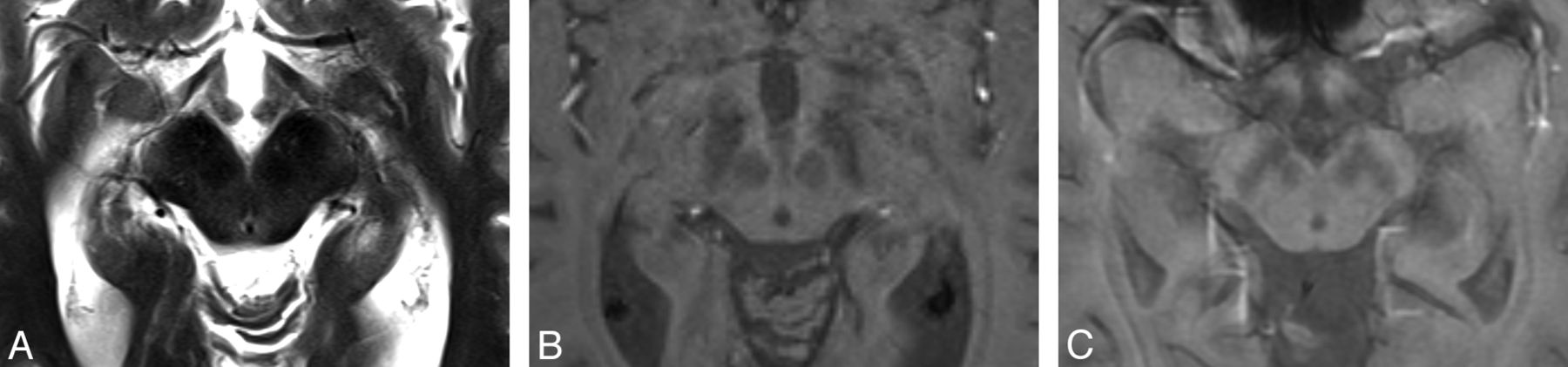
  • Fig 3.
    • Download figure
    • Open in new tab
    • Download powerpoint
    Fig 3.

    Axial T2-weighted MR imaging sequence in a patient with PD (A) demonstrating “smudging” of the hypointensity in the substantia nigra toward the red nucleus in the midbrain (right subtlely more marked than the left, concordant with the contralateral clinical features). It is more easily visualized in the corresponding susceptibility-weighted image (B). Axial T2*-weighted MR imaging sequence also demonstrates the “smudging” sign as shown in another patient with PD (C) (left more than right concordant with worse contralateral clinical features). The “smudging” sign is nonspecific and seen in progressive supranuclear palsy, multiple system atrophy, and corticobasal degeneration. It is a subtle sign and often not used in routine clinical practice.

  • Fig 4.
    • Download figure
    • Open in new tab
    • Download powerpoint
    Fig 4.

    On these inversion-recovery T1-weighted images in which deep gray matter signal is suppressed, the substantia nigra in a patient with severe PD (A) appears both substantially shrunk and with altered contrast in comparison with a healthy control (B). There is a correlation between the substantia nigra area with the Unified Parkinson Disease Rating Scale score.23 There is also a group difference between those with PD and controls; however, this metric has not been proved to be useful for individuals. Images courtesy of Dr Ludovico Minati, Scientific Department, Istituto Di Ricovero e Cura a Carattere Scientifico Foundation Neurologic Institute, Carlo Besta, Milan, Italy.

  • Fig 5.
    • Download figure
    • Open in new tab
    • Download powerpoint
    Fig 5.

    T2-weighted fast spin-echo MR image (A) and corresponding transcranial sonography images (B and C) of midbrain axial sections at the orbitomeatal line. B, Transcranial sonography image of the axial midbrain section in a healthy person depicting the hypoechoic midbrain (outlined) surrounded by the hyperechogenic basal cisterns. In the anatomic area of the ipsilateral substantia nigra, only small hyperechoic patches are visible (small arrows); the planimetrically measured area is <0.20 cm2. The raphe is detected as a highly echogenic, continuous line; the aqueduct is shown at the dorsal part of the midbrain. C, Transcranial sonography of a patient with Parkinson disease showing a bilateral hyperechogenic substantia nigra (area ≥0.20 cm2). Small arrows indicate the substantia nigra. Images courtesy of Dr Rita CL Fernandes, Department of Neurodegeneration at Tübingen University Hospital and Hertie Institute of Clinical Brain Research, Tübingen, Germany.
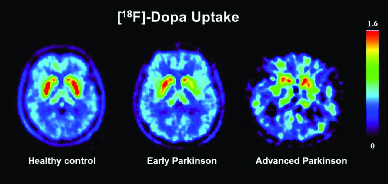
  • Fig 6.
    • Download figure
    • Open in new tab
    • Download powerpoint
    Fig 6.

    Axial 18F-DOPA PET images through the striatum. The patient with early Parkinson disease shows an asymmetric reduction in putaminal radiotracer uptake. With further disease progression, both putamina show a substantial reduction in radiotracer uptake. Images courtesy of Professor Philippe Remy, l'Hôpital Henri Mondor, Creteil, France.
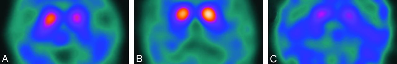
  • Fig 7.
    • Download figure
    • Open in new tab
    • Download powerpoint
    Fig 7.

    Axial 123I-FP-CIT DaT-SPECT sections depicting the different patterns of abnormality seen in PD as described by Catafau and Tolosa2—type 1: asymmetric activity with reduced putaminal uptake in 1 hemisphere (A); type 2: symmetric reduction in putaminal uptake in both hemispheres (B); and type 3: virtual absence of uptake in the putamina and caudate nuclei despite high gain as demonstrated by ample background activity (C).

Tables

  • Figures
  • Clinical indications for DaT-SPECT imaging

    Indications
    Only 1 of 3 cardinal clinical signsa with or without asymmetry
    2 Signs without bradykinesia
    Poor response to L-dopa
    Lack of disease progression
    • ↵a Cardinal signs are resting tremor, rigidity, and bradykinesia. (An alternative classification adds a fourth cardinal sign as postural instability.)

PreviousNext
Back to top

In this issue

American Journal of Neuroradiology: 36 (2)
American Journal of Neuroradiology
Vol. 36, Issue 2
1 Feb 2015
  • Table of Contents
  • Index by author
  • Complete Issue (PDF)
Advertisement
Print
Download PDF
Email Article

Thank you for your interest in spreading the word on American Journal of Neuroradiology.

NOTE: We only request your email address so that the person you are recommending the page to knows that you wanted them to see it, and that it is not junk mail. We do not capture any email address.

Enter multiple addresses on separate lines or separate them with commas.
The Role of Functional Dopamine-Transporter SPECT Imaging in Parkinsonian Syndromes, Part 1
(Your Name) has sent you a message from American Journal of Neuroradiology
(Your Name) thought you would like to see the American Journal of Neuroradiology web site.
CAPTCHA
This question is for testing whether or not you are a human visitor and to prevent automated spam submissions.
Cite this article
T.C. Booth, M. Nathan, A.D. Waldman, A.-M. Quigley, A.H. Schapira, J. Buscombe
The Role of Functional Dopamine-Transporter SPECT Imaging in Parkinsonian Syndromes, Part 1
American Journal of Neuroradiology Feb 2015, 36 (2) 229-235; DOI: 10.3174/ajnr.A3970

Citation Manager Formats

  • BibTeX
  • Bookends
  • EasyBib
  • EndNote (tagged)
  • EndNote 8 (xml)
  • Medlars
  • Mendeley
  • Papers
  • RefWorks Tagged
  • Ref Manager
  • RIS
  • Zotero
0 Responses
Respond to this article
Share
Bookmark this article
The Role of Functional Dopamine-Transporter SPECT Imaging in Parkinsonian Syndromes, Part 1
T.C. Booth, M. Nathan, A.D. Waldman, A.-M. Quigley, A.H. Schapira, J. Buscombe
American Journal of Neuroradiology Feb 2015, 36 (2) 229-235; DOI: 10.3174/ajnr.A3970
del.icio.us logo Twitter logo Facebook logo Mendeley logo
  • Tweet Widget
  • Facebook Like
  • Google Plus One
Purchase

Jump to section

  • Article
    • Abstract
    • ABBREVIATIONS:
    • Footnotes
    • REFERENCES
  • Figures & Data
  • Info & Metrics
  • Responses
  • References
  • PDF

Related Articles

  • No related articles found.
  • PubMed
  • Google Scholar

Cited By...

  • Pitfalls and Artifacts of 123I-Ioflupane SPECT in Parkinsonian Syndromes: A Quality Improvement Teaching Tool
  • Crossref (57)
  • Google Scholar

This article has been cited by the following articles in journals that are participating in Crossref Cited-by Linking.

  • An Explainable Machine Learning Model for Early Detection of Parkinson&#039;s Disease using LIME on DaTSCAN Imagery
    Pavan Rajkumar Magesh, Richard Delwin Myloth, Rijo Jackson Tom
    Computers in Biology and Medicine 2020 126
  • Dopamine and Dopamine Receptors in Alzheimer&#039;s Disease: A Systematic Review and Network Meta-Analysis
    Xiongfeng Pan, Atipatsa C. Kaminga, Shi Wu Wen, Xinyin Wu, Kwabena Acheampong, Aizhong Liu
    Frontiers in Aging Neuroscience 2019 11
  • Importance of low diagnostic Accuracy for early Parkinson&#039;s disease
    Thomas G. Beach, Charles H. Adler
    Movement Disorders 2018 33 10
  • Targeting mitophagy in Parkinson&#039;s disease
    Emily H. Clark, Aurelio Vázquez de la Torre, Tamaki Hoshikawa, Thomas Briston
    Journal of Biological Chemistry 2021 296
  • Linking Essential Tremor to the Cerebellum—Neuroimaging Evidence
    Antonio Cerasa, Aldo Quattrone
    The Cerebellum 2016 15 3
  • From simultaneous to synergistic MR‐PET brain imaging: A review of hybrid MR‐PET imaging methodologies
    Zhaolin Chen, Sharna D. Jamadar, Shenpeng Li, Francesco Sforazzini, Jakub Baran, Nicholas Ferris, Nadim Jon Shah, Gary F. Egan
    Human Brain Mapping 2018 39 12
  • Augmented Intelligence in Healthcare: A Pragmatic and Integrated Analysis
    Aryan Mohanty, Sushruta Mishra
    2022 1024
  • Dopamine Transporter Neuroimaging as an Enrichment Biomarker in Early Parkinson&#039;s Disease Clinical Trials: A Disease Progression Modeling Analysis
    Daniela J. Conrado, Timothy Nicholas, Kuenhi Tsai, Sreeraj Macha, Vikram Sinha, Julie Stone, Brian Corrigan, Massimo Bani, Pierandrea Muglia, Ian A. Watson, Volker D. Kern, Elena Sheveleva, Kenneth Marek, Diane T. Stephenson, Klaus Romero
    Clinical and Translational Science 2018 11 1
  • Neuromelanin MRI is useful for monitoring motor complications in Parkinson’s and PARK2 disease
    Taku Hatano, Ayami Okuzumi, Koji Kamagata, Kensuke Daida, Daisuke Taniguchi, Masaaki Hori, Hiroyo Yoshino, Shigeki Aoki, Nobutaka Hattori
    Journal of Neural Transmission 2017 124 4
  • Imaging analysis of Parkinson’s disease patients using SPECT and tractography
    Seong-Jin Son, Mansu Kim, Hyunjin Park
    Scientific Reports 2016 6 1

More in this TOC Section

  • An Atlas of Neonatal Neurovascular Imaging Anatomy as Depicted with Microvascular Imaging: The Intracranial Arteries
  • An Atlas of Neonatal Neurovascular Imaging Anatomy as Depicted with Microvascular Imaging: The Intracranial Veins
  • Clinical Translation of Hyperpolarized 13C Metabolic Probes for Glioma Imaging
Show more Review articles

Similar Articles

Advertisement

Indexed Content

  • Current Issue
  • Accepted Manuscripts
  • Article Preview
  • Past Issues
  • Editorials
  • Editor's Choice
  • Fellows' Journal Club
  • Letters to the Editor
  • Video Articles

Cases

  • Case Collection
  • Archive - Case of the Week
  • Archive - Case of the Month
  • Archive - Classic Case

More from AJNR

  • Trainee Corner
  • Imaging Protocols
  • MRI Safety Corner
  • Book Reviews

Multimedia

  • AJNR Podcasts
  • AJNR Scantastics

Resources

  • Turnaround Time
  • Submit a Manuscript
  • Submit a Video Article
  • Submit an eLetter to the Editor/Response
  • Manuscript Submission Guidelines
  • Statistical Tips
  • Fast Publishing of Accepted Manuscripts
  • Graphical Abstract Preparation
  • Imaging Protocol Submission
  • Evidence-Based Medicine Level Guide
  • Publishing Checklists
  • Author Policies
  • Become a Reviewer/Academy of Reviewers
  • News and Updates

About Us

  • About AJNR
  • Editorial Board
  • Editorial Board Alumni
  • Alerts
  • Permissions
  • Not an AJNR Subscriber? Join Now
  • Advertise with Us
  • Librarian Resources
  • Feedback
  • Terms and Conditions
  • AJNR Editorial Board Alumni

American Society of Neuroradiology

  • Not an ASNR Member? Join Now

© 2025 by the American Society of Neuroradiology All rights, including for text and data mining, AI training, and similar technologies, are reserved.
Print ISSN: 0195-6108 Online ISSN: 1936-959X

Powered by HighWire