
Article Figures & Data
Figures
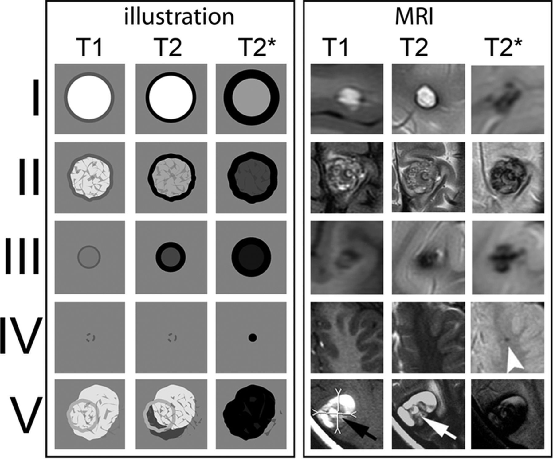
- Fig 1.
CCM types according to the Zabramski classification. Graphic illustration (left 3 rows) and corresponding MR images (right 3 rows) of CCMs according to the MR imaging classification of Zabramski et al.3 Type IV CCM: arrowhead indicates a small T2* lesion. Type V: arrows indicate parts of the actual CCMs that are visible in the center of the hemorrhage; however, the CCM is not fully distinguishable from hemorrhage.
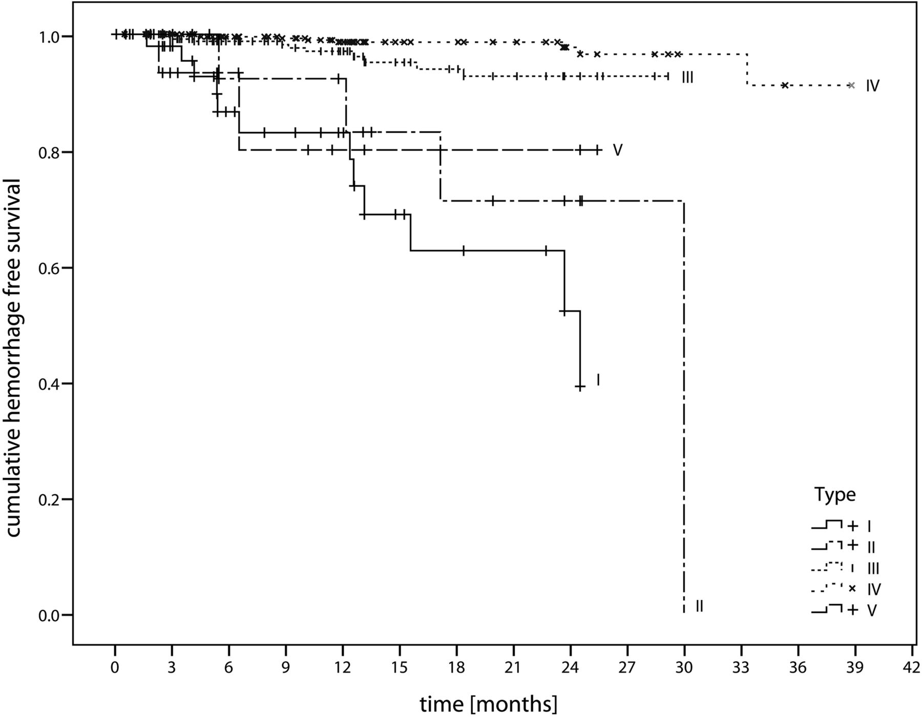
- Fig 2.
Hemorrhage-free survival. Kaplan-Meier diagram illustrates hemorrhage-free survival depending on the Zabramski CCM type.
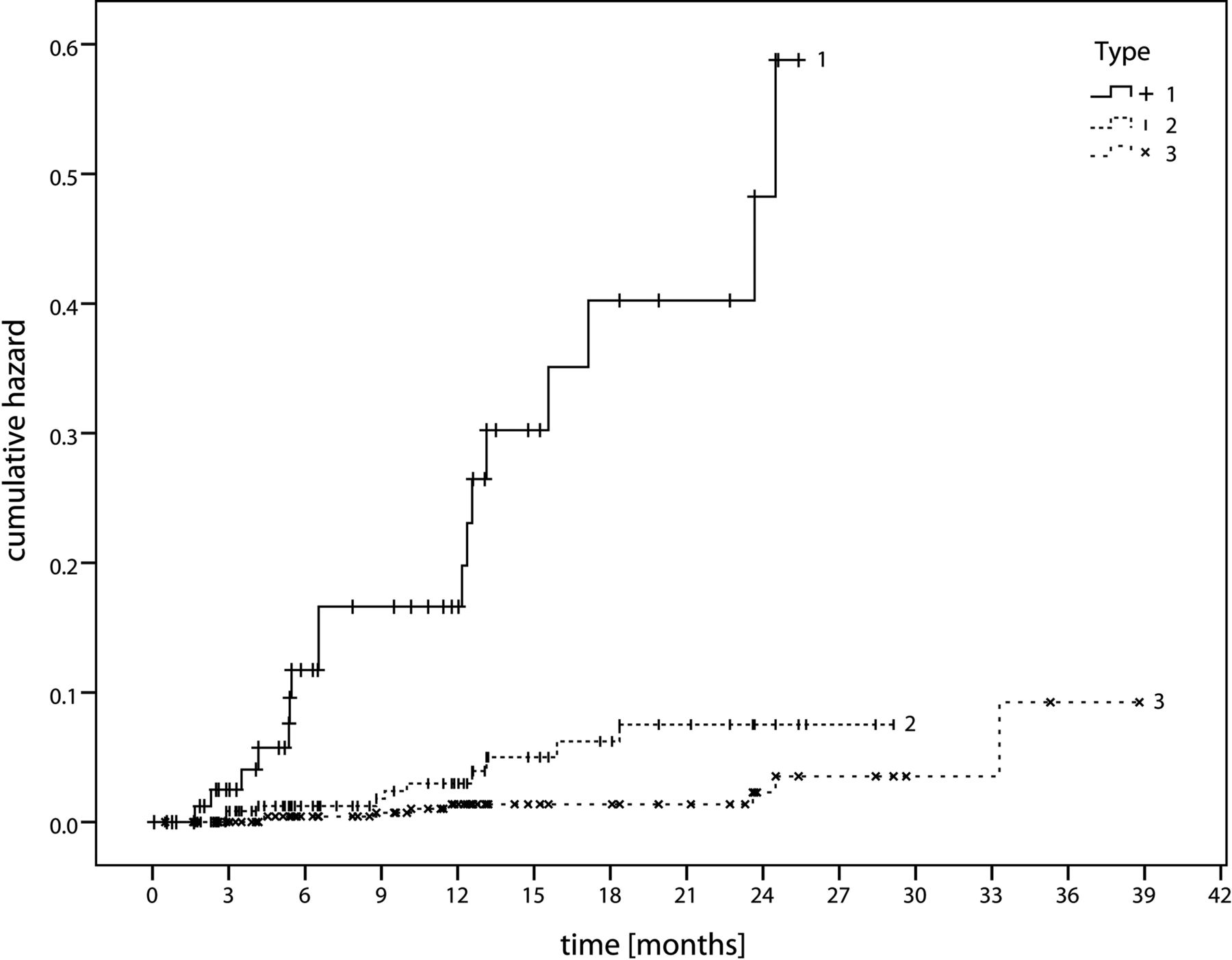
- Fig 3.
Cumulative hazard. Diagram illustrates the cumulative hazard for hemorrhage according to our proposed CCM classification, 1) CCM with signs of acute or subacute hemorrhage, 2) CCM without signs of acute or subacute hemorrhage, and 3) dot-sized CCMs.


Tables
Lesion Type MRI Signal Characteristics Pathologic Characteristics Type I T1: hyperintense core Subacute hemorrhage, surrounded by a rim of hemosiderin-stained macrophages and gliotic brain T2: hyper- or hypointense core with surrounding hypointense rim Type II T1: reticulated mixed-signal core Loculated areas of hemorrhage and thrombosis of varying ages, surrounded by gliotic, hemosiderin-stained brain; in large lesions, areas of calcification may be seen T2: reticulated mixed-signal core with surrounding hypointense rim Type III T1: iso- or hypointense core Chronic resolved hemorrhage, with hemosiderin staining within and around the lesion T2: hypointense with a hypointense rim that magnifies the size of the lesion GE: hypointense with greater magnification than T2 Type IV T1: poorly seen or not visualized at all Two lesions in the category were pathologically documented as telangiectasias T2: poorly seen or not visualized at all GE: punctate hypointense lesions CCM Type Mean Hemorrhage-Free Survival Estimator Standard Error 95% CI Lower Upper I 18.82 1.52 15.84 21.80 II 24.92 2.91 19.21 30.63 III 27.88 0.38 27.13 28.63 IV 37.78 0.40 37.00 38.57 V 21.34 2.67 16.10 26.59 I, II, V 22.63 1.50 19.68 25.57 All 36.06 0.59 34.91 37.21 ↵a Demonstrating mean hemorrhage-free survival in months depending on the extended CCM type of Zabramski et al.3
Study No. of Patients No. of Lesions Hemorrhage Assessment Hemorrhage Rate without Prior Hemorrhage Hemorrhage Rate with Prior Hemorrhage Al-Shahi Salman et al1 134 NI MRI/clinical 2.4% 29.5% Kondziolka et al8 122 NI MRI/clinical 0.6% 4.5% Moriarity et al15 68 228 MRI/clinical 3.1%b NI Porter et al16 173 NI MRI/clinical 1.6%b NI Robinson et al17 57 66 MRI/clinical 0.7%b,c NI


Jump to section
Related Articles
Cited By...
- No citing articles found.