Advertisement

Main menu

  • Home
  • Content
    • Current Issue
    • Accepted Manuscripts
    • Article Preview
    • Past Issue Archive
    • Video Articles
    • AJNR Case Collection
    • Case of the Week Archive
    • Case of the Month Archive
    • Classic Case Archive
  • Special Collections
    • AJNR Awards
    • Low-Field MRI
    • Alzheimer Disease
    • ASNR Foundation Special Collection
    • Photon-Counting CT
    • View All
  • Multimedia
    • AJNR Podcasts
    • AJNR SCANtastic
    • Trainee Corner
    • MRI Safety Corner
    • Imaging Protocols
  • For Authors
    • Submit a Manuscript
    • Submit a Video Article
    • Submit an eLetter to the Editor/Response
    • Manuscript Submission Guidelines
    • Statistical Tips
    • Fast Publishing of Accepted Manuscripts
    • Graphical Abstract Preparation
    • Imaging Protocol Submission
    • Author Policies
  • About Us
    • About AJNR
    • Editorial Board
    • Editorial Board Alumni
  • More
    • Become a Reviewer/Academy of Reviewers
    • Subscribers
    • Permissions
    • Alerts
    • Feedback
    • Advertisers
    • ASNR Home

User menu

  • Alerts
  • Log in

Search

  • Advanced search
American Journal of Neuroradiology

American Journal of Neuroradiology

ASHNR American Society of Functional Neuroradiology ASHNR American Society of Pediatric Neuroradiology ASSR
  • Alerts
  • Log in

Advanced Search

  • Home
  • Content
    • Current Issue
    • Accepted Manuscripts
    • Article Preview
    • Past Issue Archive
    • Video Articles
    • AJNR Case Collection
    • Case of the Week Archive
    • Case of the Month Archive
    • Classic Case Archive
  • Special Collections
    • AJNR Awards
    • Low-Field MRI
    • Alzheimer Disease
    • ASNR Foundation Special Collection
    • Photon-Counting CT
    • View All
  • Multimedia
    • AJNR Podcasts
    • AJNR SCANtastic
    • Trainee Corner
    • MRI Safety Corner
    • Imaging Protocols
  • For Authors
    • Submit a Manuscript
    • Submit a Video Article
    • Submit an eLetter to the Editor/Response
    • Manuscript Submission Guidelines
    • Statistical Tips
    • Fast Publishing of Accepted Manuscripts
    • Graphical Abstract Preparation
    • Imaging Protocol Submission
    • Author Policies
  • About Us
    • About AJNR
    • Editorial Board
    • Editorial Board Alumni
  • More
    • Become a Reviewer/Academy of Reviewers
    • Subscribers
    • Permissions
    • Alerts
    • Feedback
    • Advertisers
    • ASNR Home
  • Follow AJNR on Twitter
  • Visit AJNR on Facebook
  • Follow AJNR on Instagram
  • Join AJNR on LinkedIn
  • RSS Feeds

AJNR Awards, New Junior Editors, and more. Read the latest AJNR updates

Research ArticleNeurointervention
Open Access

A Comparison of 4D DSA with 2D and 3D DSA in the Analysis of Normal Vascular Structures in a Canine Model

C. Sandoval-Garcia, K. Royalty, B. Aagaard-Kienitz, S. Schafer, P. Yang and C. Strother
American Journal of Neuroradiology October 2015, 36 (10) 1959-1963; DOI: https://doi.org/10.3174/ajnr.A4359
C. Sandoval-Garcia
aFrom the Departments of Neurological Surgery (C.S.-G., B.A.-K.)
  • Find this author on Google Scholar
  • Find this author on PubMed
  • Search for this author on this site
K. Royalty
bBiomedical Engineering (K.R)
dSiemens (K.R., S.S.), Hoffman Estates, Illinois
  • Find this author on Google Scholar
  • Find this author on PubMed
  • Search for this author on this site
B. Aagaard-Kienitz
aFrom the Departments of Neurological Surgery (C.S.-G., B.A.-K.)
  • Find this author on Google Scholar
  • Find this author on PubMed
  • Search for this author on this site
S. Schafer
dSiemens (K.R., S.S.), Hoffman Estates, Illinois
  • Find this author on Google Scholar
  • Find this author on PubMed
  • Search for this author on this site
P. Yang
cRadiology (P.Y., C.S.), University of Wisconsin School of Medicine and Public Health, Madison, Wisconsin
eDepartment of Neurosurgery (P.Y.), Changhai Hospital, Second Military Medical University, Shanghai, China.
  • Find this author on Google Scholar
  • Find this author on PubMed
  • Search for this author on this site
  • ORCID record for P. Yang
C. Strother
cRadiology (P.Y., C.S.), University of Wisconsin School of Medicine and Public Health, Madison, Wisconsin
  • Find this author on Google Scholar
  • Find this author on PubMed
  • Search for this author on this site
  • Article
  • Figures & Data
  • Info & Metrics
  • Responses
  • References
  • PDF
Loading

Article Figures & Data

Figures

  • Tables
  • Fig 1.
    • Download figure
    • Open in new tab
    • Download powerpoint
    Fig 1.

    Example of the vasculature on anteroposterior 2D DSA projections for each catheter position: proximal (A), middle (B), and distal (C). These show the decrease in the vascular complexity as the injection site is moved from proximal to most distal. The magnification factor for the images increases, moving from proximal to most distal.
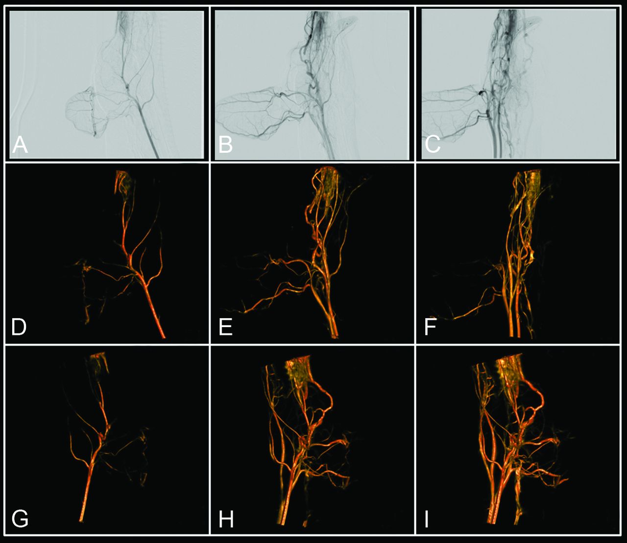
  • Fig 2.
    • Download figure
    • Open in new tab
    • Download powerpoint
    Fig 2.

    Illustration of 4D DSA reconstruction. No x-ray delay between contrast injection and rotational image acquisition results in contrast flow or inflow being visible in the rotational projections (A–C). Through a 2-step reconstruction process, this flow information is encoded into the 3D DSA for every projection, effectively creating a 4D DSA. This allows viewing of the contrast bolus passage at any desired angle at any time during the bolus passage. D–F, View of the bolus passage at 3 projection angles at 3 different time points. G–I, View of the bolus passage at 3 angles not present in the projections, again at 3 different time points. The figures correspond to sample angle projections selected to match the 2D acquisitions, even though once 4D images are reconstructed, views are available from any angle.
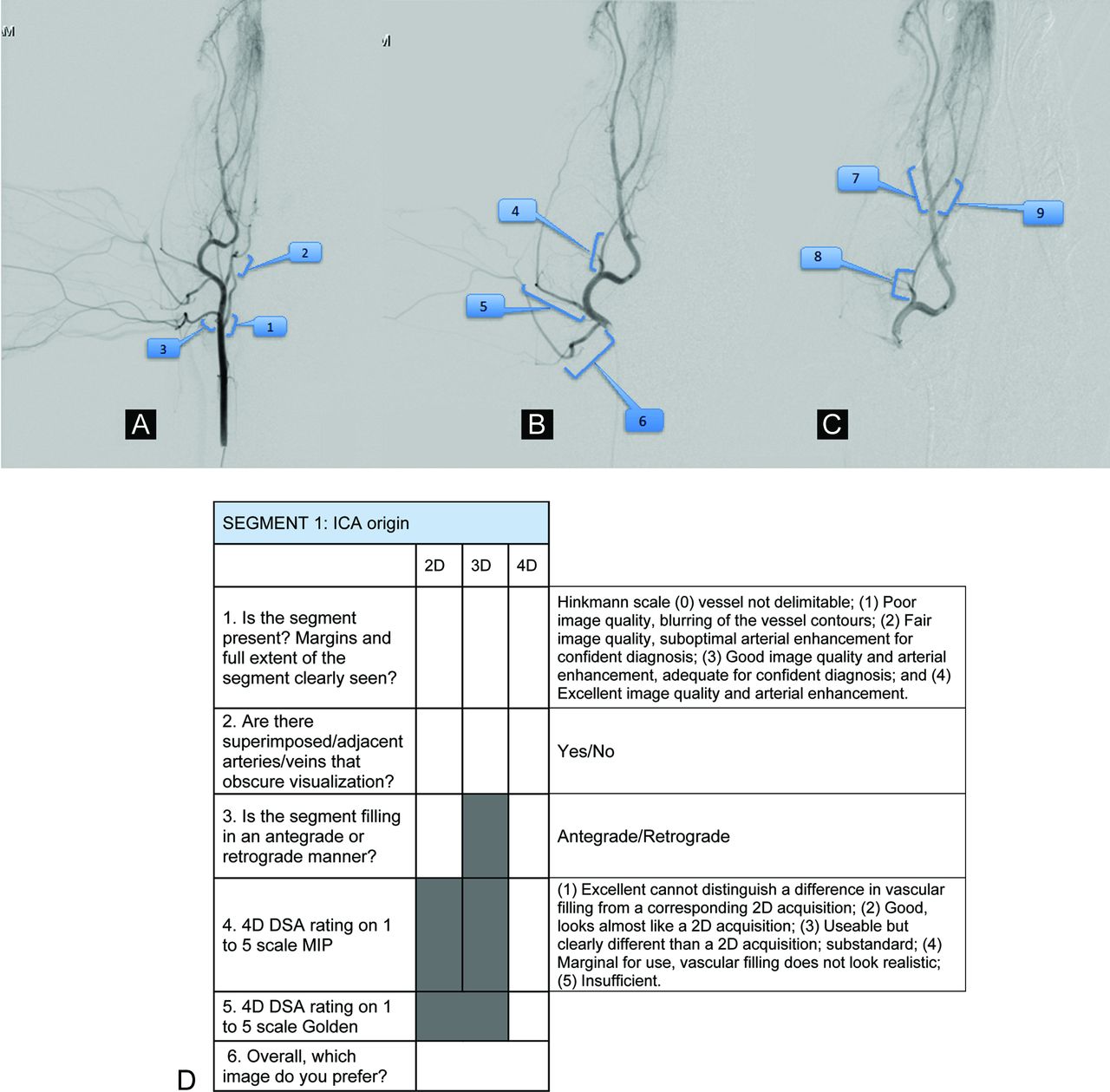
  • Fig 3.
    • Download figure
    • Open in new tab
    • Download powerpoint
    Fig 3.

    A–C, Example of the pictorial reference form provided to the image reviewers identifying the vascular segments to be evaluated: proximal (A), middle (B), and distal (C) catheter positions on anteroposterior projections. D, Evaluation form and scales used for scoring of the images.

Tables

  • Figures
    • View popup
    Table 1:

    Injection parameters used for the various acquisitions

    ProximalMiddleDistal
    2D DSA3D DSA4D DSA2D DSA3D DSA4D DSA2D DSA3D DSA4D DSA
    Contrast volume (mL)8–920208–911118–989
    Concentration (%)505050505050505050
    Flow rate (mL/s)HI33HI1.5–2.51.5–2.5HI1–1.51–1.5
    Injection duration (s)HI6.66.6HI66HI7.37.3
    Injection delay (s)100100100
    X-ray delay (s)010010010
    • Note:—HI indicates hand injection.

    • View popup
    Table 2:

    Raw data from each of the 2 observers for all 44 vessel segmentsa

    Reviewer 1Reviewer 2
    2D3D4DPreferred2D3D4DPreferred
    3444D2444D
    3344D4444D
    4244D0344D
    4242D3444D
    4244D2344D
    4244D3344D
    4342D3444D
    4332D4234D
    4244D4334D
    4444D3344D
    4444D2344D
    4444D2344D
    4332D4444D
    4342D2444D
    4344D2444D
    4332D4444D
    4423D4433D
    4344D4433D
    4242D4444D
    4134D4444D
    4342D4444D
    4442D4444D
    4342D4344D
    4344D4444D
    4442D4444D
    4342D4332D
    3222D2012D
    4444D1444D
    4244D2244D
    4344D3344D
    4444D4344D
    4444D3244D
    4344D2244D
    4444D4444D
    4344D3344D
    3344D4144D
    4342D3444D
    4244D3244D
    4442D4444D
    4342D4444D
    4444D3444D
    4344D3344D
    3442D3424D
    4432D4444D
    • ↵a The first 3 columns show the rating for each of the 3 modalities as described in the “Materials and Methods” and the scoring form (Fig 3B). The last column shows the preferred modality.

PreviousNext
Back to top

In this issue

American Journal of Neuroradiology: 36 (10)
American Journal of Neuroradiology
Vol. 36, Issue 10
1 Oct 2015
  • Table of Contents
  • Index by author
  • Complete Issue (PDF)
Advertisement
Print
Download PDF
Email Article

Thank you for your interest in spreading the word on American Journal of Neuroradiology.

NOTE: We only request your email address so that the person you are recommending the page to knows that you wanted them to see it, and that it is not junk mail. We do not capture any email address.

Enter multiple addresses on separate lines or separate them with commas.
A Comparison of 4D DSA with 2D and 3D DSA in the Analysis of Normal Vascular Structures in a Canine Model
(Your Name) has sent you a message from American Journal of Neuroradiology
(Your Name) thought you would like to see the American Journal of Neuroradiology web site.
CAPTCHA
This question is for testing whether or not you are a human visitor and to prevent automated spam submissions.
Cite this article
C. Sandoval-Garcia, K. Royalty, B. Aagaard-Kienitz, S. Schafer, P. Yang, C. Strother
A Comparison of 4D DSA with 2D and 3D DSA in the Analysis of Normal Vascular Structures in a Canine Model
American Journal of Neuroradiology Oct 2015, 36 (10) 1959-1963; DOI: 10.3174/ajnr.A4359

Citation Manager Formats

  • BibTeX
  • Bookends
  • EasyBib
  • EndNote (tagged)
  • EndNote 8 (xml)
  • Medlars
  • Mendeley
  • Papers
  • RefWorks Tagged
  • Ref Manager
  • RIS
  • Zotero
0 Responses
Respond to this article
Share
Bookmark this article
A Comparison of 4D DSA with 2D and 3D DSA in the Analysis of Normal Vascular Structures in a Canine Model
C. Sandoval-Garcia, K. Royalty, B. Aagaard-Kienitz, S. Schafer, P. Yang, C. Strother
American Journal of Neuroradiology Oct 2015, 36 (10) 1959-1963; DOI: 10.3174/ajnr.A4359
del.icio.us logo Twitter logo Facebook logo Mendeley logo
  • Tweet Widget
  • Facebook Like
  • Google Plus One
Purchase

Jump to section

  • Article
    • Abstract
    • ABBREVIATION:
    • Materials and Methods
    • Results
    • Discussion
    • Conclusions
    • Footnotes
    • REFERENCES
  • Figures & Data
  • Info & Metrics
  • Responses
  • References
  • PDF

Related Articles

  • No related articles found.
  • PubMed
  • Google Scholar

Cited By...

  • 4D-DSA: Development and Current Neurovascular Applications
  • Quantitative and Qualitative Comparison of 4D-DSA with 3D-DSA Using Computational Fluid Dynamics Simulations in Cerebral Aneurysms
  • In-room assessment of intravascular velocity from time-resolved rotational angiography in patients with arteriovenous malformation: a pilot study
  • Time-resolved 3D rotational angiography: display of detailed neurovascular anatomy in patients with intracranial vascular malformations
  • 4D DSA for Dynamic Visualization of Cerebral Vasculature: A Single-Center Experience in 26 Cases
  • Application of Time-Resolved 3D Digital Subtraction Angiography to Plan Cerebral Arteriovenous Malformation Radiosurgery
  • Comparison of the Diagnostic Utility of 4D-DSA with Conventional 2D- and 3D-DSA in the Diagnosis of Cerebrovascular Abnormalities
  • Crossref (17)
  • Google Scholar

This article has been cited by the following articles in journals that are participating in Crossref Cited-by Linking.

  • 4D DSA for Dynamic Visualization of Cerebral Vasculature: A Single-Center Experience in 26 Cases
    S. Lang, P. Gölitz, T. Struffert, J. Rösch, K. Rössler, M. Kowarschik, C. Strother, A. Doerfler
    American Journal of Neuroradiology 2017 38 6
  • 4D-DSA: Development and Current Neurovascular Applications
    K.L. Ruedinger, S. Schafer, M.A. Speidel, C.M. Strother
    American Journal of Neuroradiology 2021 42 2
  • Time-resolved 3D rotational angiography: display of detailed neurovascular anatomy in patients with intracranial vascular malformations
    Stephanie Lescher, Sonja Gehrisch, Sigrun Klein, Joachim Berkefeld
    Journal of NeuroInterventional Surgery 2017 9 9
  • Application of Time-Resolved 3D Digital Subtraction Angiography to Plan Cerebral Arteriovenous Malformation Radiosurgery
    K.-K. Chen, W.-Y. Guo, H.-C. Yang, C.-J. Lin, C.-H.F. Wu, S. Gehrisch, M. Kowarschik, Y.-T. Wu, W.-Y. Chung
    American Journal of Neuroradiology 2017 38 4
  • Comparison of the Diagnostic Utility of 4D-DSA with Conventional 2D- and 3D-DSA in the Diagnosis of Cerebrovascular Abnormalities
    C. Sandoval-Garcia, P. Yang, T. Schubert, S. Schafer, S. Hetzel, A. Ahmed, C. Strother
    American Journal of Neuroradiology 2017 38 4
  • A new time-resolved 3D angiographic technique (4D DSA): Description, and assessment of its reliability in Spetzler–Martin grading of cerebral arteriovenous malformations
    Julien Ognard, Elsa Magro, Jildaz Caroff, Douraied Ben Salem, Sebastien Andouard, Michel Nonent, Jean-Christophe Gentric
    Journal of Neuroradiology 2018 45 3
  • 4D Digital Subtraction Angiography for the Temporal Flow Visualization of Intracranial Aneurysms and Vascular Malformations
    Naoki Kato, Ichiro Yuki, Shunsuke Hataoka, Chihebeddine Dahmani, Katharina Otani, Yukiko Abe, Shota Kakizaki, Gota Nagayama, Fumiaki Maruyama, Ayako Ikemura, Issei Kan, Tomonobu Kodama, Toshihiro Ishibashi, Yuichi Murayama
    Journal of Stroke and Cerebrovascular Diseases 2020 29 12
  • Application of 4-Dimensional Digital Subtraction Angiography for Dural Arteriovenous Fistulas
    Visish M. Srinivasan, Gouthami Chintalapani, Edward A.M. Duckworth, Peter Kan
    World Neurosurgery 2016 96
  • In-room assessment of intravascular velocity from time-resolved rotational angiography in patients with arteriovenous malformation: a pilot study
    Chung Jung Lin, Huai Che Yang, Ai Chi Chien, Wan Yuo Guo, Chih Chun Wu, Sheng Che Hung, Ko Kung Chen, Hsiu Mei Wu, Chao Bao Luo, Wei Fa Chu, Jia Sheng Hong, Chun Shien Frank Wu
    Journal of NeuroInterventional Surgery 2018 10 6
  • Time-resolved 3D Rotational Angiography (4D DSA) of the Lenticulostriate Arteries: Display of Normal Anatomic Variants and Collaterals in Cases with Chronic Obstruction of the MCA
    S. Kammerer, M. Mueller-Eschner, J. Berkefeld, S. Tritt
    Clinical Neuroradiology 2017 27 4

More in this TOC Section

  • Contour Neurovascular System: Five Year Follow Up
  • Effect of SARS-CoV2 on Endovascular Thrombectomy
  • Optimizing Voxel Size in 3D Rotational Angiography
Show more NEUROINTERVENTION

Similar Articles

Advertisement

Indexed Content

  • Current Issue
  • Accepted Manuscripts
  • Article Preview
  • Past Issues
  • Editorials
  • Editor's Choice
  • Fellows' Journal Club
  • Letters to the Editor
  • Video Articles

Cases

  • Case Collection
  • Archive - Case of the Week
  • Archive - Case of the Month
  • Archive - Classic Case

More from AJNR

  • Trainee Corner
  • Imaging Protocols
  • MRI Safety Corner
  • Book Reviews

Multimedia

  • AJNR Podcasts
  • AJNR Scantastics

Resources

  • Turnaround Time
  • Submit a Manuscript
  • Submit a Video Article
  • Submit an eLetter to the Editor/Response
  • Manuscript Submission Guidelines
  • Statistical Tips
  • Fast Publishing of Accepted Manuscripts
  • Graphical Abstract Preparation
  • Imaging Protocol Submission
  • Evidence-Based Medicine Level Guide
  • Publishing Checklists
  • Author Policies
  • Become a Reviewer/Academy of Reviewers
  • News and Updates

About Us

  • About AJNR
  • Editorial Board
  • Editorial Board Alumni
  • Alerts
  • Permissions
  • Not an AJNR Subscriber? Join Now
  • Advertise with Us
  • Librarian Resources
  • Feedback
  • Terms and Conditions
  • AJNR Editorial Board Alumni

American Society of Neuroradiology

  • Not an ASNR Member? Join Now

© 2025 by the American Society of Neuroradiology All rights, including for text and data mining, AI training, and similar technologies, are reserved.
Print ISSN: 0195-6108 Online ISSN: 1936-959X

Powered by HighWire