
Article Figures & Data
Figures
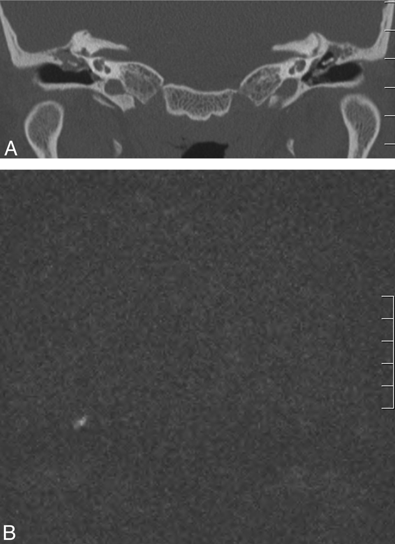
- Fig 1.
Case 3. A 39-year-old woman with no prior otologic diagnosis. A, Coronal CT scan shows right tympanic membrane thickening with occupation of Prussak space. B, Coronal HASTE shows no hyperintensity. In surgery, a granulomatous mass was identified without evidence of cholesteatoma (not shown).
- Fig 2.
Case 5. A 29-year-old man with a history of chronic bilateral otitis media with effusion with a tympanic ventilation T-tube on the left side. A, Coronal CT scan shows bilateral tympanic thickening and middle ear opacities, with erosion of the scutum; a T-tube can be observed on the left side. B, Coronal HASTE shows a 13-mm right-sided hyperintensity in relation to the epitympanum. The cholesteatoma was found intraoperatively (not shown).
- Fig 3.
Case 13. A 58-year-old woman with bilateral chronic ear disease (right tympanic retraction pocket and left external ear canal stenosis). A, Coronal CT shows a right-sided tympanic retraction pocket with partial thickening of the tympanic membrane and erosion of the scutum, with an epitympanic middle ear mass. On the left side, there is external ear canal occupation by a soft-tissue attenuation mass, with epitympanic opacity. B, Coronal HASTE image negative for cholesteatoma. Surgery confirmed the presence of a retraction pocket without invasion of skin into the middle ear on the right side (not shown).


Tables
Patient characteristics and MRI and surgical findings
Patient No. Sex Age (yr) Side HASTE Size (mm) Surgery 1 F 27 L + 3 + 2 M 21 R – 0 – 3 F 39 R – 0 – 4 F 35 L – 0 – 5 M 29 R + 13 + 6 M 43 R + 6 + 7 F 39 L + 5 + 8 F 55 L + 6 + 9 M 49 R – 0 + 10 F 42 R + 5 + 11 F 60 L + 10 + 12 M 9 L + 15 + 13 F 58 R – 0 – 14 F 48 L + 4 + 15 M 24 L – 0 – 16 F 43 R – 0 – Note:—L indicates left; R, right; +, positive finding; —, negative finding.


Jump to section
Related Articles
Cited By...
- No citing articles found.