Advertisement

Main menu

  • Home
  • Content
    • Current Issue
    • Accepted Manuscripts
    • Article Preview
    • Past Issue Archive
    • Video Articles
    • AJNR Case Collection
    • Case of the Week Archive
    • Case of the Month Archive
    • Classic Case Archive
  • Special Collections
    • Advancing NeuroMRI with High-Relaxivity Contrast Agents
    • AJNR Awards
    • Low-Field MRI
    • Alzheimer Disease
    • ASNR Foundation Special Collection
    • Photon-Counting CT
    • View All
  • Multimedia
    • AJNR Podcasts
    • AJNR SCANtastic
    • Trainee Corner
    • MRI Safety Corner
    • Imaging Protocols
  • For Authors
    • Submit a Manuscript
    • Submit a Video Article
    • Submit an eLetter to the Editor/Response
    • Manuscript Submission Guidelines
    • Statistical Tips
    • Fast Publishing of Accepted Manuscripts
    • Graphical Abstract Preparation
    • Imaging Protocol Submission
    • Author Policies
  • About Us
    • About AJNR
    • Editorial Board
    • Editorial Board Alumni
  • More
    • Become a Reviewer/Academy of Reviewers
    • Subscribers
    • Permissions
    • Alerts
    • Feedback
    • Advertisers
    • ASNR Home

User menu

  • Alerts
  • Log in

Search

  • Advanced search
American Journal of Neuroradiology

American Journal of Neuroradiology

ASHNR American Society of Functional Neuroradiology ASHNR American Society of Pediatric Neuroradiology ASSR
  • Alerts
  • Log in

Advanced Search

  • Home
  • Content
    • Current Issue
    • Accepted Manuscripts
    • Article Preview
    • Past Issue Archive
    • Video Articles
    • AJNR Case Collection
    • Case of the Week Archive
    • Case of the Month Archive
    • Classic Case Archive
  • Special Collections
    • Advancing NeuroMRI with High-Relaxivity Contrast Agents
    • AJNR Awards
    • Low-Field MRI
    • Alzheimer Disease
    • ASNR Foundation Special Collection
    • Photon-Counting CT
    • View All
  • Multimedia
    • AJNR Podcasts
    • AJNR SCANtastic
    • Trainee Corner
    • MRI Safety Corner
    • Imaging Protocols
  • For Authors
    • Submit a Manuscript
    • Submit a Video Article
    • Submit an eLetter to the Editor/Response
    • Manuscript Submission Guidelines
    • Statistical Tips
    • Fast Publishing of Accepted Manuscripts
    • Graphical Abstract Preparation
    • Imaging Protocol Submission
    • Author Policies
  • About Us
    • About AJNR
    • Editorial Board
    • Editorial Board Alumni
  • More
    • Become a Reviewer/Academy of Reviewers
    • Subscribers
    • Permissions
    • Alerts
    • Feedback
    • Advertisers
    • ASNR Home
  • Follow AJNR on Twitter
  • Visit AJNR on Facebook
  • Follow AJNR on Instagram
  • Join AJNR on LinkedIn
  • RSS Feeds

AJNR Awards, New Junior Editors, and more. Read the latest AJNR updates


Improved Turnaround Times | Median time to first decision: 12 days

Research ArticleNeurointervention
Open Access

Analysis of Hemodynamics and Aneurysm Occlusion after Flow-Diverting Treatment in Rabbit Models

J.R. Cebral, F. Mut, M. Raschi, S. Hodis, Y.-H. Ding, B.J. Erickson, R. Kadirvel and D.F. Kallmes
American Journal of Neuroradiology August 2014, 35 (8) 1567-1573; DOI: https://doi.org/10.3174/ajnr.A3913
J.R. Cebral
aFrom the Center for Computational Fluid Dynamics (J.R.C., F.M., M.R.), College of Sciences, George Mason University, Fairfax, Virginia
  • Find this author on Google Scholar
  • Find this author on PubMed
  • Search for this author on this site
F. Mut
aFrom the Center for Computational Fluid Dynamics (J.R.C., F.M., M.R.), College of Sciences, George Mason University, Fairfax, Virginia
  • Find this author on Google Scholar
  • Find this author on PubMed
  • Search for this author on this site
M. Raschi
aFrom the Center for Computational Fluid Dynamics (J.R.C., F.M., M.R.), College of Sciences, George Mason University, Fairfax, Virginia
  • Find this author on Google Scholar
  • Find this author on PubMed
  • Search for this author on this site
S. Hodis
bDepartments of Radiology (S.H., Y.-H.D., B.J.E., R.K., D.F.K.)
  • Find this author on Google Scholar
  • Find this author on PubMed
  • Search for this author on this site
Y.-H. Ding
bDepartments of Radiology (S.H., Y.-H.D., B.J.E., R.K., D.F.K.)
  • Find this author on Google Scholar
  • Find this author on PubMed
  • Search for this author on this site
B.J. Erickson
bDepartments of Radiology (S.H., Y.-H.D., B.J.E., R.K., D.F.K.)
  • Find this author on Google Scholar
  • Find this author on PubMed
  • Search for this author on this site
R. Kadirvel
bDepartments of Radiology (S.H., Y.-H.D., B.J.E., R.K., D.F.K.)
  • Find this author on Google Scholar
  • Find this author on PubMed
  • Search for this author on this site
D.F. Kallmes
bDepartments of Radiology (S.H., Y.-H.D., B.J.E., R.K., D.F.K.)
cNeurosurgery (D.F.K.), Mayo Clinic, Rochester, Minnesota.
  • Find this author on Google Scholar
  • Find this author on PubMed
  • Search for this author on this site
  • Article
  • Figures & Data
  • Supplemental
  • Info & Metrics
  • Responses
  • References
  • PDF
Loading

Article Figures & Data

Figures

  • Tables
  • Fig 1.
    • Download figure
    • Open in new tab
    • Download powerpoint
    Fig 1.

    Imaging, modeling, and data analysis.
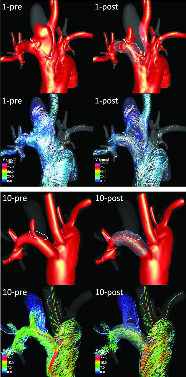
  • Fig 2.
    • Download figure
    • Open in new tab
    • Download powerpoint
    Fig 2.

    Visualization of peak systole flow structures pre- and post-treatment in an incompletely occluded aneurysm (case 1, top) and in a completely occluded aneurysm (case 10, bottom). Each panel shows 20 cm/s velocity isosurfaces (top) and flow streamlines (bottom) before (left) and after (right) treatment.
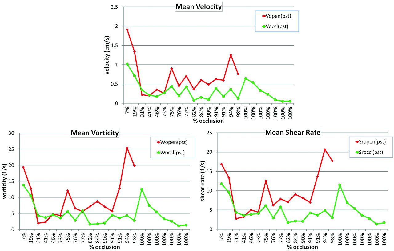
  • Fig 3.
    • Download figure
    • Open in new tab
    • Download powerpoint
    Fig 3.

    Posttreatment hemodynamic variables averaged over open and occluded regions.

Tables

  • Figures
    • View popup
    Table 1:

    Aneurysm occlusion, geometry, and device dataa

    CaseAneurysm Occlusion (%)PPA Diameter (mm)DPA Diameter (mm)Device Size (mm)Aneurysm Volume (cm3)Aneurysm Size (cm)Neck Size (Cm)Neck Area (Cm2)
    17%3.233.183.75 × 100.361.900.930.45
    219%3.282.664.5 × 120.171.130.860.48
    331%2.963.943.75 × 100.131.200.790.34
    441%3.723.543.75 × 120.061.170.650.27
    [5]46%3.473.113.50 × 120.121.310.600.22
    [6]73%3.193.403.25 × 100.080.740.530.14
    775%3.413.894.75 × 100.191.430.610.30
    [8]76%3.113.343.50 × 120.131.200.570.22
    977%2.863.794.75 × 100.031.490.640.31
    [10]82%3.263.023.50 × 100.080.940.470.14
    1184%3.974.184.00 × 100.201.330.610.25
    [12]90%3.443.113.50 × 100.121.200.420.12
    1391%3.073.583.50 × 120.321.420.700.34
    1491%3.954.244.00 × 100.081.010.530.09
    1594%3.103.034.75 × 120.251.420.650.28
    1698%3.573.574.25 × 100.060.910.340.07
    [17]100%3.253.484.75 × 100.151.170.600.25
    18100%3.823.253.50 × 100.010.390.240.04
    19100%3.702.504.25 × 100.030.620.300.07
    20100%4.243.964.75 × 100.030.590.410.12
    21100%3.793.434.75 × 120.121.090.560.19
    22100%3.652.894.00 × 100.020.530.310.05
    23100%3.533.793.50 × 120.070.740.630.17
    • Note:—PPA indicates proximal parent artery; DPA, distal parent artery.

    • ↵a Cases between brackets were sacrificed before 1 week and were not included in the analysis.

    • View popup
    Table 2:

    Statistics of aneurysm geometrical data

    ValueAneurysm Volume (cm3)Aneurysm Size (cm)Neck Size (cm)Neck Area (cm2)
    Average over patent group0.161.390.750.36
    Average over occluded group0.110.960.490.16
    Ratio (patent/occluded)1.411.441.542.25
    P value.2655.0200a.0015a.0006a
    AUC0.510.670.700.73
    • Note:—AUC indicates area under the curve.

    • ↵a Statistically significant differences (P < .05).

    • View popup
    Table 3:

    Ratio of mean posttreatment hemodynamic variables in open-over-occluded regionsa

    CaseRatio (v)Ratio (ω)Ratio <γ̇>
    11.881.401.43
    21.871.221.40
    30.640.450.63
    40.920.620.89
    [5]2.031.041.29
    [6]0.951.221.07
    72.052.152.05
    [8]2.382.312.11
    91.660.961.35
    [10]4.424.384.04
    113.905.064.16
    [12]4.963.563.83
    131.611.271.63
    143.383.583.72
    153.465.874.24
    166.067.155.86
    Mean2.642.642.48
    Mean open1.601.261.36
    Mean occluded3.974.413.93
    P value.0139b.0046b.0023b
    • ↵a Cases between brackets were sacrificed before 1 week and were not included in the analysis.

    • ↵b Statistically significant differences (P < .05).

    • View popup
    Table 4:

    Accuracy of logistic regression models of local occlusion

    RegionAccuracyCorrect/TotalRange (%)
    Sac84 %28,054/33,254(82.6, 84.5)
    Dome94 %9649/10,299(92.7, 94.0)
    Body92 %12,232/13,274(91.5, 92.4)
    Neck73 %11,060/15,050(71.8, 73.9)
    • View popup
    Table 5:

    Accuracy of different statistical models of local occlusion

    ModelAccuracyRMS Error
    Logistic regression86%0.32
    Neural network90%0.27
    Support vector machine85%0.38
    • Note:—RMS indicates root mean square.

PreviousNext
Back to top

In this issue

American Journal of Neuroradiology: 35 (8)
American Journal of Neuroradiology
Vol. 35, Issue 8
1 Aug 2014
  • Table of Contents
  • Index by author
  • Complete Issue (PDF)
Advertisement
Print
Download PDF
Email Article

Thank you for your interest in spreading the word on American Journal of Neuroradiology.

NOTE: We only request your email address so that the person you are recommending the page to knows that you wanted them to see it, and that it is not junk mail. We do not capture any email address.

Enter multiple addresses on separate lines or separate them with commas.
Analysis of Hemodynamics and Aneurysm Occlusion after Flow-Diverting Treatment in Rabbit Models
(Your Name) has sent you a message from American Journal of Neuroradiology
(Your Name) thought you would like to see the American Journal of Neuroradiology web site.
CAPTCHA
This question is for testing whether or not you are a human visitor and to prevent automated spam submissions.
Cite this article
J.R. Cebral, F. Mut, M. Raschi, S. Hodis, Y.-H. Ding, B.J. Erickson, R. Kadirvel, D.F. Kallmes
Analysis of Hemodynamics and Aneurysm Occlusion after Flow-Diverting Treatment in Rabbit Models
American Journal of Neuroradiology Aug 2014, 35 (8) 1567-1573; DOI: 10.3174/ajnr.A3913

Citation Manager Formats

  • BibTeX
  • Bookends
  • EasyBib
  • EndNote (tagged)
  • EndNote 8 (xml)
  • Medlars
  • Mendeley
  • Papers
  • RefWorks Tagged
  • Ref Manager
  • RIS
  • Zotero
0 Responses
Respond to this article
Share
Bookmark this article
Analysis of Hemodynamics and Aneurysm Occlusion after Flow-Diverting Treatment in Rabbit Models
J.R. Cebral, F. Mut, M. Raschi, S. Hodis, Y.-H. Ding, B.J. Erickson, R. Kadirvel, D.F. Kallmes
American Journal of Neuroradiology Aug 2014, 35 (8) 1567-1573; DOI: 10.3174/ajnr.A3913
del.icio.us logo Twitter logo Facebook logo Mendeley logo
  • Tweet Widget
  • Facebook Like
  • Google Plus One
Purchase

Jump to section

  • Article
    • Abstract
    • ABBREVIATIONS:
    • Materials and Methods
    • Results
    • Discussion
    • Conclusions
    • Acknowledgments
    • Footnotes
    • REFERENCES
  • Figures & Data
  • Supplemental
  • Info & Metrics
  • Responses
  • References
  • PDF

Related Articles

  • PubMed
  • Google Scholar

Cited By...

  • Correlation of Flow Diverter Malapposition at the Aneurysm Neck with Incomplete Aneurysm Occlusion in Patients with Small Intracranial Aneurysms: A Single-Center Experience
  • Comparison of the Pipeline embolisation device alone or combined with coiling for treatment of different sizes of intracranial aneurysms
  • Evaluation of Outcome Prediction of Flow Diversion for Intracranial Aneurysms
  • Predictive score for complete occlusion of intracranial aneurysms treated by flow-diverter stents using machine learning
  • Implementation of computer simulation to assess flow diversion treatment outcomes: systematic review and meta-analysis
  • Role of distal cerebral vasculature in vessel constriction after aneurysm treatment with flow diverter stents
  • Analysis of Flow Dynamics and Outcomes of Cerebral Aneurysms Treated with Intrasaccular Flow-Diverting Devices
  • Endothelialization following Flow Diversion for Intracranial Aneurysms: A Systematic Review
  • Multicenter Experience with FRED Jr Flow Re-Direction Endoluminal Device for Intracranial Aneurysms in Small Arteries
  • Relationship between aneurysm occlusion and flow diverting device oversizing in a rabbit model
  • Hemodynamic analysis of fast and slow aneurysm occlusions by flow diversion in rabbits
  • RNA-Sequencing Analysis of Messenger RNA/MicroRNA in a Rabbit Aneurysm Model Identifies Pathways and Genes of Interest
  • Crossref
  • Google Scholar

This article has not yet been cited by articles in journals that are participating in Crossref Cited-by Linking.

More in this TOC Section

  • Cavernous dural AVF treated by transfacial route
  • Prophylactic Endovascular Treatments for CBS
  • Impact of Stenosis Degree on Outcomes of Stenting
Show more Neurointervention

Similar Articles

Advertisement

Indexed Content

  • Current Issue
  • Accepted Manuscripts
  • Article Preview
  • Past Issues
  • Editorials
  • Editor's Choice
  • Fellows' Journal Club
  • Letters to the Editor
  • Video Articles

Cases

  • Case Collection
  • Archive - Case of the Week
  • Archive - Case of the Month
  • Archive - Classic Case

More from AJNR

  • Trainee Corner
  • Imaging Protocols
  • MRI Safety Corner
  • Book Reviews

Multimedia

  • AJNR Podcasts
  • AJNR Scantastics

Resources

  • Turnaround Time
  • Submit a Manuscript
  • Submit a Video Article
  • Submit an eLetter to the Editor/Response
  • Manuscript Submission Guidelines
  • Statistical Tips
  • Fast Publishing of Accepted Manuscripts
  • Graphical Abstract Preparation
  • Imaging Protocol Submission
  • Evidence-Based Medicine Level Guide
  • Publishing Checklists
  • Author Policies
  • Become a Reviewer/Academy of Reviewers
  • News and Updates

About Us

  • About AJNR
  • Editorial Board
  • Editorial Board Alumni
  • Alerts
  • Permissions
  • Not an AJNR Subscriber? Join Now
  • Advertise with Us
  • Librarian Resources
  • Feedback
  • Terms and Conditions
  • AJNR Editorial Board Alumni

American Society of Neuroradiology

  • Not an ASNR Member? Join Now

© 2025 by the American Society of Neuroradiology All rights, including for text and data mining, AI training, and similar technologies, are reserved.
Print ISSN: 0195-6108 Online ISSN: 1936-959X

Powered by HighWire