
Article Figures & Data
Figures
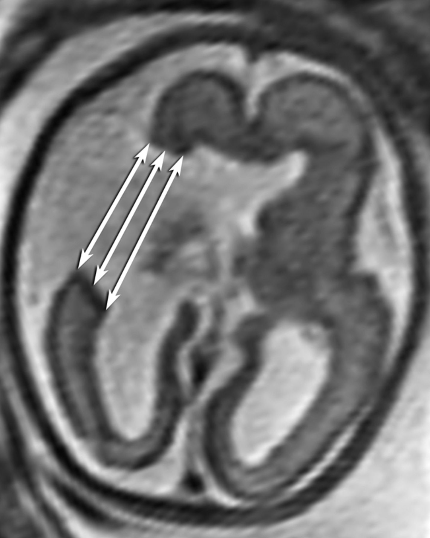
- Fig 1.
Measurement of the outer, middle, and inner widths of the schizencephalic cleft.
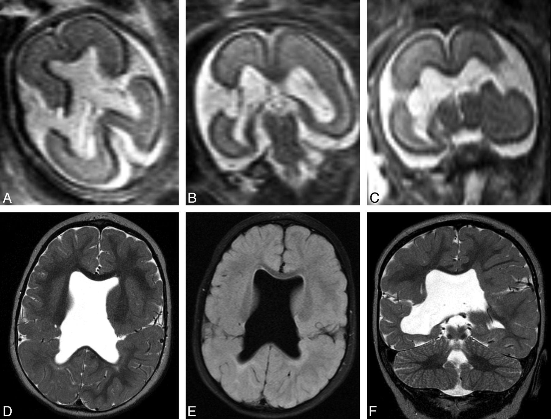
- Fig 2.
Prenatal imaging at 22 weeks' gestational age demonstrates bilateral wide-open clefts on axial (A) and coronal HASTE (B and C) imaging. Postnatal imaging at 15 months of age demonstrates interval closure of both defects, with apposed lips that contain intervening vessels. Note the complete absence of the septum pellucidum. Axial T2 (D), axial FLAIR (D and E), and coronal T2 (F) images are shown.
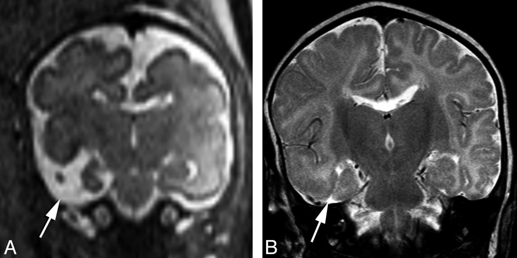
- Fig 3.
Prenatal coronal HASTE imaging at 26 weeks' gestational age demonstrating a right temporal open cleft communicating with the temporal horn (arrow), with a faint membrane covering (A). Postnatal coronal T2 imaging at 2 months of age demonstrates interval closure of defect lips, which are now apposed to each other and closed (B).
- Fig 4.
Prenatal imaging at 29 weeks' gestational age. Axial HASTE image (A) reveals an open cleft with a membrane along the roof of the cleft (arrow), which remained open in postnatal imaging 1 month after birth (B).


Tables
Subject Clefts Prenatal: Visualization of Cortex Lining the 2 Margins of the Cleft Postnatal: Visualization of Cortex Lining the 2 Margins of the Cleft Prenatal: Membrane along the Cleft Postnatal: Membrane along the Cleft Prenatal: Evidence of Hemorrhage Postnatal: Evidence of Hemorrhage 1 R Partial-partial Complete-complete Present Absent Bilateral ventricular hemosiderin staining Absent L Complete-partial Complete-complete Absent Absent Right choroid plexus hemorrhage 2 L Complete-partial Complete-complete Absent Absent Absent Absent L Complete-complete Complete-complete Absent Absent 3 R Complete-partial Complete-complete Present Absent Absent Absent L Complete-complete Complete-complete Present Absent 4 R Complete-partial Complete-complete Absent Absent Absent Absent L Complete-partial Complete-complete Absent Absent 5 R Complete-complete Complete-complete Present Absent Left lateral ventricle and choroid plexus gross hemorrhage Bilateral ventricular staining, left choroid plexus staining L Complete-partial Complete-complete Present Absent 6 L Complete-complete Complete-complete Absent Absent Hemosiderin staining in the cleft Bilateral ventricular and cleft staining 7 R Complete-partial Complete-complete Absent Absent Absent Left choroid plexus hemorrhage L Complete-partial Complete-complete Absent Absent 8 L Complete-partial Complete-complete Present Partial Bilateral ventricular, choroid plexus, and cleft hemosiderin staining Bilateral ventricular, choroid plexus, and cleft hemosiderin staining 9 R Complete-partial Complete-complete Present Absent Bilateral ventricular hemosiderin staining Bilateral ventricular hemosiderin staining L Complete-partial Complete-complete Present Absent 10 R Complete-complete Complete-complete Present Absent Bilateral ventricular hemosiderin staining Bilateral ventricular hemosiderin staining R Complete-complete Complete-complete Present Present Note:—R indicates right; L, left.
- Table 2:
Comparison of the dimensions of the clefts and ipsilateral ventricle between different types of open cleftsa
Measurement Normalized Prenatal Diameters (Open Clefts That Closed) Normalized Prenatal Diameters (Open Clefts That Remained Open) P Value Ipsilateral ventricle Mean = 0.16 ± 0.045 Mean = 0.14 ± 0.039 .24 Cleft width (outer) Mean = 0.17 ± 0.14 Mean = 0.27 ± 0.13 .11 Cleft width (inner) Mean = 0.14 ± 0.88 Mean = 0.20 ± 0.12 .14 Cleft width (mid) Mean = 0.12 ± 0.08 Mean = 0.23 ± 0.13 .06 ↵a Measurements are all normalized to the mean of the maximum anteroposterior and transverse inner diameters of the skull.


