
Article Figures & Data
Figures
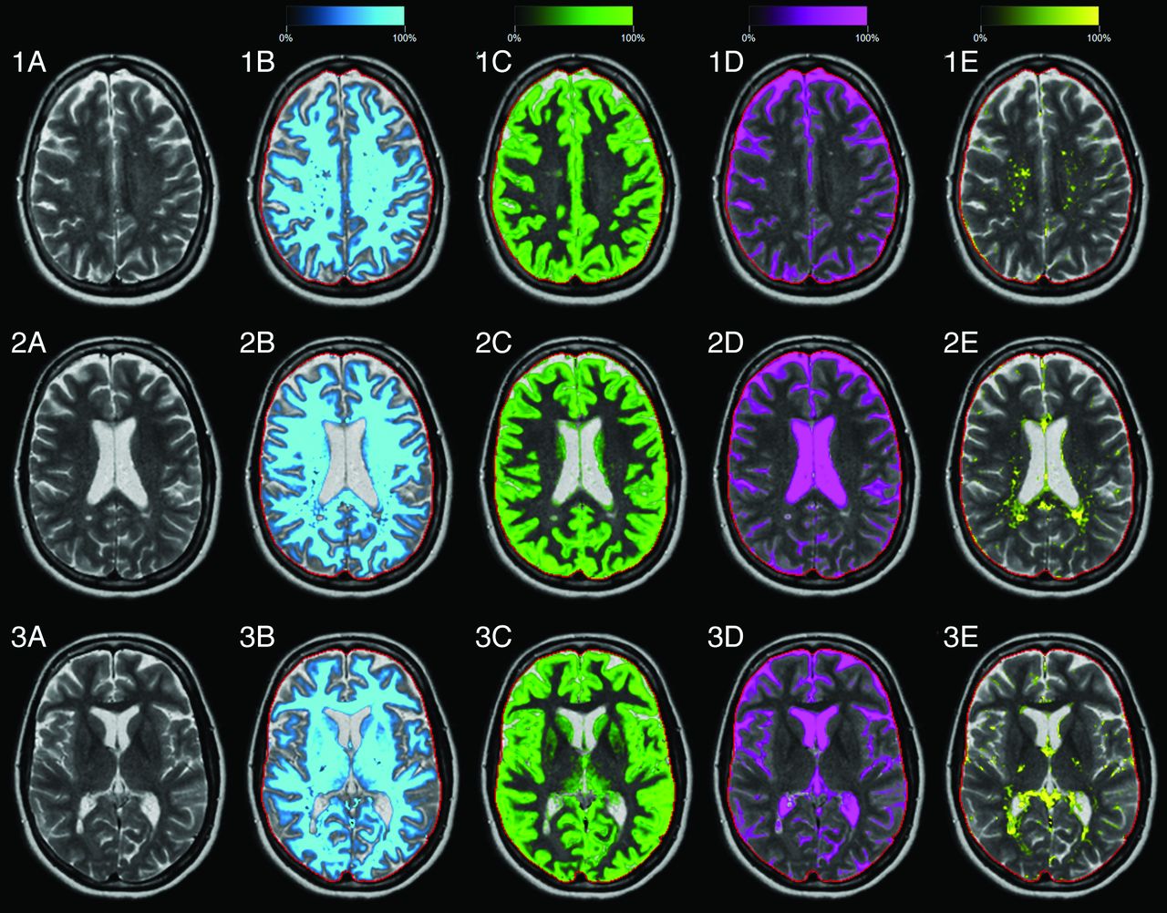
- Fig 1.
Typical images of the automatic segmentation software of a patient with MS (39-year-old woman, EDSS = 4.0). Three slices are shown, numbers 19, 16, and 13 of the 30 acquired slices. A, T2-weighted image. B, White matter segmentation, in which the intensity of the light-blue color overlay corresponds to the calculated white matter partial volume per voxel. The red line indicates the intracranial volume. Similar images are shown for gray matter in green (C), CSF in pink (D), and non-WM/GM/CSF in yellow (E).
- Fig 2.
Brain-tissue fraction results of the first measurement: the brain parenchymal fraction, white matter fraction, gray matter fraction, CSF fraction, and the non-WM/GM/CSF fraction of the intracranial volume, as a function of subject age. Markers are zero for the control group and plus for the MS group. The colors are similar to the segmentation overlay colors of Fig 1.
- Fig 3.
Bland-Altman plots for WM fraction, GM fraction, CSF fraction, remaining, unclassified non-WM/GM/CSF tissue fraction, and brain parenchymal fraction of all subjects with the mean tissue fraction against the difference in tissue fraction between measurements 1 and 2. The dotted lines indicate the mean difference ± 2 SDs, on the left for the controls and on the right for the MS group. The scaling on both axes is identical; the colors are identical to those in Fig 2.


Tables
- Table 1:
Fully automatic measurements of the WMV, GMV, CSFV, NV, BPV, and ICV volumes of the control group and the MS groupa
WMV (mL) GMV (mL) CSFV (mL) NV (mL) BPV (mL) ICV (mL) Contr1 581 ± 67 639 ± 58 156 ± 45 28 ± 7 1247 ± 119 1404 ± 119 Contr2 581 ± 69 637 ± 53 156 ± 47 31 ± 12 1250 ± 119 1406 ± 120 MS1 452 ± 88 617 ± 48 247 ± 60 52 ± 22 1122 ± 99 1369 ± 99 MS2 464 ± 109 600 ± 47 231 ± 60 60 ± 21 1124 ± 98 1355 ± 97 Diff Contr1-Contr2 0 ± 11 −1 ± 13 0 ± 4 4 ± 12 2 ± 6 2 ± 7 Diff MS1-MS2 12 ± 37 −17 ± 40 −16 ± 7c 8 ± 11b 2 ± 9 −13 ± 8c Diff Contr1-MS1 −129 ± 104c −22 ± 68 91 ± 71c 24 ± 18c −126 ± 133c −35 ± 117 Diff Contr2-MS2 −117 ± 113c −37 ± 67b 75 ± 70c 29 ± 17c −126 ± 136c −50 ± 116 Note:—Diff indicates difference; Contr, control group; MS, MS patient group; 1, measurement 1; 2, measurement 2.
↵a For each tissue volume, the mean value and SD are given. The mean difference and SD of the first measurement and the second measurement and between controls and patients with MS are also provided. The MS group received Gd in the second measurement.
↵b P < .05 (significant difference).
↵c P < .005 (significant difference).
- Table 2:
Fully automatic measurements of the normalized WMF, GMF, CSFF, NF, and BPF of the control group and the MS groupa
WMF (%) GMF (%) CSFF (%) NF (%) BPF (%) Contr1 41.4 ± 2.6 45.5 ± 1.9 11.2 ± 3.1 2.0 ± 0.5 88.8 ± 3.1 Contr2 41.3 ± 2.4 45.4 ± 1.8 11.1 ± 3.1 2.2 ± 0.9 88.9 ± 3.1 MS1 32.9 ± 5.4 45.2 ± 3.0 18.0 ± 4.1 3.8 ± 1.7 82.0 ± 4.1 MS2 34.0 ± 6.8 44.5 ± 4.6 17.1 ± 4.2 4.4 ± 1.6 82.9 ± 4.2 Diff Contr1-Contr2 −0.1 ± 0.9 −0.1 ± 0.8 0.0 ± 0.3 0.2 ± 0.8 0.0 ± 0.3 Diff MS1-MS2 1.1 ± 2.9 −0.7 ± 3.2 −1.0 ± 0.5c 0.6 ± 0.8b 1.0 ± 0.5c Diff Contr1-MS1 −8.4 ± 6.1c −0.4 ± 3.4 6.9 ± 5.1c 1.9 ± 1.5c −6.9 ± 5.1c Diff Contr2-MS2 −7.3 ± 6.9c −0.9 ± 4.3 6.0 ± 5.1c 2.2 ± 1.4c −6.0 ± 5.1c Note:—Diff indicates difference; Contr, control group; MS, MS patient group; 1, measurement 1; 2, measurement 2; WMF, WM fraction; GMF, GM fraction; CSFF, CSF fraction; NF, remaining, unclassified non-WM/GM/CSF tissue fraction; BPF, brain parenchymal fraction.
↵a Each tissue fraction corresponds to the tissue volume divided by the ICV. The mean value and SD are given as well as the mean difference and SD of the first measurement and the second measurement and between the controls and patients with MS.
↵b P < .05 (significant difference).
↵c P < .005 (significant difference).
- Table 3:
Linear regression of the normalized WMF, GMF, CSFF, NF, and BPF of the control and the MS groups as a function of agea
WMF GMF CSFF NF BPF Contr1-age (%/yr) −0.12 (−0.21 to −0.03)b −0.03 (−0.11–0.05) 0.17 (0.07–0.27)c −0.01 (−0.03–0.01) −0.17 (−0.27 to −0.07)c Contr2-age (%/yr) −0.10 (−0.19 to −0.01)b −0.05 (−0.12–0.03) 0.17 (0.07–0.27)c −0.02 (−0.05–0.02) −0.17 (−0.2 to −0.07)c MS1-age (%/yr) −0.02 (−0.26–0.22) −0.03 (−0.16–0.10) 0.05 (−0.13–0.23) 0.00 (−0.08–0.07) −0.05 (−0.23, −0.13) MS2-age (%/yr) −0.01 (−0.31–0.30) −0.05 (−0.25–0.15) 0.04 (−0.14–0.23) 0.02 (−0.06–0.09) −0.04 (−0.23–0.14) MS1-EDSS (%/unit) −1.16 (−2.28 to −0.05)b −0.17 (−0.87–0.54) 0.93 (−0.19–2.04) 0.23 (−0.14–0.62) −1.08 (−1.90 to −0.26)b MS2-EDSS (%/unit) −1.34 (−2.81–0.13) 0.09 (−1.01–1.19) 0.93 (−0.19–2.06) 0.19 (−0.20–0.58) −1.06 (−1.92 to −0.20)b Note:—Contr indicates control group; MS, MS patient group; 1, measurement 1; 2, measurement 2; WMF, WM fraction; GMF, GM fraction; CSFF, CSF fraction; NF, remaining, unclassified non-WM/GM/CSF tissue fraction; BPF, brain parenchymal fraction.
↵a The 95% confidence interval is given between parentheses. For the MS group, the linear regression of the age-corrected fractions with EDSS is provided.
↵b P < .05 (significant difference).
↵c P < .005 (significant difference).


