
Article Figures & Data
Figures
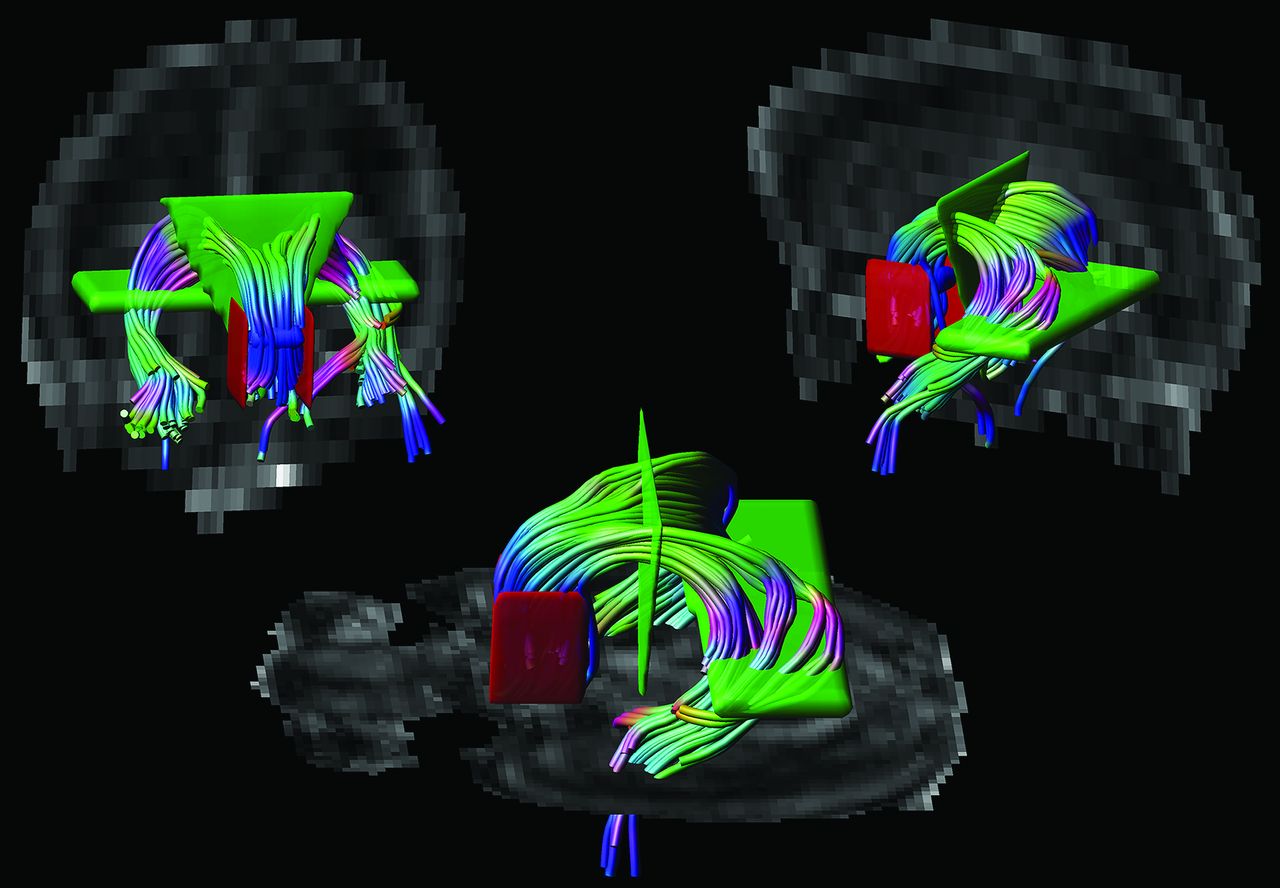
- Fig 1.
Placement of ROIs. Tractography of the fornix was performed by placing 1 “OR” ROI (in blue), 2 “AND” ROIs (in green), and 2 “NOT” ROIs (in red) on color-coded fractional anisotropy maps.
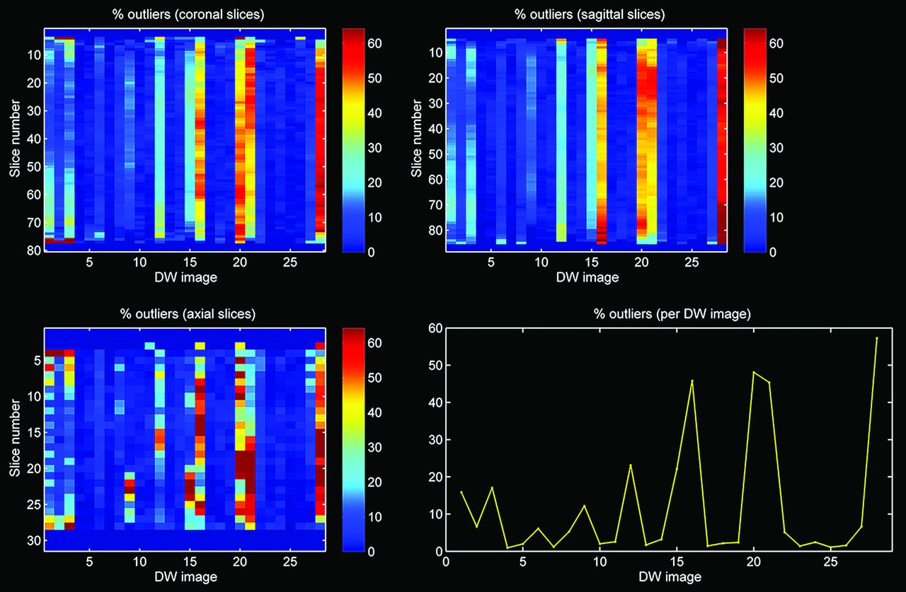
- Fig 2.
Outlier profile of DTI data with high percentage of outliers.
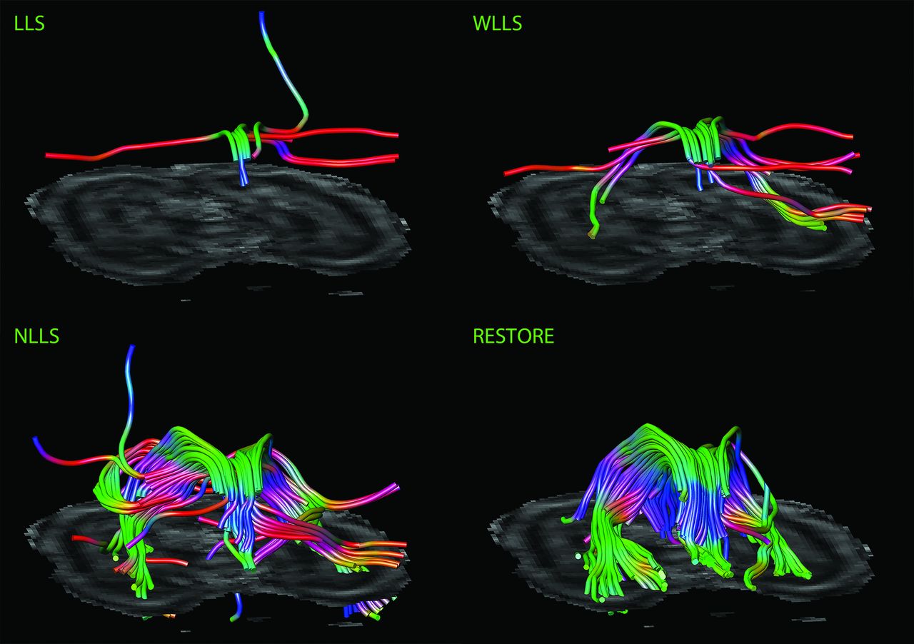
- Fig 3.
Impact of diffusion tensor estimation method on tract reconstruction of poor-quality DTI data. Characteristic representations illustrate the effect of tensor estimation methodology on reconstruction of the fornix with high percentage of data outliers (>10%). Note that reconstruction is not possible with the use of the linear least squares (LLS) and weighted linear least-squares (WLLS) methods and appears to be slightly possible with nonlinear least squares (NLLS) but is very well performed if the robust estimation of tensors by outlier rejection (RESTORE) approach is used.
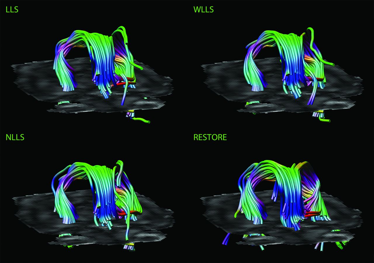
- Fig 4.
Impact of diffusion tensor estimation method on tract reconstruction of good-quality DTI data. Characteristic representations illustrate the effect of the tensor estimation on fiber tracking of the fornix with low percentage of data outliers (<10%). Note the more accurate tract reconstruction with the use of the robust estimation of tensors by outlier rejection approach.
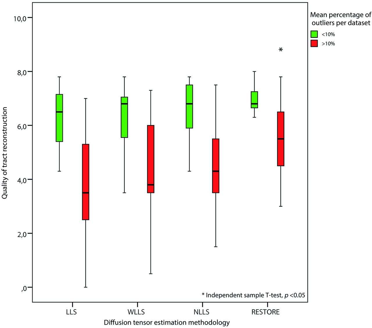
- Fig 5.
Impact of DTI data quality on tract reconstruction of the fornix. Quality of the reconstructed fornix was significantly higher by use of the robust estimation of tensors by outlier rejection technique; this was particularly evident for datasets with high percentages of outliers in the diffusion-weighted images (>10%).
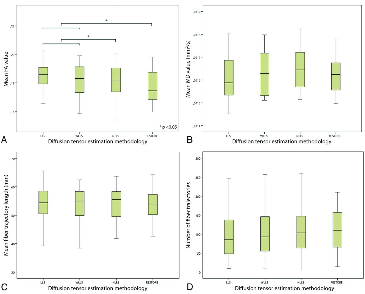
- Fig 6.
Impact of diffusion tensor estimation method on tract parameters. Tract parameters, such as fractional anisotropy (FA) (A), mean diffusivity (B), mean fiber trajectory length (C), and number of fiber trajectories (D) were affected by the tensor estimation method; mean FA value was significantly lower with use of the nonlinear least squares and robust estimation of tensors by outlier rejection techniques (paired sample t test, P < .05).
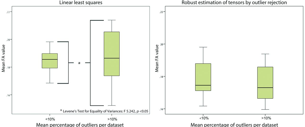
- Fig 7.
Impact of data quality on variability of tract parameters. Diffusion-weighted images with high outlier percentages (>10%) resulted in a significantly increased variability of mean fractional anisotropy values compared with data with fewer data outliers (<10%) if linear least squares was used (Levene test for equality of variances, P < .05). With application of robust estimation of tensors by outlier rejection, there was no difference in variability with regard to data quality.


Tables
Scoring system for visual evaluation of tract reconstruction of the fornix
0 Points 1 Point 2 Points Shape of the fornixa No recognition of shape Partially abnormal shape Normal shape Orientation of fibersa Complete disorientation Partially abnormal orientation Normal orientation Symmetry of crura One missing crus Partially asymmetric Normal symmetry Presence of non-realistic fibersa Outweighing the total number of fibers Less than the number of realistic fibers None No. of fiber trajectories <10 10–100 >100 ↵a Shape, orientation, and presence of non-realistic fibers with regard to description of anatomy by Nieuwenhuys et al, The Human Central Nervous System, 2008.20


Jump to section
Related Articles
Cited By...
- Limbic System White Matter in Children and Adolescents with ADHD: A Longitudinal Diffusion MRI Analysis
- Brain-wide structural and functional disruption in mice with oligodendrocyte-specific Nf1 deletion is rescued by inhibition of nitric oxide synthase
- Brain-wide structural and functional disruption in mice with oligodendrocyte specific Nf1 deletion is rescued by inhibition of NOS