
Article Figures & Data
Figures
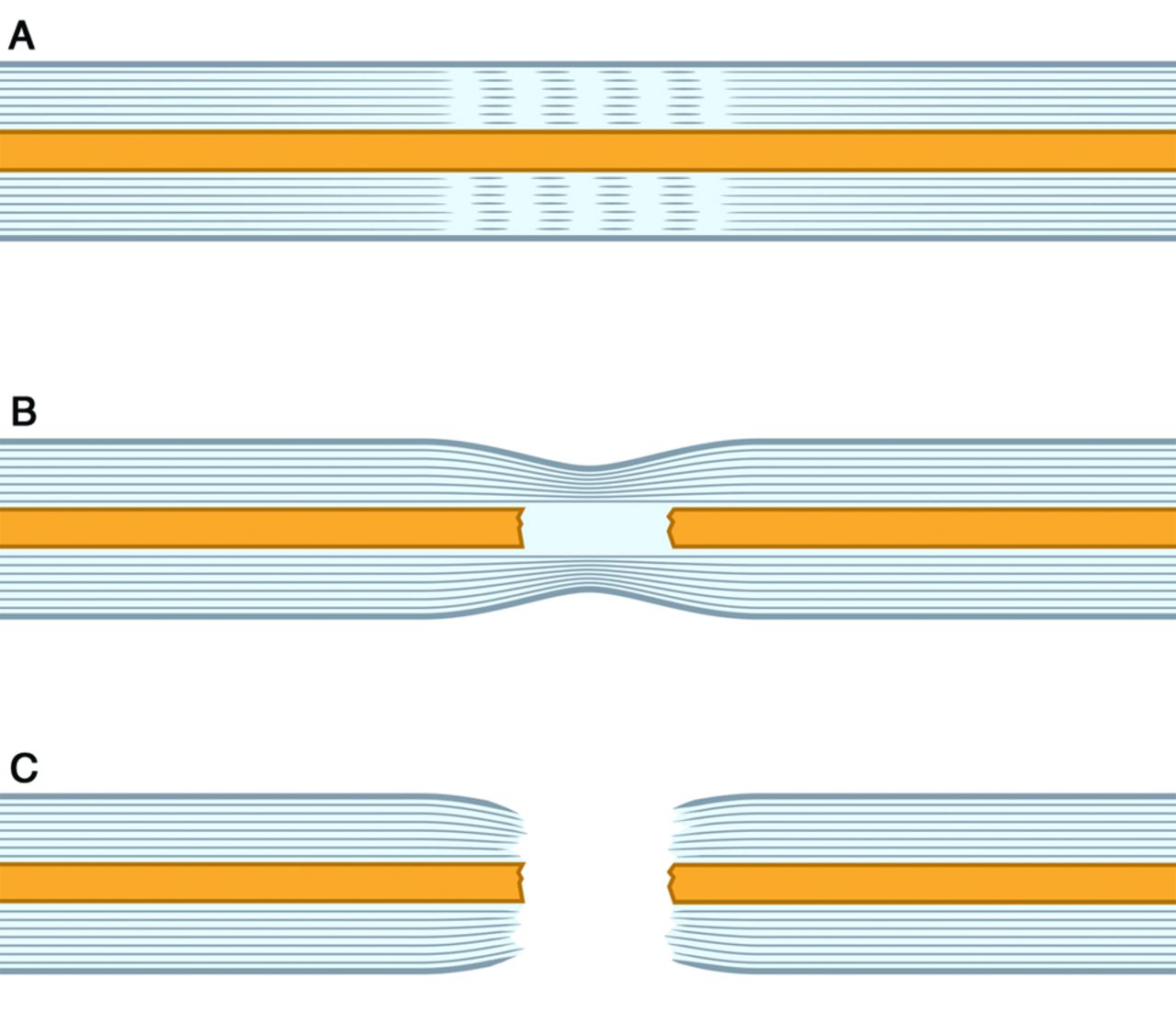
- Fig 1.
Schematic diagram showing different patterns of injuries affecting the nerve, including neuropraxia (A), mild axonotmesis (B), and neurotmesis (C).
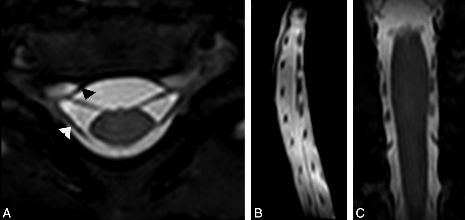
- Fig 2.
A, Axial high-resolution MR imaging in a 6-month-old girl with clinically suspected right-sided brachial plexus palsy showing intact right ventral (arrowhead) and dorsal nerve roots on both sides. Compare with normal left-sided nerve roots. Sagittal (B) and (C) coronal reformatted images from the axial data again show normal ventral and dorsal nerve roots at each vertebral level.
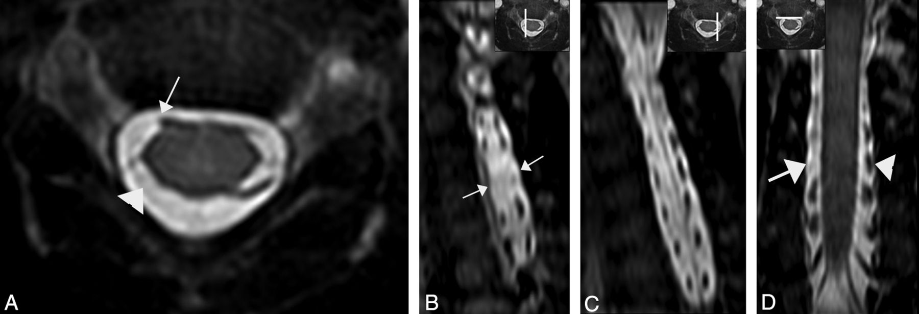
- Fig 3.
A, Axial high-resolution MR imaging in a 5-month-old girl with clinically suspected right-sided brachial plexus palsy shows avulsion injury of the right C5 ventral nerve root (arrow). The right dorsal nerve root is also avulsed (arrowhead). Sagittal (B) reformatted images on the right side show absent ventral and dorsal C5 nerve roots (arrows), compared with the normal nerve roots on left side (C). Coronal reformatted image (D) at the level of ventral nerve roots show absent right C5 ventral nerve root from avulsion (arrow), compared with the normal left-sided nerve root (arrowhead).
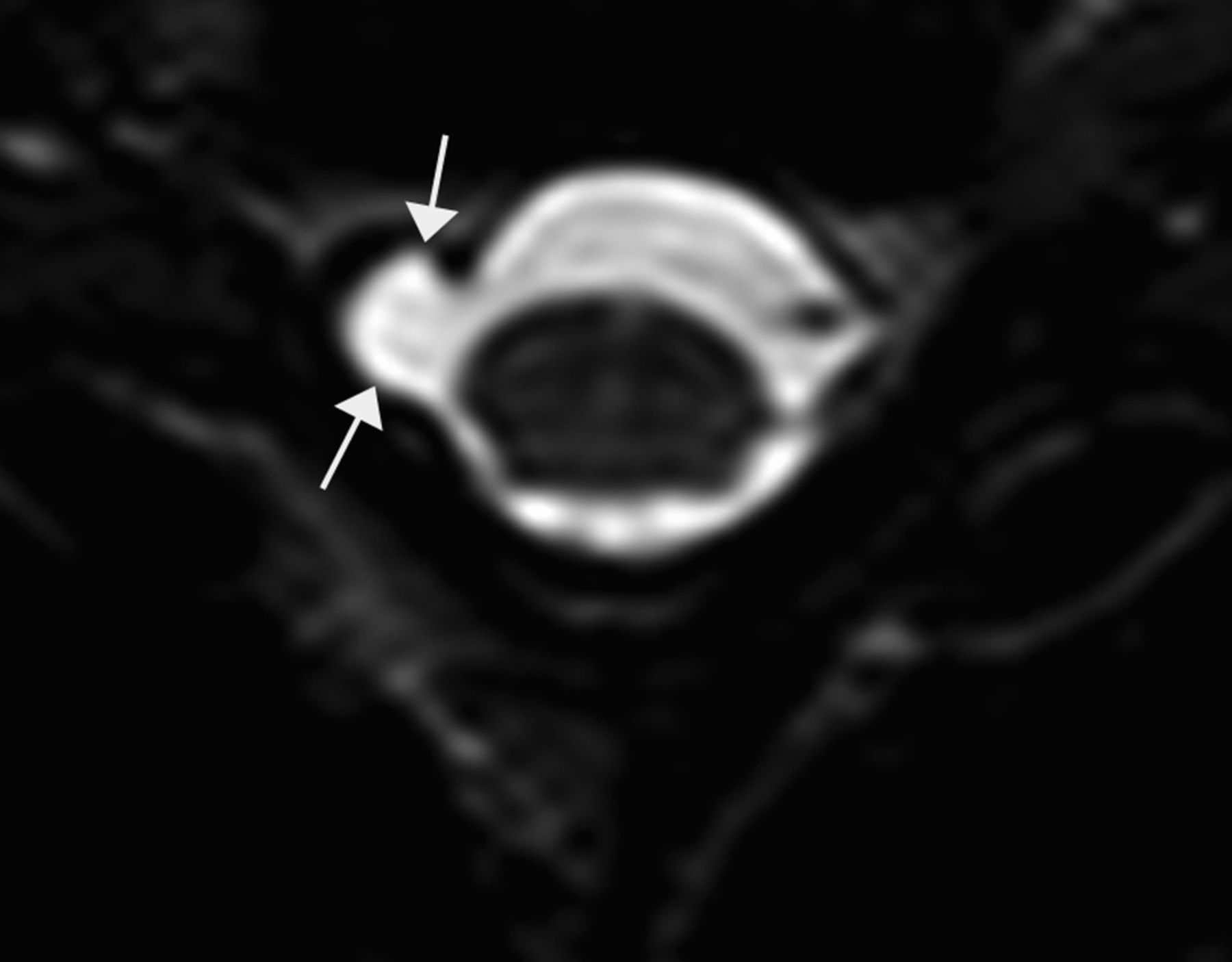
- Fig 4.
Axial high-resolution MR imaging in a 4-month-old boy with clinically suspected right-sided brachial plexus palsy shows a pseudomeningocele at right C5–6 level (arrow). Note absent nerve roots on right side suggestive of nerve root avulsion injury. Compare with normal ventral and dorsal nerve roots on the left side.


Tables
Comparison of MRI and surgical findings at each cervical level
Cervical Level Patient Affected Side C5 C6 C7 C8 T1 MRI Surgery MRI Surgery MRI Surgery MRI Surgery MRI Surgery 1 Right N N N N N N N N/A N N/A 2 Left N N N A N N N N/A N N/A 3 Right A A A A A N A N/A N N/A 4 Left N N N A N N N N/A N N/A 5 Right N N A A A A A A N N 6 Right N N A A A N N N/A N N/A Note:—N indicates normal; A, avulsion.


