
Article Figures & Data
Figures
- Fig 1.
Graphic representation of mean image noise for the pons, globe, masseter, and air within the maxillary sinus for each of the tested CT series: soft-tissue algorithm, filtered back-projection, low dose; bone algorithm, filtered back-projection, low dose; Veo model-based iterative reconstruction, low dose; soft-tissue algorithm, filtered back-projection, standard dose; and bone algorithm, filtered back-projection, standard dose.
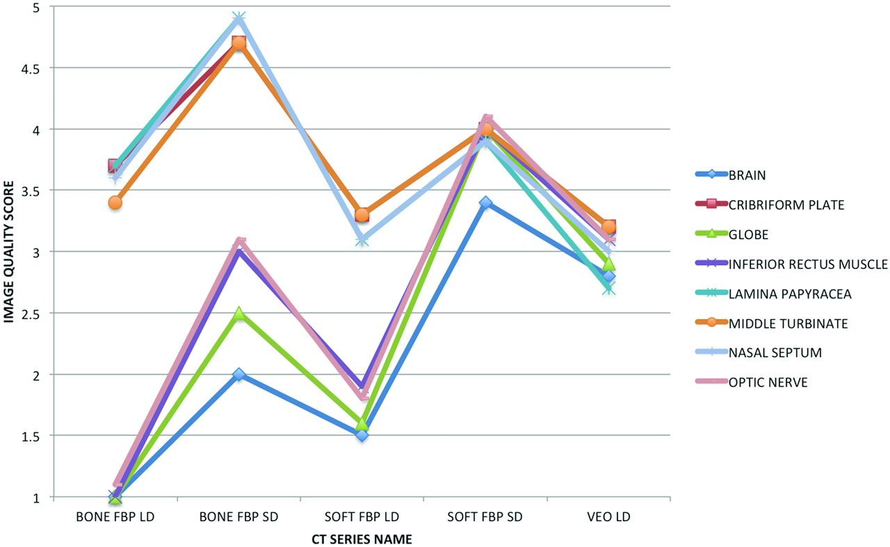
- Fig 2.
Graphic representation of mean image quality for the brain, cribriform plate, globe, inferior rectus muscle, lamina papyracea, middle turbinate, nasal septum, and optic nerve for each of the tested CT series: soft-tissue algorithm, filtered back-projection, low dose; bone algorithm, filtered back-projection, low dose; Veo model-based iterative reconstruction, low dose; soft-tissue algorithm, filtered back-projection, standard dose; and bone algorithm, filtered back-projection, standard dose.
- Fig 3.
Axial noncontrast sinus CT (window level, 40 HU; window width, 400 HU) through the level of the orbits for the following imaging series: soft-tissue algorithm, filtered back-projection, standard dose (A); bone algorithm, filtered back-projection, standard dose (B); soft-tissue algorithm, filtered back-projection, low dose (C); bone algorithm, filtered back-projection, low dose (D); and Veo model-based iterative reconstruction, low dose (E). The decreased image noise secondary to Veo allows improved visualization of orbital and intracranial soft-tissue structures relative to the other low-dose protocols but not to the level of the standard-dose sinus CT.
- Fig 4.
Coronal noncontrast sinus CT (window level, 800 HU; window width, 3500 HU) through the level of the maxillary sinuses for the following imaging series: soft-tissue algorithm, filtered back-projection, standard dose (A); bone algorithm, filtered back-projection, standard dose (B); soft-tissue algorithm, filtered back-projection, low dose (C); bone algorithm, filtered back-projection, low dose (D); and Veo model-based iterative reconstruction, low dose (E). The noise reduction achieved with Veo results in loss of edge enhancement, making thin bones such as the nasal septum (white arrow) and left lamina papyracea (white arrowhead) difficult to see.


Tables
Pons (n = 20) Globe (n = 20) Masseter (n = 20) Maxillary Sinus (n = 20) Total (n = 80) SOFT FBP LD 74.9 (10.60)b 55.6 (8.79)b 52.6 (8.79)b 45.3 (13.16)b 57.1 (15.06)b BONE FBP LD 278.8 (36.73)b 173.0 (19.66)b 175.3 (27.03)b 105.5 (15.64)b 183.1 (67.38)b VEO LD 18.9 (2.98) 12.4 (1.37) 14.3 (2.16) 12.3 (2.11) 14.5 (3.49) SOFT FBP SD 24.7 (5.14)b 19.5 (3.06)b 19.1 (3.35)b 17.5 (2.48)b 20.2 (4.48)b BONE FBP SD 97.4 (14.47)b 66.9 (7.78)b 66.3 (8.96)b 50.1 (6.42)b 70.2 (19.73)b Brain (n = 40) Globe (n = 40) Inferior Rectus Muscle (n = 40) Optic Nerve (n = 40) Cribriform Plate (n = 40) Lamina Papyracea (n = 40) Middle Turbinate (n = 40) Nasal Septum (n = 40) Total (n = 320) SOFT FBP LD 1.5 (0.51)b 1.6 (0.55)b 1.9 (0.43)b 1.8 (0.52)b 3.3 (0.47) 3.1 (0.38)b 3.3 (0.44) 3.1 (0.38) 2.4 (0.90)b BONE FBP LD 1.0 (0.00)b 1.0 (0.16)b 1.0 (0.16)b 1.1 (0.27)b 3.7 (0.62)b 3.7 (0.56)b 3.4 (0.58) 3.6 (0.59)b 2.3 (1.35)b VEO LD 2.8 (0.41) 2.9 (0.35) 3.1 (0.32) 3.1 (0.32) 3.2 (0.48) 2.7 (0.53) 3.2 (0.53) 3.0 (0.45) 3.0 (0.46) SOFT FBP SD 3.4 (0.55)b 4.0 (0.42)b 4.0 (0.58)b 4.1 (0.40)b 4.0 (0.51)b 3.9 (0.47)b 4.0 (0.55)b 3.9 (0.56)b 3.9 (0.54)b BONE FBP SD 2.0 (0.28)b 2.5 (0.51)b 3.0 (0.53) 3.1 (0.45) 4.7 (0.47)b 4.9 (0.33)b 4.7 (0.46)b 4.9 (0.36)b 3.7 (1.19)b


